Review
. 2015 Oct 5;370(1679):20150029.
doi: 10.1098/rstb.2015.0029.

Lipopolysaccharide transport to the cell surface: biosynthesis and extraction from the inner membrane

Affiliations
Review

Lipopolysaccharide transport to the cell surface: biosynthesis and extraction from the inner membrane

Brent W Simpson et al. Philos Trans R Soc Lond B Biol Sci. .

Abstract

The cell surface of most Gram-negative bacteria is covered with lipopolysaccharide (LPS). The network of charges and sugars provided by the dense packing of LPS molecules in the outer leaflet of the outer membrane interferes with the entry of hydrophobic compounds into the cell, including many antibiotics. In addition, LPS can be recognized by the immune system and plays a crucial role in many interactions between bacteria and their animal hosts. LPS is synthesized in the inner membrane of Gram-negative bacteria, so it must be transported across their cell envelope to assemble at the cell surface. Over the past two decades, much of the research on LPS biogenesis has focused on the discovery and understanding of Lpt, a multi-protein complex that spans the cell envelope and functions to transport LPS from the inner membrane to the outer membrane. This paper focuses on the early steps of the transport of LPS by the Lpt machinery: the extraction of LPS from the inner membrane. The accompanying paper (May JM, Sherman DJ, Simpson BW, Ruiz N, Kahne D. 2015 Phil. Trans. R. Soc. B 370, 20150027. (doi:10.1098/rstb.2015.0027)) describes the subsequent steps as LPS travels through the periplasm and the outer membrane to its final destination at the cell surface.

Keywords: glycolipid; lpx; membrane biogenesis; permeability barrier.


Figures

Figure 1.
Figure 1.
Transport of LPS across the cell envelope. After synthesis, lipid A-core oligosaccharide (LPS) molecules are flipped across the IM in an ATP-dependent manner by MsbA. At the outer leaflet, the O antigen is ligated to the outer core to form full-length LPS (not shown). LPS is transported across the periplasm and OM, and assembled at the cell surface by LptB2FGCADE. LptB2FG form an ABC transporter that uses ATP hydrolysis to extract LPS from the IM and push it along a periplasmic bridge built of homologous domains in LptCAD. It is unclear whether LptF, LptG or both, interact with LptC. At the OM, the LptDE form a plug-and-barrel translocon that inserts LPS into the outer leaflet. Structural composition of E. coli LPS is shown on the left. Kdo, 3-deoxy-D-manno-oct-2-ulosonic acid; Hep, l-glycero-d-manno-heptose; Etn, ethanolamine; P, phosphate; Glu, d-glucose; Gal, d-galactose.
Figure 2.
Figure 2.
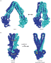
MsbA undergoes structural conformational changes proposed to mediate LPS flipping. (a) Crystal structures of MsbA (monomers in blue and cyan) suggest that the NBDs readily separate and dimerize in its inward-facing state (PDB accession no. 3B5W, 3B5X) [31]. (b) MsbA bound to a non-hydrolysable ATP analogue (AMPPNP in yellow spheres) shows an outward-facing state and associated twisting of the transmembrane helices (PDB accession no. 3B5X, 3B5Y) [31].
Figure 3.
Figure 3.
Structures of LptB reveal site of interaction with TMD partners and conformational changes induced by ATP hydrolysis. (a) Crystal structure of the LptB dimer (monomers in dark and light grey, nucleotides in yellow spheres) shows the groove (highlighted in brown) that is involved in interactions with LptFG at the membrane interface (symmetry mate derived from PDB accession no. 4P33) [47]. (b) Superimposed structures of an ATP-bound (green) and an ADP-bound (blue) LptB monomer show conformational changes associated with ATP hydrolysis (PDB accession no. 4P32, 4P33) [47].

Similar articles

Cited by

References

    1. Silhavy TJ, Kahne D, Walker S. 2010. The bacterial cell envelope. Cold Spring Harb. Perspect. Biol. 2, a000414 (10.1101/cshperspect.a000414) - DOI - PMC - PubMed
    1. Trent MS, Stead CM, Tran AX, Hankins JV. 2006. Diversity of endotoxin and its impact on pathogenesis. J. Endotoxin Res. 12, 205–223. (10.1179/096805106X118825) - DOI - PubMed
    1. Whitfield C, Trent MS. 2014. Biosynthesis and export of bacterial lipopolysaccharides. Annu. Rev. Biochem. 83, 99–128. (10.1146/annurev-biochem-060713-035600) - DOI - PubMed
    1. Nikaido H. 2003. Molecular basis of bacterial outer membrane permeability revisited. Microbiol. Mol. Biol. Rev. 67, 593–656. (10.1128/MMBR.67.4.593-656.2003) - DOI - PMC - PubMed
    1. Leive L. 1965. Release of lipopolysaccharide by EDTA treatment of E. coli. Biochem. Biophys. Res. Commun. 21, 290–296. (10.1016/0006-291X(65)90191-9) - DOI - PubMed

Publication types

MeSH terms

LinkOut - more resources