. 2015 Sep 8;16(9):21643-57.
doi: 10.3390/ijms160921643.

SOX2 Promotes the Epithelial to Mesenchymal Transition of Esophageal Squamous Cells by Modulating Slug Expression through the Activation of STAT3/HIF-α Signaling

Affiliations

SOX2 Promotes the Epithelial to Mesenchymal Transition of Esophageal Squamous Cells by Modulating Slug Expression through the Activation of STAT3/HIF-α Signaling

Hui Gao et al. Int J Mol Sci. .

Abstract

The transcription factor sex determining region (Y SRY)-box 2 (SOX2) is known to play a crucial role in the maintenance of self renewal or pluripotency of undifferentiated embryonic and neuronal stem cells. An elevated expression of SOX2 has been correlated with poor prognosis of esophageal squamous cell carcinoma (ESCC). We sought to investigate the mechanism(s) by which SOX2 modulates the ESCC metastasis. The SOX2 coding DNA sequence was inserted into pCMV vector and stably transfected in ESCC cells (Eca-109). The effect of SOX2 over expression was evaluated on cell migration, invasion and epithelial to mesenchymal transition (EMT). We also measured the expression of Slug to explore if this transcription factor is involved in SOX2-mediated regulation of cell migration/invasion and EMT. In addition, we determined the role of STAT3/HIF-1α to further probe the mechanism of SOX2-mediated metastasis via Slug. Our results demonstrated that SOX2 over expressing Eca-109 cells showed an enhanced cell migration/invasion. Moreover, these cells exhibited the EMT characteristics, that is, a significantly suppressed expression of the epithelial cells marker with a concomitant enhancement of those of the mesenchymal markers. An increased expression of Slug in SOX2 over expressing cells suggested the involvement of this transcription factor in SOX2-regulated metastasis. Whereas the expressions of STAT3/HIF-1α were found to be up-regulated in SOX2 expressing cells, blockade of these transcription factors resulted in the inhibition of Slug expression at both protein and mRNA levels.

Conclusion: These results suggest that SOX2 promoted the metastasis of ESCC, at least in part, by modulating Slug expression through the activation of STAT3/HIF-1α signaling.

Keywords: EMT (epithelial to mesenchymal transition); SOX2; Slug; esophageal squamous cell carcinoma; metastasis.


Figures

Figure 1
Figure 1
Over expression of SOX2 in Eca-109 cells and effect of SOX2 over expression on migration and invasion. (A) The SOX2 mRNA expression measured by qRT-PCR; (B) Western blot showing the SOX2 protein expression; (C) Cell migration assessed by Transwell chamber assay without Matrigel; (D) Cell invasion assessed by Transwell with Matrigel (200× magnification). Data are represented as mean ± SD of three separately conducted experiments. ** indicates p < 0.01. The Western blot and the images of Transwell chamber assay represent three independently conducted experiments.
Figure 2
Figure 2
SOX2 over expression promotes the EMT process. (A) Phase-contrast microscopy images showing the effect of SOX2 over expression on cell morphology; (B) Western blot showing the effect of SOX2 on the expressions of epithelial and mesenchymal cells markers (E-cadherin, N-cadherin, vimentin, fibronectin and α-SMA). Data are represented as mean ± SD of three independently conducted experiments. ** indicates p < 0.01. The microscopic images (20× magnification) and Western blots are representatives of three independent experiments (The other two western blot image were provided as Figure S1).
Figure 3
Figure 3
(A) The Slug mRNA expression measured by qRT-PCR; (B) Western blot showing the expression of Slug protein; (C) Western blot showing the effect of Slug knockdown on the expression of epithelial and mesenchymal markers in SOX2 overexpressing cells; (D,E) Effect of Slug knockdown on cell migration and invasion assessed by Transwell chamber assay without or with Matrigel, respectively (200× magnification). Data are represented as mean ± SD of three independently conducted experiments. ** indicates p < 0.01. Transwell chamber assay images and Western blots represent three independent experiments (The other two Western blot image were provided as Figure S2).
Figure 3
Figure 3
(A) The Slug mRNA expression measured by qRT-PCR; (B) Western blot showing the expression of Slug protein; (C) Western blot showing the effect of Slug knockdown on the expression of epithelial and mesenchymal markers in SOX2 overexpressing cells; (D,E) Effect of Slug knockdown on cell migration and invasion assessed by Transwell chamber assay without or with Matrigel, respectively (200× magnification). Data are represented as mean ± SD of three independently conducted experiments. ** indicates p < 0.01. Transwell chamber assay images and Western blots represent three independent experiments (The other two Western blot image were provided as Figure S2).
Figure 4
Figure 4
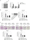
(A) Western blot showing the expression of STAT3/HIF-1α in Eca109/pCMV-SOX2 cells; (B) Western blot showing the effect of STAT3 or HIF-1α knockdown with specific siRNA on Slug expression; (C) The effect of STAT3 or HIF-1α knockdown with specific siRNA on migration and invasion of Eca109/pCMV-SOX2 cells evaluated by Transwell assay; (D) Western blots showing the effect of STAT3 or HIF-1α knockdown with specific siRNA on EMT (200× magnification). Data are represented as mean ± SD of three independently conducted experiments, ** indicates p < 0.01. The images of Transwell chamber assay and Western blots are representatives of three independent experiments.
Figure 4
Figure 4
(A) Western blot showing the expression of STAT3/HIF-1α in Eca109/pCMV-SOX2 cells; (B) Western blot showing the effect of STAT3 or HIF-1α knockdown with specific siRNA on Slug expression; (C) The effect of STAT3 or HIF-1α knockdown with specific siRNA on migration and invasion of Eca109/pCMV-SOX2 cells evaluated by Transwell assay; (D) Western blots showing the effect of STAT3 or HIF-1α knockdown with specific siRNA on EMT (200× magnification). Data are represented as mean ± SD of three independently conducted experiments, ** indicates p < 0.01. The images of Transwell chamber assay and Western blots are representatives of three independent experiments.

References

    1. Sakai N.S., Samia-Aly E., Barbera M., Fitzgerald R.C. A review of the current understanding and clinical utility of miRNAs in esophageal cancer. Semin. Cancer Biol. 2013;23:512–521. doi: 10.1016/j.semcancer.2013.08.005. - DOI - PubMed
    1. Jemal A., Bray F., Center M.M., Ferlay J., Ward E., Forman D. Global cancer statistics. Cancer J. Clin. 2011;61:69–90. doi: 10.3322/caac.20107. - DOI - PubMed
    1. Sagar P.M., Gauperaa T., Sue-Ling H., McMahon M.J., Johnston D. An audit of the treatment of cancer of the oesophagus. Gut. 1994;35:941–945. doi: 10.1136/gut.35.7.941. - DOI - PMC - PubMed
    1. Kim T., Grobmyer S.R., Smith R., Ben-David K., Ang D., Vogel S.B., Hochwald S.N. Esophageal cancer—The five year survivors. J. Surg. Oncol. 2011;103:179–183. doi: 10.1002/jso.21784. - DOI - PubMed
    1. Pevny L.H., Lovell-Badge R. SOX genes find their feet. Curr. Opin. Genet. Dev. 1997;7:338–344. doi: 10.1016/S0959-437X(97)80147-5. - DOI - PubMed

Publication types

MeSH terms

Substances