. 2015 Nov 15;309(10):H1740-52.
doi: 10.1152/ajpheart.00557.2015. Epub 2015 Sep 14.

Central-peripheral neural network interactions evoked by vagus nerve stimulation: functional consequences on control of cardiac function

Affiliations

Central-peripheral neural network interactions evoked by vagus nerve stimulation: functional consequences on control of cardiac function

Jeffrey L Ardell et al. Am J Physiol Heart Circ Physiol. .

Abstract

Using vagus nerve stimulation (VNS), we sought to determine the contribution of vagal afferents to efferent control of cardiac function. In anesthetized dogs, the right and left cervical vagosympathetic trunks were stimulated in the intact state, following ipsilateral or contralateral vagus nerve transection (VNTx), and then following bilateral VNTx. Stimulations were performed at currents from 0.25 to 4.0 mA, frequencies from 2 to 30 Hz, and a 500-μs pulse width. Right or left VNS evoked significantly greater current- and frequency-dependent suppression of chronotropic, inotropic, and lusitropic function subsequent to sequential VNTx. Bradycardia threshold was defined as the current first required for a 5% decrease in heart rate. The threshold for the right vs. left vagus-induced bradycardia in the intact state (2.91 ± 0.18 and 3.47 ± 0.20 mA, respectively) decreased significantly with right VNTx (1.69 ± 0.17 mA for right and 3.04 ± 0.27 mA for left) and decreased further following bilateral VNTx (1.29 ± 0.16 mA for right and 1.74 ± 0.19 mA for left). Similar effects were observed following left VNTx. The thresholds for afferent-mediated effects on cardiac parameters were 0.62 ± 0.04 and 0.65 ± 0.06 mA with right and left VNS, respectively, and were reflected primarily as augmentation. Afferent-mediated tachycardias were maintained following β-blockade but were eliminated by VNTx. The increased effectiveness and decrease in bradycardia threshold with sequential VNTx suggest that 1) vagal afferents inhibit centrally mediated parasympathetic efferent outflow and 2) the ipsilateral and contralateral vagi exert a substantial buffering capacity. The intact threshold reflects the interaction between multiple levels of the cardiac neural hierarchy.

Keywords: afferent; autonomic nervous system; intrinsic cardiac nervous system; parasympathetic; vagus nerve stimulation.


Figures

Fig. 1.
Fig. 1.
Representative hemodynamic response to right cervical vagus bioelectric stimulation in an anesthetized animal with intact vagus nerves. Vagus nerve stimulation (VNS) was delivered at 10 Hz, 500-μs pulse width, and 2.50 mA for 14 s. LV, left ventricle; LVP, LV pressure; dp/dt, 1st derivative of LVP; BP, blood pressure; EKG, lead II electrocardiogram.
Fig. 2.
Fig. 2.
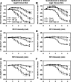
Evoked changes in chronotropic (A and B) and LV inotropic and lusitropic (C–F) function in response to right cervical vagus (RCV) bioelectric stimulation prior to and following sequential cervical vagus transection rostral to the stimulating electrode. VNS was delivered at 10 Hz and 500-μs pulse width for 14 s. Responses reflect percent change from baseline during VNS as a function of stimulus intensity. Left: responses in the intact state, after right vagus (RV) transection, and then following bilateral cervical vagus transection. Right: responses in the intact state, after left vagus (LV) transection, and then following bilateral cervical vagus transection. Note that all augmenting responses to VNS were eliminated when ipsilateral vagi were transected. *P < 0.004 vs. intact. #P < 0.0001, unilateral vs. bilateral vagus transection.
Fig. 3.
Fig. 3.
Baseline and right cervical vagus-evoked heart rate levels in the intact state and following sequential unilateral [right (RVx, A) and left (LVx, B)] and bilateral [RVxLVx (A) and LVxRVx (B)] cervical vagotomy. Vagus nerves were cut rostral to the stimulating electrode. Note that all positive chronotropic responses to right cervical vagus stimulation were eliminated when the right vagus nerve was transected and that negative chronotropic responses to right cervical vagus stimulation where progressively increased from unilateral to bilateral vagotomy conditions. *P < 0.001 vs. intact. #P < 0.005, unilateral vs. bilateral vagus transection.
Fig. 4.
Fig. 4.
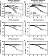
Evoked changes in chronotropic (A and B) and LV inotropic and lusitropic (C–F) function in response to left cervical vagus (LCV) bioelectric stimulation prior to and following sequential cervical vagus transection rostral to the stimulating electrode. A, C, and E: effects of ipsilateral (left) vs. bilateral vagus transection. B, D, and F: effects of contralateral vs. bilateral vagus transection. *P < 0.002 vs. intact. #P < 0.0001, unilateral vs. bilateral vagus transection.
Fig. 5.
Fig. 5.
VNS bradycardia threshold is reduced by sequential vagal transection. Threshold is defined as 5% decrease in heart rate. A and C: average current required in intact state, following unilateral transection of the right or left cervical vagus, and following bilateral vagus transection. Vagi were transected rostral to the stimulation point. B and D: percent change from intact controls for baseline threshold following unilateral right, left, and bilateral vagus transection. +P < 0.04 vs. intact. *P < 0.001 vs. intact. #P < 0.002, unilateral vs. bilateral vagus transection.
Fig. 6.
Fig. 6.
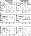
Evoked changes in chronotropic (A and D), LV inotropic (B and E), and lusitropic (C and F) function in response to left cervical VNS prior to and following sequential cervical vagus transections rostral to the stimulating electrode. VNS was delivered at 500-μs pulse width for 14 s at an intensity of 1.2 times bradycardiac threshold as determined in the intact state. Responses reflect percent change from baseline during VNS as a function of stimulus frequency. A–C: responses in the intact state, after left vagus transection, and then after bilateral cervical vagus transection. D–F: responses in the intact state, after right vagus transection, and then after bilateral cervical vagus transection. Across all conditions: *P < 0.001 vs. intact; #P < 0.0001, unilateral vs. bilateral vagus transection. +P < 0.0001, intact vs. bilateral decentralization (paired comparison).
Fig. 7.
Fig. 7.
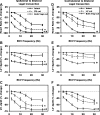
Evoked changes in chronotropic (A and D), LV inotropic (B and E), and lusitropic (C and F) function in response to right cervical VNS prior to and following sequential cervical vagus transections rostral to the stimulating electrode. See details in Fig. 6 legend. A–C: effects of ipsilateral (right) vs. bilateral vagus transection. D–F: effects of contralateral vs. bilateral vagal transection. Across all conditions: *P < 0.0001 vs. intact; #P < 0.0001, unilateral vs. bilateral vagus transection. +P < 0.0001, intact vs. bilateral decentralization (paired comparison).
Fig. 8.
Fig. 8.
Evoked changes in heart rate, LV end-systolic pressure (LVSP), LV −dp/dt, and LV +dp/dt in response to right cervical vagus stimulation (10 Hz) prior to and following sequential timolol (1 mg/kg), bilateral cervical vagus transection (vagotomy), and atropine (1 mg/kg). Responses reflect percent change from baseline during VNS as a function of stimulus current intensity. *P < 0.02 vs. control. #P < 0.001 vs. timolol. +P < 0.0001 vs. timolol + vagotomy.
Fig. 9.
Fig. 9.
Evoked changes in heart rate, LV end-systolic pressure, LV −dp/dt, and LV +dp/dt in response to left cervical vagus nerve stimulation (10 Hz) prior to and following sequential timolol (1 mg/kg), bilateral cervical vagus transection (vagotomy), and atropine (1 mg/kg). See details in Fig. 8 legend. *P < 0.03 vs. control. #P < 0.003 vs. timolol. +P < 0.0001 vs. timolol + vagotomy.
Fig. 10.
Fig. 10.
Schematic summary of proposed neural interactions within the hierarchy for cardiac control engaged by VNS. Centrally mediated changes in preganglionic input to the intrinsic cardiac nervous system (ICNS) were evoked at current intensities ∼20% of bradycardiac threshold. Dashed lines, preganglionic projections; dotted-dashed lines, afferent projections. NTS, nucleus tractus solitarius; NA, nucleus ambiguus; BSRF, brain stem reticular formation; IML, intermediolateral cell column; DRG, dorsal root ganglion; MCG, middle cervical ganglion; LCN, local circuit neuron; β, β-adrenergic receptor; M2, muscarinic receptor; Gs and Gi, G-coupled proteins; AC, adenylate cyclase.

References

    1. Andresen MC, Kunze DL, Mendelowitz D. Central nervous system regulation of the heart. In: Basic and Clinical Neurocardiology, edited by Armour JA and Ardell JL. New York: Oxford University Press, 2004, p. 187–219.
    1. Ardell JL, Beaumont E, Nier H, Southerland EM, Griffin S, KenKnight BH, Armour JA. Vagal nerve stimulation elicits heart rate responses that are dependent on relative activation of both central and peripheral limbs of the autonomic nervous system: defining the neural fulcrum (Abstract). Heart Rhythm. 2014.
    1. Ardell JL, Butler CK, Smith FM, Hopkins DA, Armour JA. Activity of in vivo atrial and ventricular neurons in chronically decentralized canine hearts. Am J Physiol Heart Circ Physiol 260: H713–H721, 1991. - PubMed
    1. Ardell JL, Cardinal R, Vermeulen M, Armour JA. Dorsal spinal cord stimulation obtunds the capacity of intrathoracic extracardiac neurons to transduce myocardial ischemia. Am J Physiol Regul Integr Comp Physiol 297: R470–R477, 2009. - PMC - PubMed
    1. Ardell JL, Randall WC. Selective vagal innervation of sinoatrial and atrioventricular nodes in canine heart. Am J Physiol Heart Circ Physiol 251: H764–H773, 1986. - PubMed

Publication types

LinkOut - more resources