. 2015 Dec 4;117(12):990-4.
doi: 10.1161/CIRCRESAHA.115.307465. Epub 2015 Sep 14.

Sympathetic Reinnervation Is Required for Mammalian Cardiac Regeneration

Affiliations

Sympathetic Reinnervation Is Required for Mammalian Cardiac Regeneration

Ian A White et al. Circ Res. .

Abstract

Rationale: Although mammalian cardiac regeneration can occur in the neonatal period, the factors involved in this process remain to be established. Because tissue and limb regeneration require concurrent reinnervation by the peripheral nervous system, we hypothesized that cardiac regeneration also requires reinnervation.

Objective: To test the hypothesis that reinnervation is required for innate neonatal cardiac regeneration.

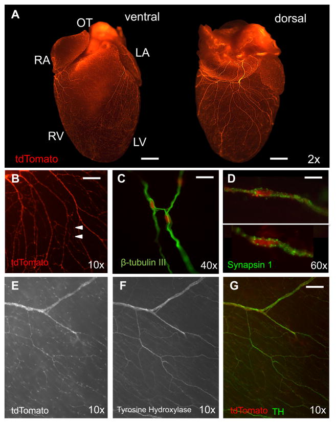
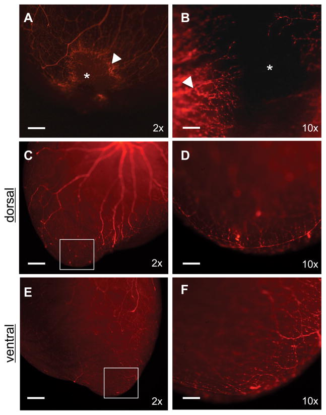
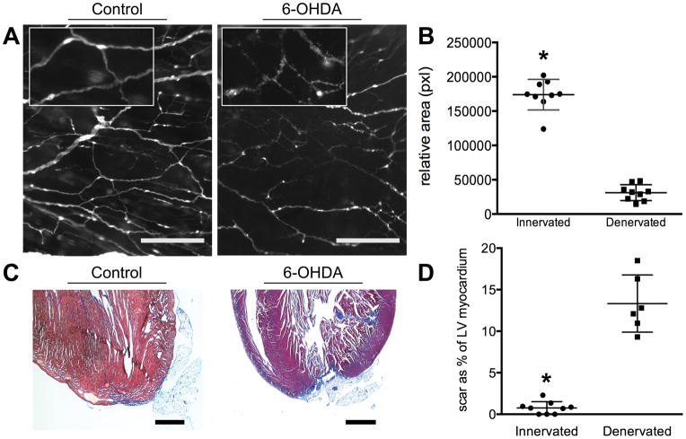
Methods and results: We crossed a Wnt1-Cre transgenic mouse with a double-tandem Tomato reporter strain to identify neural crest-derived cell lineages including the peripheral autonomic nerves in the heart. This approach facilitated the precise visualization of subepicardial autonomic nerves in the ventricles using whole mount epifluorescence microscopy. After resection of the left ventricular apex in 2-day-old neonatal mice, sympathetic nerve structures, which envelop the heart under normal conditions, exhibited robust regrowth into the regenerating myocardium. Chemical sympathectomy inhibited sympathetic regrowth and subsequent cardiac regeneration after apical resection significantly (scar size as cross-sectional percentage of viable left ventricular myocardium, n=9; 0.87%±1.4% versus n=6; 14.05±4.4%; P<0.01).

Conclusions: These findings demonstrate that the profound regenerative capacity of the neonatal mammalian heart requires sympathetic innervation. As such, these data offer significant insights into an underlying basis for inadequate adult regeneration after myocardial infarction, a situation where nerve growth is hindered by age-related influences and scar tissue.

Keywords: mice, transgenic; myocardium; regeneration; sympathectomy; sympathetic nervous system.


Figures

Figure 1
Figure 1. Subepicardial distribution of post-ganglionic sympathetic nerve fibers in the mouse heart
A, Crossing Wnt1-Cre transgenic mice with tdTomato reporter mice identifies nerve fibers throughout the entire subepicardium from the base of the heart to the apex of the ventricles (bar, 1 mm). B, The fibers are heavily varicosed (arrows) (bar, 200 μm). C, They stain positive for the neurofilament marker β-tubulin III (bar, 50 μm), the presynaptic marker synapsin 1 (D) (bar, 20 μm) and the sympathetic nerve fiber marker TH (E–G) (bar, 200 μm).
Figure 2
Figure 2. Concurrent reinnervation and cardiac regeneration
A, B, At day 14 post-resection the apex is in the process of regenerating (*) as nerves regrow into the injury (arrow marks site of hyperinnervation at injury border) (bars, 500 μm and 100 μm, respectively). C–F. After 21 days the apex has regenerated and fully reinnervated (bar, 500 μm). Boxes from C and E are expanded with greater magnification in D and F (bar, 100 μm). All images are of Wnt1-Cre;tdTomato hearts.
Figure 3
Figure 3. Cardiac innervation is required for myocardial regeneration
A, Administering 6-OHDA to neonates induces Wallerian degeneration of sympathetic nerve fibers. Inset demonstrates degeneratio of nerve structure (epifluorescence, Wnt1-Cre;tdTomato. bar, 200 μm). B, Densitometry of Wnt1-Cre;tdTomato nerves from 21-day-old mouse hearts that are innervated compared to denervated (n=9, 1.74×10⁵±7447pixels; n=9, 3.11×10⁴±3863pixels; p<0.01). C, In the presence of sympathetic nerves the apex can regenerate with minimal fibrosis; visualized histologically with Masson’s Trichrome stain (blue). Small amounts of adipose are adherent to the apex on occasion. Following denervation with 6-OHDA innate regeneration is blocked and extensive scar and adipocyte deposition can be observed (bar, 1 mm). D, Scar area as a percentage of left ventricular (LV) myocardium in cross-section (n=9, 0.87±1.4% vs. n=6, 14.05±4.4%; p<0.01).

Comment in

References

    1. Bryant DM, O’Meara CC, Ho NN, Gannon J, Cai L, Lee RT. A systematic analysis of neonatal mouse heart regeneration after apical resection. J Mol Cell Cardiol. 2015;79:315–318. - PMC - PubMed
    1. Porrello ER, Mahmoud AI, Simpson E, Hill JA, Richardson JA, Olson EN, Sadek HA. Transient regenerative potential of the neonatal mouse heart. Science. 2011;331:1078–1080. - PMC - PubMed
    1. Singh BN, Koyano-Nakagawa N, Garry JP, Weaver CV. Heart of newt: A recipe for regeneration. J Cardiovasc Transl Res. 2010;3:397–409. - PubMed
    1. Poss KD, Wilson LG, Keating MT. Heart regeneration in zebrafish. Science. 2002;298:2188–2190. - PubMed
    1. Lam NT, Currie PD, Lieschke GJ, Rosenthal NA, Kaye DM. Nerve growth factor stimulates cardiac regeneration via cardiomyocyte proliferation in experimental heart failure. PLoS One. 2012;7:e53210. - PMC - PubMed

Publication types

Substances