. 2015 Sep 29;112(39):12205-10.
doi: 10.1073/pnas.1511724112. Epub 2015 Sep 14.

EBS7 is a plant-specific component of a highly conserved endoplasmic reticulum-associated degradation system in Arabidopsis

Affiliations

EBS7 is a plant-specific component of a highly conserved endoplasmic reticulum-associated degradation system in Arabidopsis

Yidan Liu et al. Proc Natl Acad Sci U S A. .

Abstract

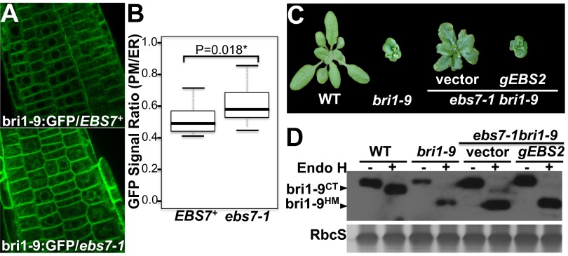
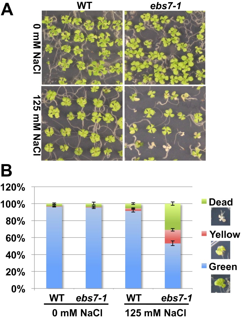
Endoplasmic reticulum (ER)-associated degradation (ERAD) is an essential part of an ER-localized protein quality-control system for eliminating terminally misfolded proteins. Recent studies have demonstrated that the ERAD machinery is conserved among yeast, animals, and plants; however, it remains unknown if the plant ERAD system involves plant-specific components. Here we report that the Arabidopsis ethyl methanesulfonate-mutagenized brassinosteroid-insensitive 1 suppressor 7 (EBS7) gene encodes an ER membrane-localized ERAD component that is highly conserved in land plants. Loss-of-function ebs7 mutations prevent ERAD of brassinosteroid insensitive 1-9 (bri1-9) and bri1-5, two ER-retained mutant variants of the cell-surface receptor for brassinosteroids (BRs). As a result, the two mutant receptors accumulate in the ER and consequently leak to the plasma membrane, resulting in the restoration of BR sensitivity and phenotypic suppression of the bri1-9 and bri1-5 mutants. EBS7 accumulates under ER stress, and its mutations lead to hypersensitivity to ER and salt stresses. EBS7 interacts with the ER membrane-anchored ubiquitin ligase Arabidopsis thaliana HMG-CoA reductase degradation 1a (AtHrd1a), one of the central components of the Arabidopsis ERAD machinery, and an ebs7 mutation destabilizes AtHrd1a to reduce polyubiquitination of bri1-9. Taken together, our results uncover a plant-specific component of a plant ERAD pathway and also suggest its likely biochemical function.

Keywords: EMS-mutagenized bri1 suppressor; ERAD; brassinosteroid receptor BRI1; ubiquitin ligase E3; unfolded protein response.


Conflict of interest statement

The authors declare no conflict of interest.

Figures

Fig. 1.
Fig. 1.
The ebs7-1 mutation suppresses bri1-9 and restores its BR sensitivity by blocking bri1-9 degradation. (A) Immunoblot analysis of BRI1/bri1-9. Total proteins of 2-wk-old seedlings were treated with or without Endo H, separated by SDS/PAGE, and analyzed by immunoblot with a BRI1 antibody. bri1-9HM and bri1-9CT denote BRI1/bri1-9 proteins carrying HM and CT N-glycans, respectively. (B) Immunoblot analysis of the bri1-9 stability. Two-week-old seedlings were treated with 180 μM CHX for the indicated hours, and extracted proteins were separated by SDS/PAGE and analyzed by immunoblot with the anti-BRI1 antibody. (CE) Pictures of 4-wk-old light-grown (C) and 5-d-old dark-grown (D) seedlings, and 2-mo-old soil-grown plants (E). (F) The BR-induced root inhibition assay. Root lengths of ∼40 7-d-old seedlings grown on BL-containing 1/2 Murashiga and Skoog (MS) medium were measured, and measurements were converted into percentage values relative to that of the same genotype grown on medium without BL. Error bars represent ± SE of three independent assays. (G) The BR-induced BES1 dephosphorylation assay. Protein extracts of 2-wk-old seedlings treated with or without 1 μM BL were separated by SDS/PAGE and analyzed by immunoblot with an anti-BES1 antibody. In A, B, and G, Coomassie blue staining of the small subunit of the ribulose-1,5-bisphosphate carboxylase/oxygenase (RuBisCo, hereafter RbcS) enzyme on a duplicate gel was used as a loading control.
Fig. S1.
Fig. S1.
Overexpression of EBS2 nullifies the suppressive effect of the ebs7-1 mutation on bri1-9 dwarfism. (A) The confocal images of root cells transgenically expressing bri1-9GFP in the EBS7+ (Left) and ebs7-1 (Right) background. (B) Quantitative analysis of the ratio of the GFP signal on the PM and in the ER. ImageJ was used to quantify the GFP signal in the PM and ER. In each picture in A, the average integrated fluorescent density of 15 cells was measured for the whole cell (ER and PM) and the cytosol (ER) to calculate the GFP[PM]/GFP[ER] ratio. The box-and-whisker plot was generated to show the difference between the EBS7+ and ebs7-1 genotypes, and the indicated P value was calculated by a one-tailed two-sample t test. It is important to note that the GFP[PM]/GFP[ER] ratios presented here are high, likely caused by tethering of the cortical ER with the PM. (C) Pictures of 4-wk-old soil-grown wild-type and bri1-9 plants and two transgenic ebs7-1 bri1-9 mutants carrying an empty vector or a genomic EBS2 transgene. (D) Immunoblot analysis of BRI1/bri1-9. Total proteins extracted from 2-wk-old Arabidopsis seedlings of the genotypes shown in B were treated with or without Endo H, separated by SDS/PAGE, and analyzed by immunoblot with an anti-BRI1 antibody. bri1-9HM and bri1-9CT denote the BRI1/bri1-9 proteins carrying the HM and CT N-glycans, respectively. Coomassie blue staining of RbcS on a duplicated gel serves as a loading control.
Fig. 2.
Fig. 2.
ebs7-1 inhibits the degradation of bri1-5 and misfolded EFR. (A and B) Pictures of 4-wk-old light-grown (A) and 5-d-old dark-grown (B) seedlings. In (B), three individual images of two seedlings (separated by white lines) were assembled together. (C) The root-growth inhibition assay of wild-type, bri1-5, and ebs7-1 bri1-5 seedlings. (D) The BES1 dephosphorylation assay. (E) Immunoblot analysis of BRI1/bri1-5 abundance. bri1-5HM and bri1-5CT denote the BRI1/bri1-5 proteins carrying the HM and CT N-glycans, respectively. (F) Immunoblot analysis of EFR. Protein extracts of 2-wk-old seedlings were separated by SDS/PAGE and analyzed by immunoblot with an anti-EFR antibody. In D and E, Coomassie blue staining of the RbcS band on a duplicate gel serves as a loading control, and in F, a nonspecific cross-reacting band indicated by the asterisk serves as a loading control.
Fig. S2.
Fig. S2.
Screen for bri1-5 suppressors identified two additional ebs7 alleles. (A) Pictures of 2-mo-old soil-grown wild-type, bri1-5, and ebs7-1 bri1-5 plants. (B) Pictures of 4-wk-old wild-type (Ws-2), bri1-5, ebs7-1 bri1-5, ebs7-2 bri1-5, and ebs7-3 bri1-5 plants. (C) Immunoblot analysis of EBS7 protein. Total proteins extracted from 2-wk-old seedlings of the genotypes shown in B were separated by SDS/PAGE and analyzed by immunoblot with an affinity-purified anti-EBS7 antibody. Coomassie blue staining of RbcS on a duplicated gel serves as a loading control. (D) The full image of the immunoblot presented in Fig. 3D. Arrowheads on the left indicate the positions of the molecular weight standards and EBS7. The asterisk denotes a nonspecific band recognized by the affinity-purified EBS7 antibody.
Fig. 3.
Fig. 3.
EBS7 encodes an ER-localized membrane protein that is highly conserved in land plants. (A) Map-based cloning of EBS7. EBS7 was mapped to an ∼850-kb region on chromosome 4. Marker names are shown above the line, and recombinant numbers are shown below the line. The EBS7 gene structure is shown with bars denoting exons and lines indicating introns. Arrowheads show the positions of three ebs7 mutations. (B) The nucleotide changes and predicted molecular defects of the three ebs7 alleles. (C) Pictures of 4-wk-old wild-type, bri1-9, and two transgenic ebs7-1 bri1-9 mutant plants. (D) Immunoblot analysis of EBS7. Total proteins of 2-wk-old seedlings were separated by SDS/PAGE and analyzed by immunoblot using an anti-EBS7 antibody. (E) Immunoblot analysis of BRI1/bri1-9. (F) The confocal images of tobacco leaf epidermal cells transiently expressing GFPEBS7 (Left), RFPHDEL (Center), and superimposition of GFP and RFP signals (Right). (G) Immunoblot analysis of EBS7. Total (T), membrane (M), and soluble (S) proteins extracted from 2-wk-old seedlings were separated by SDS/PAGE and analyzed by immunoblot with antibodies against EBS2, EBS5, and EBS7. In D and G, the asterisk indicates a nonspecific band serving as a loading control.
Fig. S3.
Fig. S3.
At4g29960 is coexpressed with at least three known/predicted ERAD genes and is ubiquitously expressed in Arabidopsis. (A) The coexpressed gene network around At4g29960, which was obtained from ATTEDII (atted.jp/) using At4g29960 (shaded yellow) as query. The red dots denote genes known/predicted to be involved in the Arabidopsis ERAD process, and black lines link strongly (thick lines) or weakly (thin lines) coexpressed genes. (B) Visualization of the expression profile of At4g29960 in different tissues of growing Arabidopsis plants, which was obtained from the Arabidopsis eFP browser web server (bar.utoronto.ca/efp_arabidopsis/cgi-bin/efpWeb.cgi) using At4g29960 as query. Reprinted with permission from ref. 34.
Fig. S4.
Fig. S4.
The sequence analyses of EBS7 homologs in land plants. (A) Sequence alignment of EBS7 and its representative plant homologs. Protein sequences of EBS7 (accession no. NP_567837), BdEBS7 (Bd: Brachypodium distachyon, XP_003577873), OsEBS7 (Os: Oryza sativa Japonica, NP_001053803), PtEBS7 (Pt: Populus trichocarpa, XP_002308117), GaEBS7 (Ga: Genlisea aurea, EPS72061), AtEBS7 (At: Amborella trichopoda, ERN12475), PsEBS7 (Ps: Picea sitchensis, ABR16454), and SmEBS7 (Sm: Selaginella moellendorffii, XP_002974885) were aligned using the ClustalW program (mobyle.pasteur.fr/cgi-bin/portal.py). Aligned sequences were color-shaded at a BoxShade 3.31 server (mobyle.pasteur.fr/cgi-bin/portal.py). Residues identical in more than six proteins are shaded red, and similar residues are shaded cyan. The positions of ebs7 mutations are indicated by green triangles, and the three predicted transmembrane segments are indicated by blue bars. (B) The phylogeny tree of plant EBS7 homologs. Names of plant species and accession numbers of the corresponding EBS7 homologs are as follows: Coffea canephora, CDP19559.1; Solanum lycopersicum, XP_004242818; Solanum tuberosum, XP_006361645; Mimulus guttatus, EYU18221.1; Mimulus guttatus, EYU38750.1; Genlisea aurea, EPS72061.1; Arabis alpina, KFK40291.1; Brassica napus, CDY03554.1; Brassica napus, CDY43545.1; Arabis alpina, KFK29555.1; Eutrema salsugineum, XP_006412781.1; Capsella rubella, XP_006284250; Arabidopsis lyrata, XP_002867378; Arabidopsis thaliana (EBS7), NP_567837.1; Cucumis melo, XP_008453570.1; Cucumis sativus, XP_004146332; Medicago truncatula, XP_003595067; Cicer arietinum, XP_004487987; Phaseolus vulgaris, ESW10771; Glycine max, XP_006597064; Glycine max, NP_001242739.1; Vitix vinifera, XP_002269262; Ricinus communis, XP_002527805; Jatropha curcas, KDP47202.1; Populus trichocarpa, XP_002324697.1; Populus trichocarpa, XP_002308117.2; Theobroma cacao, XP_007014411.1; Morus notabilis, EXB30996.1; Fragaria vesca subsp. vesca, XP_004296487.1; Malus domestica, XP_008354453; Prunus persica, EMJ12895; Prunus mume XP_008223885.1; Eucalyptus grandis, KCW78661.1; Citrus sinensis, KDO61891.1;Citrus clementina, XP_006453265; Setaria italica, XP_004976735; Zea mays, NP_001142664.1; Sorghum bicolor, XP_002447061; Zea mays, NP_001131686.1; Oryza brachyantha, XP_006653743; Oryza sativa Japonica Group, NP_001053803; Oryza sativa Indica Group, i-CAH66780; Aegilops tauschii, EMT30053; Fragaria vesca, XP_004296487; Triticum urartu, EMS50749; Brachypodium distachyon, XP_003577873; Phoenix dactylifera, XP_008781761.1; Amborella trichopoda, ERN12475; Picea sitchensis, ABR16454; Selaginella moellendorffii, XP_002974885; Physcomitrella patens, XP_001756855.1.
Fig. S5.
Fig. S5.
Analysis of stable Arabidopsis transgenic lines expressing GFP-tagged EBS7. (A) Pictures of representative transgenic ebs7-1 bri1-9 seedlings carrying GFP–EBS7, EBS7–GFP, or an empty vector, which were grown on 1/2 MS medium containing 100 μg/mL kanamycin. (B) Confocal images of root cells of a GFP–EBS7 transgenic line stained with 1 μM ER-Tracker Red dye and superimposition of the green and red fluorescent signals.
Fig. 4.
Fig. 4.
The ebs7-1 mutant exhibits constitutive UPR activation and is hypersensitive to ER stress. (A) Immunoblot analysis of ER proteins. Total proteins of 2-wk-old seedlings treated with or without 5 μg/mL TM were separated by SDS/PAGE and assayed by immunoblot with antibodies against binding immunoglobulin proteins (BiPs), protein disulfide isomerases (PDIs), calnexin/calreticulins (CNX/CRTs), EBS5, EBS6, and EBS7. Coomassie blue staining of RbcS on a duplicated gel serves as the loading control. (B) Pictures of 2-wk-old seedlings grown on 1/2 MS medium containing 0 or 1 mM DTT. (C) Quantification analysis of seedling sensitivity to DTT. “Green”, “yellow”, or “dead” seedlings were counted, and the percentages of each type of seedlings were calculated and are shown in the bar graph. Error bars represent ± SD for three independent assays of ∼50 seedlings/each.
Fig. S6.
Fig. S6.
The ebs7-1 mutation causes hypersensitivity to salt stress. (A) Pictures of 2-wk-old wild-type and ebs7-1 seedlings grown on 1/2 MS medium supplemented without (Upper) or with (Lower) 125 mM NaCl. (B) Quantification analysis of sensitivity to salt stress. Green, yellow, or dead seedlings were counted, and the percentages of each type of seedlings were calculated and are shown in the bar graph. Error bars represent ± SD for three independent assays of ∼50 seedlings/each.
Fig. 5.
Fig. 5.
EBS7 interacts with AtHrd1a and affects its stability. (A) CoIP of bri1-9 and EBS5. Total proteins and anti-GFP immunoprecipitates obtained from wild-type or transgenic plants were separated by SDS/PAGE and analyzed by immunoblot with antibodies against GFP, ubiquitin, or EBS5. (B) CoIP of EBS7, AtHrd1a, and EBS5. Total proteins and anti-GFP immunoprecipitates obtained from wild-type or transgenic Arabidopsis plants were separated by SDS/PAGE and analyzed by immunoblot with anti-GFP, anti-EBS7, and anti-EBS5 antibodies. (C and D) Immunoblot analysis of Hrd1aGFP. Total proteins extracted from 2-wk-old seedlings treated with 180 μM CHX (C) or 80 μM MG132 (D) for varying durations were separated by SDS/PAGE and assayed by immunoblot with an anti-GFP antibody. Coomassie blue staining of RbcS on a duplicate gel serves as a loading control.
Fig. S7.
Fig. S7.
Analysis of EBS7 interaction with Hrd1a and EBS5. (A) Pictures of 12-d-old wild-type seedlings, the bri1-9 single mutant, an hrd1a hrd1b bri1-9 triple mutant, and its transgenic line expressing a p35S:Hrd1a–GFP transgene. (B) CoIP of Hrd1a and EBS7. Total proteins and anti-EBS7 immunoprecipitates from wild-type or AtHrd1a–GFPexpressing transgenic Arabidopsis seedlings were separated by SDS/PAGE and analyzed by immunoblot with anti-EBS7 or anti-GFP antibodies. (C) Schematic diagram of the truncated C-Hrd1a and N-EBS7 fragments used for a yeast two-hybrid assay testing AtHrd1a-EBS7 interaction. The blue bars represent putative transmembrane segments, and the pink triangle denotes the RING domain of the AtHrd1a E3 ligase. (D) Yeast colonies coexpressing C-Hrd1a fused with the activation domain (AD) of the yeast GAL4 protein and the GAL4’s DNA-binding (BD) domain only, Gal4BD-fused C-Hrd1a, or Gal4BD-fused N-EBS7. (E) CoIP of EBS7 and EBS5. Total proteins and anti-EBS7 immunoprecipitates obtained from wild, ebs5-5, or ebs7-1 plants were separated by SDS/PAGE, transferred to a membrane, and analyzed by immunoblot with antibodies made against EBS5 or EBS7.
Fig. S8.
Fig. S8.
EBS7 is not likely to be a functional homolog of the yeast Usa1 or the mammalian HERP. (A) Immunoblot analysis of the protein abundance of CPY* in the yeast BY4742 and Δusa1p strains carrying the indicated expression plasmids. Coomassie blue staining of an unknown major yeast protein on a duplicate gel serves as the loading control. (B) Pictures of two representative 4-wk-old soil-grown transgenic ebs7-1 bri1-9 plants carrying the empty vector (Left) or the p35S:HERP–GFP transgene (Right).

Similar articles

Cited by

References

    1. Ruggiano A, Foresti O, Carvalho P. Quality control: ER-associated degradation: Protein quality control and beyond. J Cell Biol. 2014;204(6):869–879. - PMC - PubMed
    1. Vembar SS, Brodsky JL. One step at a time: Endoplasmic reticulum-associated degradation. Nat Rev Mol Cell Biol. 2008;9(12):944–957. - PMC - PubMed
    1. Hebert DN, Molinari M. Flagging and docking: Dual roles for N-glycans in protein quality control and cellular proteostasis. Trends Biochem Sci. 2012;37(10):404–410. - PMC - PubMed
    1. Denic V, Quan EM, Weissman JS. A luminal surveillance complex that selects misfolded glycoproteins for ER-associated degradation. Cell. 2006;126(2):349–359. - PubMed
    1. Gauss R, Jarosch E, Sommer T, Hirsch C. A complex of Yos9p and the HRD ligase integrates endoplasmic reticulum quality control into the degradation machinery. Nat Cell Biol. 2006;8(8):849–854. - PubMed

Publication types

MeSH terms