

Biochemically engineered stromal cell-derived factor 1-alpha analog increases perfusion in the ischemic hind limb
- PMID: 26372192
- PMCID: PMC5453729
- DOI: 10.1016/j.jvs.2015.06.140
Biochemically engineered stromal cell-derived factor 1-alpha analog increases perfusion in the ischemic hind limb
Abstract
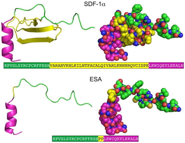
Background: Despite promising therapeutic innovation over the last decade, peripheral arterial disease remains a prevalent morbidity, as many patients are still challenged with peripheral ischemia. We hypothesized that delivery of engineered stromal cell-derived factor 1-alpha (ESA) in an ischemic hind limb will yield significant improvement in perfusion.
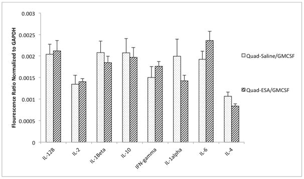
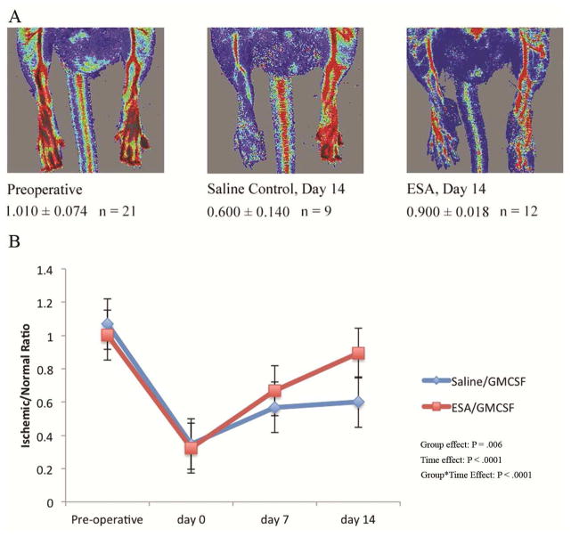
Methods: Male rats underwent right femoral artery ligation, and animals were randomized to receive a 100 μL injection of saline (n = 9) or 6 μg/kg dosage of equal volume of ESA (n = 12) into the ipsilateral quadriceps muscle. Both groups of animals were also given an intraperitoneal injection of 40 μg/kg of granulocyte macrophage colony-stimulating factor (GMCSF). Perfusion was quantified using a laser Doppler imaging device preoperatively, and on postoperative days 0, 7, and 14. Immunohistochemistry was performed to quantify angiogenesis on day 14, and an mRNA profile was evaluated for angiogenic and inflammatory markers.
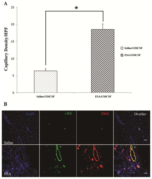
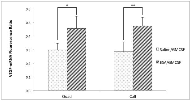
Results: Compared with the saline/GMCSF group at day 14, the ESA/GMCSF-injected animals had greater reperfusion ratios (Saline/GMCSF, 0.600 ± 0.140 vs ESA/GMCSF, 0.900 ± 0.181; group effect P = .006; time effect P < .0001; group×time effect P < .0001), elevated capillary density (10×; Saline/GMCSF, 6.40 ± 2.01 vs ESA/GMCSF, 18.55 ± 5.30; P < .01), and increased mRNA levels of vascular endothelial growth factor-A (Saline/GMCSF [n = 6], 0.298 ± 0.205 vs ESA/GMCSF [n = 8], 0.456 ± 0.139; P = .03).
Conclusions: Delivery of ESA significantly improves perfusion in a rat model of peripheral arterial disease via improved neovasculogenesis, a finding which may prove beneficial in the treatment strategy for this debilitating disease.
Copyright © 2016 Society for Vascular Surgery. Published by Elsevier Inc. All rights reserved.
Figures
Comment in
-
Invited commentary.J Vasc Surg. 2016 Oct;64(4):1099-100. doi: 10.1016/j.jvs.2016.07.100. J Vasc Surg. 2016. PMID: 27666448 No abstract available.
Similar articles
-
Combination of stromal-derived factor-1alpha and vascular endothelial growth factor gene-modified endothelial progenitor cells is more effective for ischemic neovascularization.J Vasc Surg. 2009 Sep;50(3):608-16. doi: 10.1016/j.jvs.2009.05.049. Epub 2009 Jul 12. J Vasc Surg. 2009. PMID: 19595531
-
Beneficial Effects of Sildenafil on Tissue Perfusion and Inflammation in a Murine Model of Limb Ischemia and Atherosclerosis.Curr Vasc Pharmacol. 2017;15(3):282-287. doi: 10.2174/1570161115666170126123258. Curr Vasc Pharmacol. 2017. PMID: 28128066
-
Thrombin promotes arteriogenesis and hemodynamic recovery in a rabbit hindlimb ischemia model.J Vasc Surg. 2009 Apr;49(4):1000-12. doi: 10.1016/j.jvs.2008.11.004. Epub 2009 Feb 14. J Vasc Surg. 2009. PMID: 19217750
-
Variations in Surgical Procedures for Inducing Hind Limb Ischemia in Mice and the Impact of These Variations on Neovascularization Assessment.Int J Mol Sci. 2019 Jul 29;20(15):3704. doi: 10.3390/ijms20153704. Int J Mol Sci. 2019. PMID: 31362356 Free PMC article. Review.
-
Therapeutic angiogenesis: controlled delivery of angiogenic factors.Ther Deliv. 2012 Jun;3(6):693-714. doi: 10.4155/tde.12.50. Ther Deliv. 2012. PMID: 22838066 Free PMC article. Review.
Cited by
-
Preparation of controlled degradation of insulin-like growth factor 1/spider silk protein nanofibrous membrane and its effect on endothelial progenitor cell viability.Bioengineered. 2021 Dec;12(1):8031-8042. doi: 10.1080/21655979.2021.1982270. Bioengineered. 2021. PMID: 34670479 Free PMC article.
-
Navigating the Crossroads of Cell Therapy and Natural Heart Regeneration.Front Cell Dev Biol. 2021 May 11;9:674180. doi: 10.3389/fcell.2021.674180. eCollection 2021. Front Cell Dev Biol. 2021. PMID: 34046410 Free PMC article. Review.
-
The Expanding Armamentarium of Innovative Bioengineered Strategies to Augment Cardiovascular Repair and Regeneration.Front Bioeng Biotechnol. 2021 Jun 1;9:674172. doi: 10.3389/fbioe.2021.674172. eCollection 2021. Front Bioeng Biotechnol. 2021. PMID: 34141702 Free PMC article. Review.
References
-
- Selvin E, Erlinger TP. Prevalence of and risk factors for peripheral arterial disease in the united states: Results from the national health and nutrition examination survey, 1999–2000. Circulation. 2004;110:738–743. - PubMed
-
- Asahara T, Murohara T, Sullivan A, Silver M, van der Zee R, Li T, et al. Isolation of putative progenitor endothelial cells for angiogenesis. Science. 1997;275:964–967. - PubMed
-
- Ho TK, Tsui J, Xu S, Leoni P, Abraham DJ, Baker DM. Angiogenic effects of stromal cell-derived factor-1 (sdf-1/cxcl12) variants in vitro and the in vivo expressions of cxcl12 variants and cxcr4 in human critical leg ischemia. J Vasc Surg. 2010;51:689–699. - PubMed
Publication types
MeSH terms
Substances
Grants and funding
LinkOut - more resources
Full Text Sources
Other Literature Sources
