. 2015 Dec;52(6):1098-101.
doi: 10.1002/mus.24912. Epub 2015 Oct 10.

Differential expression of HDAC and HAT genes in atrophying skeletal muscle

Affiliations

Differential expression of HDAC and HAT genes in atrophying skeletal muscle

Adam W Beharry et al. Muscle Nerve. 2015 Dec.

Abstract

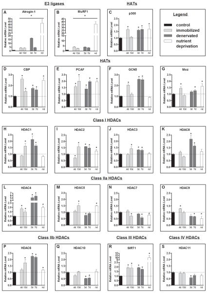
Introduction: Histone deacetylase (HDAC) proteins, which counter the activity of histone acetyltransferases (HATs), are necessary for normal muscle atrophy in response to several pathophysiological conditions. Despite this, it remains unknown whether a common or unique transcriptional profile of HDAC and HAT genes exist during the progression of muscle atrophy.

Methods: Muscles were harvested from cast immobilized, denervated, or nutrient deprived animals for quantitative reverse transcriptase-polymerase chain reaction analysis of HDAC and HAT gene expression.

Results: The mRNA levels of Hdac2, Hdac4, Hdac6, Sirt1, p300, Cbp, and Pcaf increased, and Hdac7 decreased in skeletal muscle in each experimental model of muscle atrophy. Hdac1 and Hdac3 were increased only in cast immobilized and denervated muscles.

Conclusions: While specific HDACs and HATs are increased in multiple models of muscle atrophy, increased expression of class I HDACs was unique to muscle disuse, reinforcing that specific HDAC inhibitors may be more effective than pan-HDAC inhibitors at countering muscle atrophy.

Keywords: denervation; histone acetyltransferase; histone deacetylase; muscle disuse; muscle wasting.


Conflict of interest statement

Conflict of Interests

No conflict of interests

Figures

Figure 1
Figure 1
Atrophy gene expression profile. Relative mRNA levels of; the E3 ligases (A) atrogin-1 (Mm_026346.3/Rn_133521.1) and (B) MuRF1 (Mm_001039048.2/Rn_080903.1), the HATs: (C) p300 (E1A binding protein p300; Mm_177821.6/Rn_576312.4), (D) Cbp (CREB (cAMP-responsive element binding protein)-binding protein; Mm_001025432.1/Rn_133381.3), (E) Pcaf (P300/CBP-associated factor; Mm_001177798.1/Rn_001107442.1), (F) Gcn5 (general control of amino-acid synthesis; Mm_029090.3/Rn_001107050.1), and (G) Moz (Monocytic leukemia zinc-finger protein; Mm_001081149.1/Rn_001100570.1); the class I Hdacs: (H–K) Hdac1 (Mm_008228.2/Rn_001025409.1), Hdac2 (Mm_008229.2/Rn_053447.1), Hdac3 (Mm_010411.2/Rn_053448.1), and Hdac8 (Mm_027382.3/Rn_001126373.2), class IIa Hdacs: (L–O) Hdac4 (Mm_207225.1/Rn_343629.4), Hdac5 (Mm_001077696/Rn_053450.1), Hdac7 (Mm_001204275.1/Rn_345868.4), Hdac9 (Mm_024124.3/Rn_001200045.1), class IIb Hdacs: (P& Q) Hdac6 (Mm_001130416.1/Rn_228753.5) and Hdac10 (Mm_199198.2/Rn_001035000.1), the class III Hdac: (R) Sirt1 (Mm_001159589.1/Rn_001080493.1) and the class IV Hdac: (S) Hdac11 (Mm_144919.2/Rn_001106610.2) from the soleus muscle of control and 4 day (4d) and 10 day (10d) immobilized mice, tibialis anterior (TA) muscle of control and 3 day (3d) and 7 day (7d) denervated rats, and nutrient deprived (nd) mice. All values were normalized to 18s. Bars represent means ± SE for 6 muscles/group. *P<0.05 vs control.

References

    1. Moresi V, Williams AH, Meadows E, Flynn JM, Potthoff MJ, McAnally J, Shelton JM, Backs J, Klein WH, Richardson JA, Bassel-Duby R, Olson EN. Myogenin and class II HDACs control neurogenic muscle atrophy by inducing E3 ubiquitin ligases. Cell. 2010;143(1):35–45. - PMC - PubMed
    1. Lee D, Goldberg AL. SIRT1 by blocking the activities of FoxO1 and 3 inhibits muscle atrophy and promotes muscle growth. J Biol Chem. 2013 - PMC - PubMed
    1. Beharry AW, Sandesara PB, Roberts BM, Ferreira LF, Senf SM, Judge AR. HDAC1 activates FoxO and is both sufficient and required for skeletal muscle atrophy. J Cell Sci. 2014;127(Pt 7):1441–1453. - PMC - PubMed
    1. Demos-Davies KM, Ferguson BS, Cavasin MA, Mahaffey JH, Williams SM, Spiltoir JI, Schuetze KB, Horn TR, Chen B, Ferrara C, Scellini B, Piroddi N, Tesi C, Poggesi C, Jeong MY, McKinsey TA. HDAC6 contributes to pathological responses of heart and skeletal muscle to chronic angiotensin-II signaling. American journal of physiology Heart and circulatory physiology. 2014;307(2):H252–258. - PMC - PubMed
    1. Senf SM, Sandesara PB, Reed SA, Judge AR. p300 Acetyltransferase activity differentially regulates the localization and activity of the FOXO homologues in skeletal muscle. Am J Physiol Cell Physiol. 2011;300(6):C1490–1501. - PMC - PubMed

Publication types