

Identification of a VxP Targeting Signal in the Flagellar Na+ /K+ -ATPase
- PMID: 26373354
- PMCID: PMC4715669
- DOI: 10.1111/tra.12332
Identification of a VxP Targeting Signal in the Flagellar Na+ /K+ -ATPase
Abstract
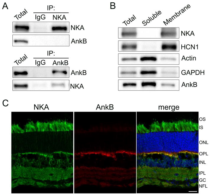
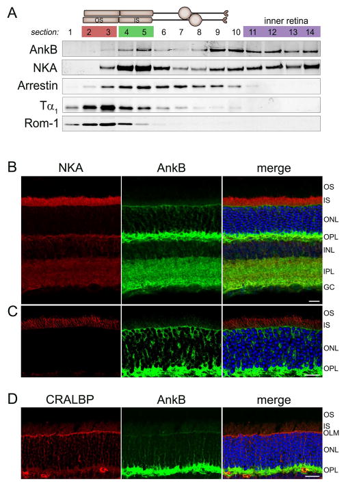
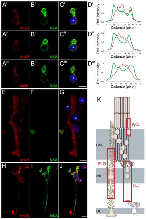
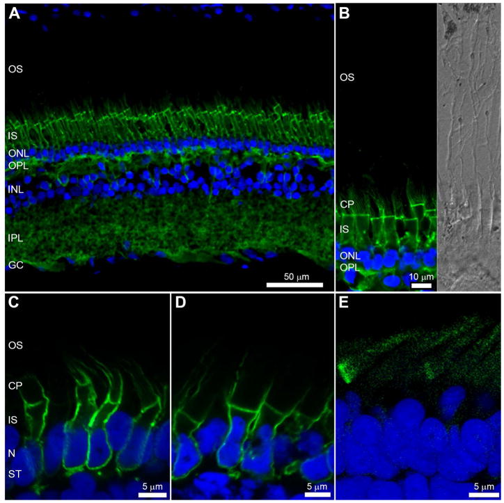
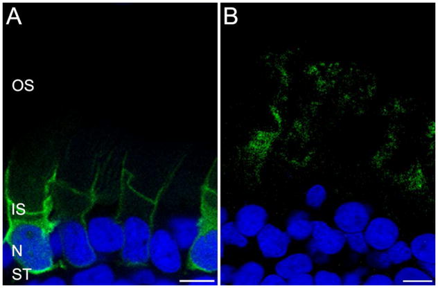
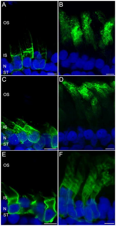
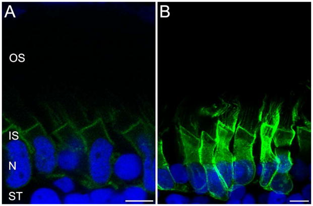
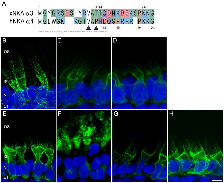
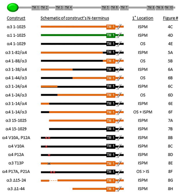
Na(+) /K(+) -ATPase (NKA) participates in setting electrochemical gradients, cardiotonic steroid signaling and cellular adhesion. Distinct isoforms of NKA are found in different tissues and subcellular localization patterns. For example, NKA α1 is widely expressed, NKA α3 is enriched in neurons and NKA α4 is a testes-specific isoform found in sperm flagella. In some tissues, ankyrin, a key component of the membrane cytoskeleton, can regulate the trafficking of NKA. In the retina, NKA and ankyrin-B are expressed in multiple cell types and immunostaining for each is striking in the synaptic layers. Labeling for NKA is also prominent along the inner segment plasma membrane (ISPM) of photoreceptors. NKA co-immunoprecipitates with ankyrin-B, but on a subcellular level colocalization of these two proteins varies dependent on the cell type. We used transgenic Xenopus laevis tadpoles to evaluate the subcellular trafficking of NKA in photoreceptors. GFP-NKA α3 and α1 are localized to the ISPM, but α4 is localized to outer segments (OSs). We identified a VxP motif responsible for the OS targeting by using a series of chimeric and mutant NKA constructs. This motif is similar to previously identified ciliary targeting motifs. Given the structural similarities between OSs and flagella, our findings shed light on the subcellular targeting of this testes-specific NKA isoform.
Keywords: ATP1a3; ATP1a4; ATPase; Muller glia; ankyrin; cilia; flagella; photoreceptors; retina; sperm; synapse; trafficking.
© 2015 John Wiley & Sons A/S. Published by John Wiley & Sons Ltd.
Conflict of interest statement
The authors have no conflict of interest to declare.
Figures
Similar articles
-
Ankyrin-B is required for coordinated expression of beta-2-spectrin, the Na/K-ATPase and the Na/Ca exchanger in the inner segment of rod photoreceptors.Exp Eye Res. 2009 Jan;88(1):57-64. doi: 10.1016/j.exer.2008.09.022. Epub 2008 Nov 1. Exp Eye Res. 2009. PMID: 19007774
-
Cellular and subcellular specification of Na,K-ATPase alpha and beta isoforms in the postnatal development of mouse retina.J Neurosci. 1999 Nov 15;19(22):9878-89. doi: 10.1523/JNEUROSCI.19-22-09878.1999. J Neurosci. 1999. PMID: 10559397 Free PMC article.
-
Disruption of Ankyrin B and Caveolin-1 Interaction Sites Alters Na+,K+-ATPase Membrane Diffusion.Biophys J. 2017 Nov 21;113(10):2249-2260. doi: 10.1016/j.bpj.2017.08.053. Epub 2017 Oct 5. Biophys J. 2017. PMID: 28988699 Free PMC article.
-
Identified and potential internalization signals involved in trafficking and regulation of Na+/K+ ATPase activity.Mol Cell Biochem. 2024 Jul;479(7):1583-1598. doi: 10.1007/s11010-023-04831-y. Epub 2023 Aug 27. Mol Cell Biochem. 2024. PMID: 37634170 Free PMC article. Review.
-
Crosstalk between dopamine receptors and the Na⁺/K⁺-ATPase (review).Mol Med Rep. 2013 Nov;8(5):1291-9. doi: 10.3892/mmr.2013.1697. Epub 2013 Sep 19. Mol Med Rep. 2013. PMID: 24065247 Review.
Cited by
-
Functional compartmentalization of photoreceptor neurons.Pflugers Arch. 2021 Sep;473(9):1493-1516. doi: 10.1007/s00424-021-02558-7. Epub 2021 Apr 20. Pflugers Arch. 2021. PMID: 33880652 Free PMC article. Review.
-
RPE Cells Engulf Microvesicles Secreted by Degenerating Rod Photoreceptors.eNeuro. 2020 May 21;7(3):ENEURO.0507-19.2020. doi: 10.1523/ENEURO.0507-19.2020. Print 2020 May/Jun. eNeuro. 2020. PMID: 32376599 Free PMC article.
-
The role of ATP1A3 gene in epilepsy: We need to know more.Front Cell Neurosci. 2023 Feb 13;17:1143956. doi: 10.3389/fncel.2023.1143956. eCollection 2023. Front Cell Neurosci. 2023. PMID: 36866063 Free PMC article. Review.
-
Retinoschisin and novel Na/K-ATPase interaction partners Kv2.1 and Kv8.2 define a growing protein complex at the inner segments of mammalian photoreceptors.Cell Mol Life Sci. 2022 Jul 25;79(8):448. doi: 10.1007/s00018-022-04409-9. Cell Mol Life Sci. 2022. PMID: 35876901 Free PMC article.
-
Disrupted Plasma Membrane Protein Homeostasis in a Xenopus Laevis Model of Retinitis Pigmentosa.J Neurosci. 2019 Jul 10;39(28):5581-5593. doi: 10.1523/JNEUROSCI.3025-18.2019. Epub 2019 May 6. J Neurosci. 2019. PMID: 31061086 Free PMC article.
References
-
- Hilgenberg LG, Su H, Gu H, O’Dowd DK, Smith MA. Alpha3Na+/K+-ATPase is a neuronal receptor for agrin. Cell. 2006;125(2):359–369. - PubMed
-
- Friedrich U, Stohr H, Hilfinger D, Loenhardt T, Schachner M, Langmann T, Weber BH. The Na/K-ATPase is obligatory for membrane anchorage of retinoschisin, the protein involved in the pathogenesis of X-linked juvenile retinoschisis. Hum Mol Genet. 2011;20(6):1132–1142. - PubMed
Publication types
MeSH terms
Substances
Grants and funding
LinkOut - more resources
Full Text Sources
Other Literature Sources
Molecular Biology Databases
Miscellaneous
