Review
. 2015 Oct;13(10):605-19.
doi: 10.1038/nrmicro3525.

Outer-membrane vesicles from Gram-negative bacteria: biogenesis and functions

Affiliations
Review

Outer-membrane vesicles from Gram-negative bacteria: biogenesis and functions

Carmen Schwechheimer et al. Nat Rev Microbiol. 2015 Oct.

Abstract

Outer-membrane vesicles (OMVs) are spherical buds of the outer membrane filled with periplasmic content and are commonly produced by Gram-negative bacteria. The production of OMVs allows bacteria to interact with their environment, and OMVs have been found to mediate diverse functions, including promoting pathogenesis, enabling bacterial survival during stress conditions and regulating microbial interactions within bacterial communities. Additionally, because of this functional versatility, researchers have begun to explore OMVs as a platform for bioengineering applications. In this Review, we discuss recent advances in the study of OMVs, focusing on new insights into the mechanisms of biogenesis and the functions of these vesicles.


Conflict of interest statement

statement The authors declare no competing interests.

Figures

Figure 1
Figure 1. The composition of the Gram-negative cell envelope
The cell envelope of Gram-negative bacteria consists of two membranes, the outer membrane and the cytoplasmic membrane. The cytoplasmic membrane is composed of a phospholipid bilayer, whereas the outer membrane comprises an interior leaflet of phospholipids and an exterior leaflet of lipopolysaccharide (LPS); LPS is composed of lipid A, the core oligosaccharide and O antigen. In between the two membranes is the periplasmic space, which contains the peptidoglycan (PG) layer and periplasmic proteins. The PG layer comprises long polymers of the repeating disaccharide N-acetylglucosamine–N-acetylmuramic acid (NAG–NAM) that are linked via peptide bridges: both traditional 4–3 (D-Ala–meso-diaminopimelic acid (mDAP)) crosslinks and non-traditional 3–3 (mDAP–mDAP) crosslinks. Envelope proteins can be soluble (periplasmic; orange and red spheres), transmembrane proteins (pink ovals and cyan cylinder) or anchored into the leaflet of either membrane via covalently attached lipid appendages (lipoproteins; green and blue ovals). Envelope stability comes from various crosslinks: the covalent crosslinking of Braun’s lipoprotein (Lpp) in the outer membrane with the PG; the non-covalent interactions between the PG and the porin outer-membrane protein A (OmpA); and the non-covalent interactions between the PG and the Tol–Pal (peptidoglycan-associated lipoprotein) complex, which is composed of TolA, TolB, TolQ, TolR and Pal, and spans the envelope from the cytoplasmic membrane across the periplasm to the outer membrane.
Figure 2
Figure 2. Outer-membrane vesicle biogenesis and cargo selection
Several factors influence the biogenesisof outer-membrane vesicles (OMVs). a | Peptidoglycan (PG) endopeptidases and other enzymes that are involved in regulating PG breakdown and synthesis govern the ability of the envelope to form crosslinks between Braun’s lipoprotein (Lpp) and PG. Therefore, OMV production is increased in areas with reduced Lpp–PG crosslinks. b | Meso-diaminopimelic acid (mDAP)–mDAP crosslinks within the PG also control the formation of crosslinks between PG and the outer membrane. Accordingly, sites where the PG lacks mDAP–mDAP crosslinks have more Lpp–PG crosslinks, which reduces OMV production. c | In areas where misfolded proteins or envelope components (such as lipopolysaccharide (LPS) or PG fragments) accumulate, crosslinks are either displaced or locally depleted, promoting bulging of these outer-membrane nanoterritories and leading to increased OMV production. d | Some areas of the outer membrane can become enriched in particular types of LPS, phospholipids and/or specific LPS-associated molecules. These lipid microdomains have a propensity to bulge outwards owing to their charge, their cargo or increased membrane fluidity, and this bulging results in increased OMV production. e | Insertion of Pseudomonas quinolone signal (PQS) into the outer leaflet of the outer membrane can also increase membrane curvature and lead to the formation on OMVs. f | Envelope components may be enriched in OMVs because of direct or indirect interactions with integral or auxiliary outer-membrane components that are prone to budding. By contrast, envelope components may be excluded from OMVs by direct or indirect interactions with integral or auxiliary envelope components that are not prone to budding. FA, fatty acid.
Figure 3
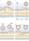
Figure 3. Functions of outer-membrane vesicles in bacterial physiology
Outer-membrane vesicles (OMVs) function in multiple pathways that promote bacterial survival. a | OMVs can serve as a mechanism to remove toxic compounds, such as misfolded proteins, from bacterial cells under stress conditions. b | Stress conditions can increase OMV production. For example, exposure to antibiotics can induce DNA breaks, which triggers an SOS response. As part of the SOS response, changes in the synthesis of lipopolysaccharide (LPS) can alter the composition of the outer membrane and increase the production of OMVs. c | OMVs can serve as sources of carbon and nitrogen, and can carry and disseminate enzymes that break down complex macromolecules to provide the cell with essential nutrients. d | OMVs can also carry iron and zinc acquisition systems that are able to bind these metals in the environment, providing the bacteria with access to these essential compounds.
Figure 4
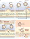
Figure 4. Functions of outer-membrane vesicles in pathogenesis
Outer-membrane vesicles (OMVs) can increase bacterial pathogenicity via multiple mechanisms. a | OMVs can increase bacterial resistance to antibiotics and phages by serving as decoy targets for these molecules, thus protecting the bacteria cell. b | OMVs can also transfer DNA between cells, including antibiotic-resistance genes, and can carry enzymes that degrade antibiotics. c | Pathogenic Gram-negative bacteria are thought to utilize OMVs to interact with host cells during infection. For example, bacteria can use OMVs to mediate the delivery of virulence factors, such as toxins, into host cells, including immune cells. d | OMVs can also cross the mucus barrier in the gut and reach the intestinal epithelium, delivering bacterial antigens to the underlying macrophages, which triggers intestinal inflammation.

References

    1. Kulp A, Kuehn MJ. Biological functions and biogenesis of secreted bacterial outer membrane vesicles. Annu Rev Microbiol. 2010;64:163–184. - PMC - PubMed
    1. Schwechheimer C, Sullivan CJ, Kuehn MJ. Envelope control of outer membrane vesicle production in Gram-negative bacteria. Biochemistry. 2013;52:3031–3040. This review highlights the challenges of elucidating mechanistic details in OMV production. - PMC - PubMed
    1. Zhou L, Srisatjaluk R, Justus DE, Doyle RJ. On the origin of membrane vesicles in Gram-negative bacteria. FEMS Microbiol Lett. 1998;163:223–228. - PubMed
    1. Hickey CA, et al. Colitogenic Bacteroides thetaiotaomicron antigens access host immune cells in a sulfatase-dependent manner via outer membrane vesicles. Cell Host Microbe. 2015;17:672–680. - PMC - PubMed
    1. Beveridge TJ. Structures of gram-negative cell walls and their derived membrane vesicles. J Bacteriol. 1999;181:4725–4733. - PMC - PubMed

Publication types

MeSH terms