. 2015 Sep 12;17(1):251.
doi: 10.1186/s13075-015-0753-8.

The inhibitory effects of a RANKL-binding peptide on articular and periarticular bone loss in a murine model of collagen-induced arthritis: a bone histomorphometric study

Affiliations

The inhibitory effects of a RANKL-binding peptide on articular and periarticular bone loss in a murine model of collagen-induced arthritis: a bone histomorphometric study

Genki Kato et al. Arthritis Res Ther. .

Abstract

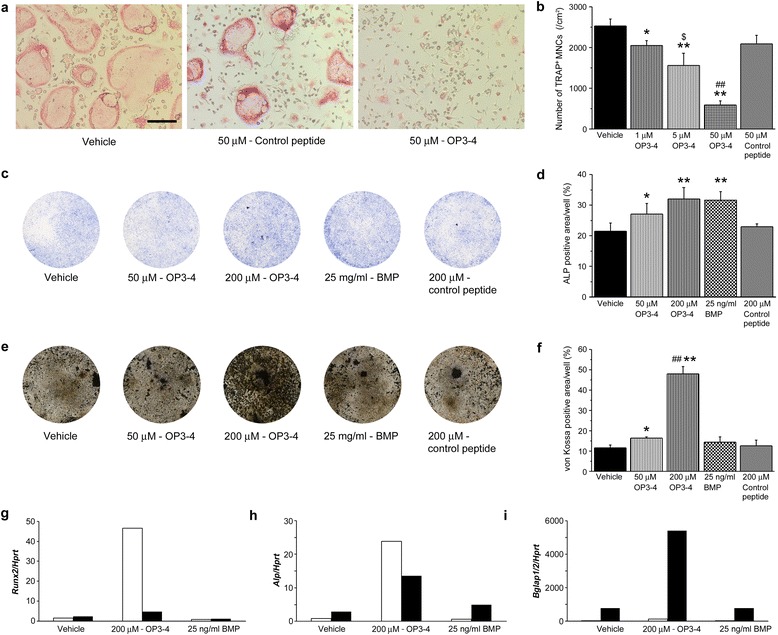
Introduction: We designed OP3-4 (YCEIEFCYLIR), a cyclic peptide, to mimic the soluble osteoprotegerin (OPG), and was proven to bind to RANKL (receptor activator of NF-κB ligand), thereby inhibiting osteoclastogenesis. We recently found that another RANKL binding peptide, W9, could accelerate bone formation by affecting RANKL signaling in osteoblasts. We herein demonstrate the effects of OP3-4 on bone formation and bone loss in a murine model of rheumatoid arthritis.

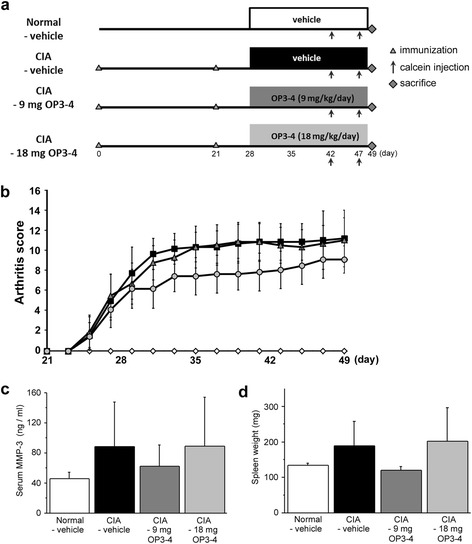
Methods: Twenty-four seven-week-old male DBA/1J mice were used to generate a murine model of collagen-induced arthritis (CIA). Then, vehicle or OP3-4 (9 mg/kg/day or 18 mg/kg/day) was subcutaneously infused using infusion pumps for three weeks beginning seven days after the second immunization. The arthritis score was assessed, and the mice were sacrificed on day 49. Thereafter, radiographic, histological and biochemical analyses were performed.

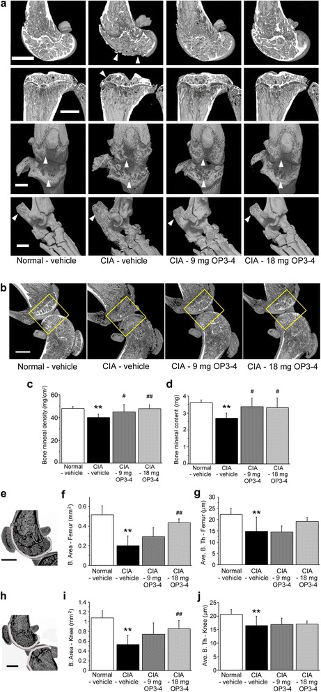
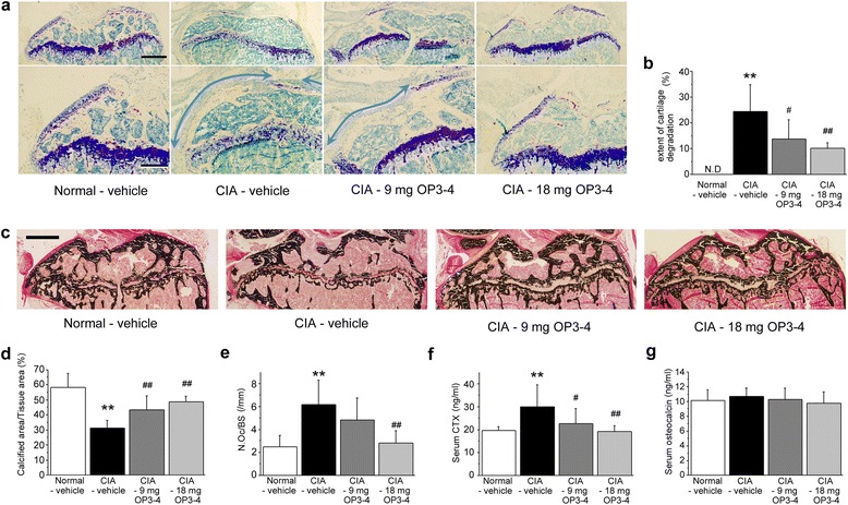
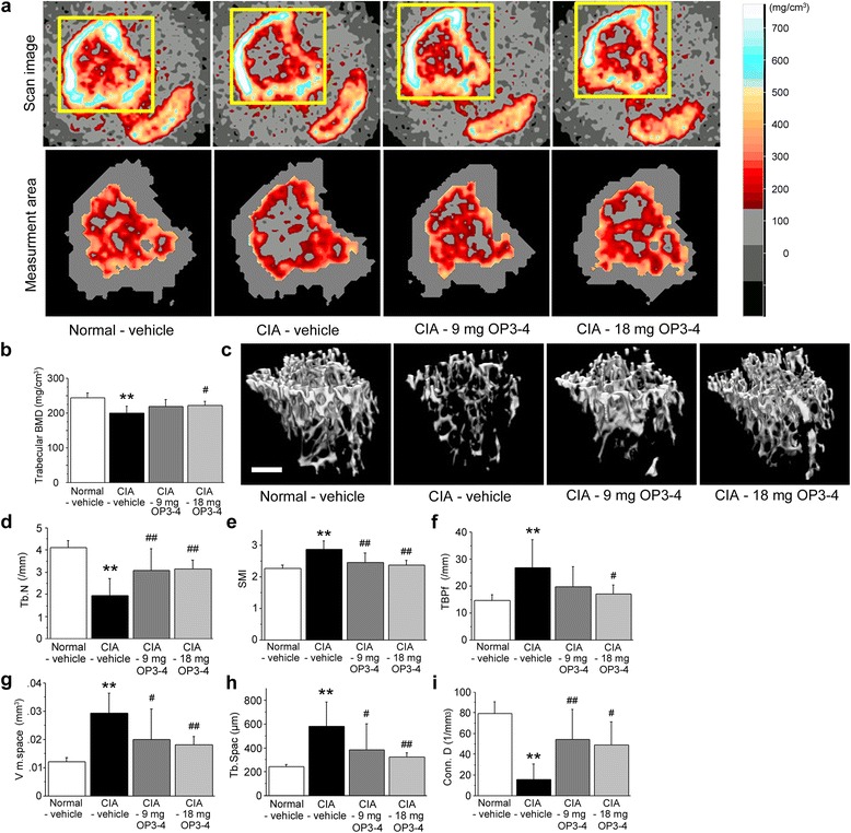
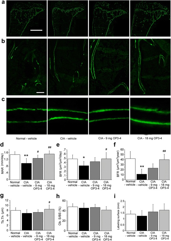
Results: The OP3-4 treatment did not significantly inhibit the CIA-induced arthritis, but limited bone loss. Micro-CT images and quantitative measurements of the bone mineral density revealed that 18 mg/kg/day OP3-4 prevented the CIA-induced bone loss at both articular and periarticular sites of tibiae. As expected, OP3-4 significantly reduced the CIA-induced serum CTX levels, a marker of bone resorption. Interestingly, the bone histomorphometric analyses using undecalcified sections showed that OP3-4 prevented the CIA-induced reduction of bone formation-related parameters at the periarticular sites.

Conclusion: The peptide that mimicked OPG prevented inflammatory bone loss by inhibiting bone resorption and stimulating bone formation. It could therefore be a useful template for the development of small molecule drugs for inflammatory bone loss.


Figures

Fig. 1
Fig. 1
Direct effects of OP3-4 on osteoclastogenesis and osteoblast differentiation. a Microscopic view of osteoclast formation in vitro. TRAP-positive cells are shown (red). Bone marrow macrophages were treated with 25 ng/ml M-CSF and 50 ng/ml RANKL for 4 days. Scale bar represents 100 μm. b Number of TRAP-positive multinucleated cells. Data presented as mean ± standard deviation (SD). *p <0.05, **p <0.01 vs. vehicle control, $ p <0.05 vs. 1 μM OP3-4, ## p <0.01 vs. 5 μM OP3-4. c ALP-positive cells in the cultures of primary osteoblastic cells on day 7. d Percentage of ALP-stained areas in each well. Data presented as mean ± SD. *p <0.05, **p <0.01 vs. vehicle control. e von Kossa-positive cells in the cultures of primary osteoblastic cells on day 21. f Percentage of von Kossa-stained areas in each well. Data presented as mean ± SD. *p <0.05, **p <0.01 vs. vehicle control; ## p <0.01 vs. 50 μM OP3-4. All cultures were performed at least three times independently, and similar data were obtained each time. Representative images of cultures and quantitative data are provided (n = 3–4/group in each independent experiment). g–i Gene expression analyses of Runx2 g, Alp h, and Bglap1/2 i by quantitative PCR were performed using the cells on day 5 (white bar) and day 14 (black bar). BMP bone morphogenetic protein
Fig. 2
Fig. 2
The in-vivo experimental protocol, the clinical score, and the inflammatory indices. a Collagen-induced arthritis (CIA) was induced by primary (day 0) and secondary (day 21) immunizations with bovine type II collagen in CFA. Infusion pumps were implanted subcutaneously in mice with CIA on day 28. b Arthritis scores (clinical severity of arthritis). The maximum possible score is 16, as described in Materials and methods. Shown are the results of vehicle-treated mice with CIA (closed squares), mice with CIA treated with 9 mg/kg/day OP3-4 peptide (closed triangles), mice with CIA treated with 18 mg/kg/day OP3-4 peptide (closed circles), and vehicle-treated nonimmunized mice (open diamond). Values are mean ± SD (n = 6 mice per group). c Serum matrix metalloproteinase (MMP)-3 levels on day 49. d The spleen weight was measured on day 49. Normal-vehicle group, nonimmunized mice receiving vehicle (20 % DMSO); CIA-vehicle group, immunized mice receiving vehicle (20 % DMSO); CIA-9 mg OP3-4 group, immunized mice receiving 9 mg/kg/day OP3-4 peptide; CIA-18 mg OP3-4 group, immunized mice receiving 18 mg/kg/day OP3-4 peptide
Fig. 3
Fig. 3
Radiographic observations of femurs, tibiae, knee joints, and ankle joints. a μCT was used to clarify the structural changes of bones. Arrowheads, erosion surface or site of the destruction of bones. OP3-4 appeared to inhibit the bone loss induced by collagen-induced arthritis (CIA). Scale bar represents 1 mm. b ROIs used for the quantitative measurements of mineralized tissue at knee joints by DXA. The square areas (2 × 2 mm) are the ROIs used for the DXA analyses. c BMD and d bone mineral content at the joint. The regions of interest shown in light gray for measuring the indices (f/g and i/j) to show the changes of e femur epiphysis and h knee joint, respectively. Data presented as mean ± SD (n = 6). **p <0.01 vs. Normal-vehicle; # p <0.05, ## p <0.01 vs. CIA-vehicle. Normal-vehicle group, nonimmunized mice receiving vehicle (20 % DMSO); CIA-vehicle group, immunized mice receiving vehicle (20 % DMSO); CIA-9 mg OP3-4 group, immunized mice receiving 9 mg/kg/day OP3-4 peptide; CIA-18 mg OP3-4 group, immunized mice receiving 18 mg/kg/day OP3-4 peptide
Fig. 4
Fig. 4
Histological observations and quantitative analyses of the articular sites of tibiae. a Representative microscopic images of toluidine blue-stained undecalcified sections. The area of metachromasia after toluidine blue staining was decreased in the CIA-vehicle group. Arrows, length of the cartilage degradation site facing the proximal end of the tibiae. Scale bars represent 500 μm and 250 μm in the upper and lower panels, respectively. b The extent of cartilage degradation = (length of cartilage degradation surface) / (total length of proximal end of tibial epiphysis, excluding anterior intercondylar area) × 100. c von Kossa-stained sections at the tibial epiphysis. d Calcified area measurements at the tibial epiphysis obtained by bone histomorphometry. Scale bar represents 1 mm. e Osteoclast number per bone surface (N.Oc/BS) at the tibial epiphysis. f Serum levels of C-terminal telopeptides of type I collagen (CTX-I) on day 49. g Serum levels of osteocalcin on day 49. Data presented as mean ± SD (n = 6). **p <0.01 vs. Normal-vehicle; # p <0.05, ## p <0.01 vs. CIA-vehicle. Normal-vehicle group, nonimmunized mice receiving vehicle (20 % DMSO); CIA-vehicle group, immunized mice receiving vehicle (20 % DMSO); CIA-9 mg OP3-4 group, immunized mice receiving 9 mg/kg/day OP3-4 peptide; CIA-18 mg OP3-4 group, immunized mice receiving 18 mg/kg/day OP3-4 peptide. CIA collagen-induced arthritis
Fig. 5
Fig. 5
OP3-4 prevented CIA-induced peripheral bone loss at the tibial metaphysis. a, b pQCT was used to measure the bone mineral density (BMD) at the secondary spongiosa of the tibiae. Upper panels, representative scan images from the pQCT analyses; lower panels, trabecular area for each group; right panel, color table for the BMD. c μCT reconstruction images of the tibial metaphysis. Scale bar represents 200 μm. d Trabecular number (Tb.N; measure of the average number of trabeculae per unit length). e Structure model index (SMI; an indicator of the structure of trabeculae). SMI will be 0 for parallel plates and 3 for cylindrical rods. f Trabecular bone pattern factor (TBPf; an indicator of morphological changes of trabecular surface). g Marrow space star volume (V m. space; an indicator of osteoporotic changes). h Trabecular spacing (Tb.Spac). i Degree of connectivity of trabeculae normalized by tissue volume (Conn.D). Data expressed as mean ± SD (n = 6) for each group. **p <0.01 vs. Normal-vehicle; # p <0.05, ## p <0.01 vs. CIA-vehicle. Normal-vehicle group, nonimmunized mice receiving vehicle (20 % DMSO); CIA-vehicle group, immunized mice receiving vehicle (20 % DMSO); CIA-9 mg OP3-4 group, immunized mice receiving 9 mg/kg/day OP3-4 peptide; CIA-18 mg OP3-4 group, immunized mice receiving 18 mg/kg/day OP3-4 peptide. CIA collagen-induced arthritis
Fig. 6
Fig. 6
Fluorescent images of undecalcified sections of the periarticular sites in tibiae. a Representative fluorescent images of the proximal tibiae. Scale bar represents 1 mm. b Representative fluorescent images of the secondary spongiosa of the tibiae. Scale bar represents 50 μm. c Magnified images of calcein double labeling of the trabecular bone. d Mineral apposition rate (MAR)/day. e Bone formation rate (BFR)/day in a bone surface reference. f BFR/year in a total tissue reference. g Trabecular thickness (Tb.Th). h Osteoblast surface per bone surface (Ob.S/BS). i Labeling surface at epiphysis. Data expressed as mean ± SD (n = 6) for each group. *p <0.05, **p <0.01 vs. Normal-vehicle; # p <0.05, ## p <0.01 vs. CIA-vehicle. Normal-vehicle group, nonimmunized mice receiving vehicle (20 % DMSO); CIA-vehicle group, immunized mice receiving vehicle (20 % DMSO); CIA-9 mg OP3-4 group, immunized mice receiving 9 mg/kg/day OP3-4 peptide; CIA-18 mg OP3-4 group, immunized mice receiving 18 mg/kg/day OP3-4 peptide. CIA collagen-induced arthritis

References

    1. Graudal N, Jurgens G. Similar effects of disease-modifying antirheumatic drugs, glucocorticoids, and biologic agents on radiographic progression in rheumatoid arthritis: meta-analysis of 70 randomized placebo-controlled or drug-controlled studies, including 112 comparisons. Arthritis Rheum. 2010;62:2852–63. doi: 10.1002/art.27592. - DOI - PubMed
    1. Seriolo B, Paolino S, Sulli A, Ferretti V, Cutolo M. Bone metabolism changes during anti-TNF-alpha therapy in patients with active rheumatoid arthritis. Ann N Y Acad Sci. 2006;1069:420–7. doi: 10.1196/annals.1351.040. - DOI - PubMed
    1. Breuil V, Euller-Ziegler L. Bisphosphonate therapy in rheumatoid arthritis. Joint Bone Spine. 2006;73:349–54. doi: 10.1016/j.jbspin.2005.10.019. - DOI - PubMed
    1. Romas E. Bone loss in inflammatory arthritis: mechanisms and therapeutic approaches with bisphosphonates. Best Pract Res Clin Rheumatol. 2005;19:1065–79. doi: 10.1016/j.berh.2005.06.008. - DOI - PubMed
    1. Somford MP, Draijer FW, Thomassen BJ, Chavassieux PM, Boivin G, Papapoulos SE. Bilateral fractures of the femur diaphysis in a patient with rheumatoid arthritis on long-term treatment with alendronate: clues to the mechanism of increased bone fragility. J Bone Miner Res. 2009;24:1736–40. doi: 10.1359/jbmr.090408. - DOI - PubMed

Publication types

MeSH terms