. 2015 Oct;8(7):438-48.
doi: 10.1007/s12265-015-9654-0. Epub 2015 Sep 15.

Intravenous Followed by X-ray Fused with MRI-Guided Transendocardial Mesenchymal Stem Cell Injection Improves Contractility Reserve in a Swine Model of Myocardial Infarction

Affiliations

Intravenous Followed by X-ray Fused with MRI-Guided Transendocardial Mesenchymal Stem Cell Injection Improves Contractility Reserve in a Swine Model of Myocardial Infarction

Eric G Schmuck et al. J Cardiovasc Transl Res. 2015 Oct.

Abstract

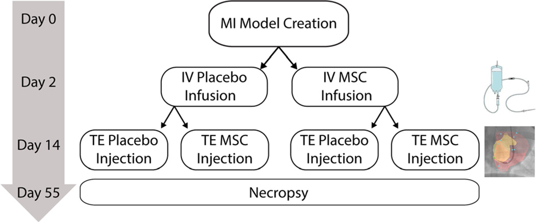
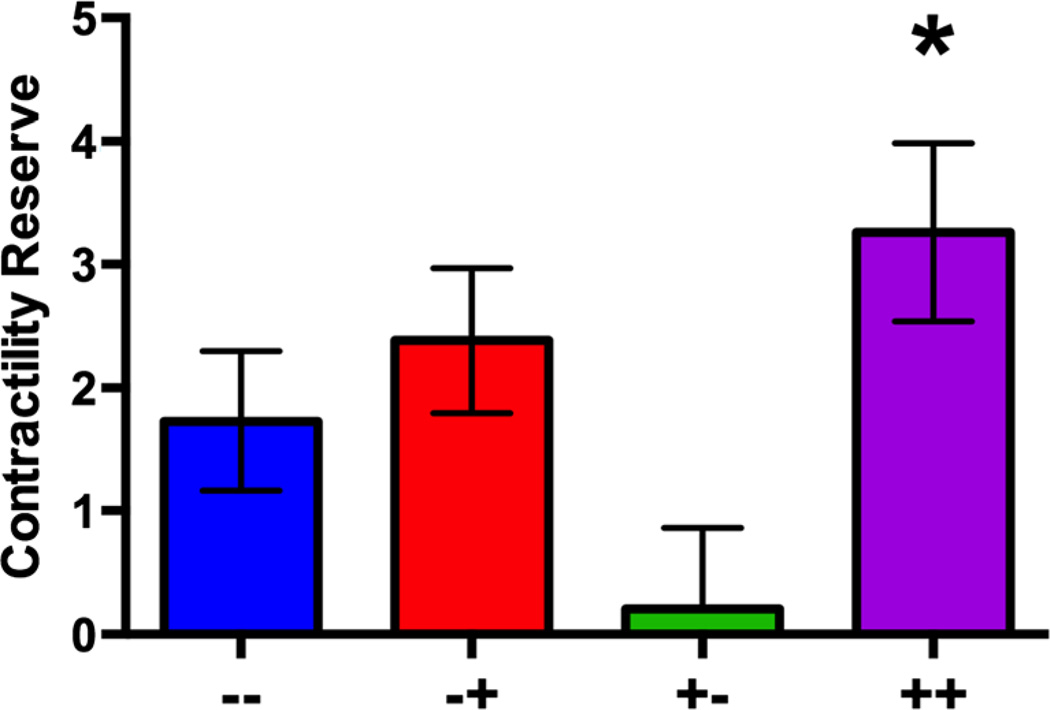
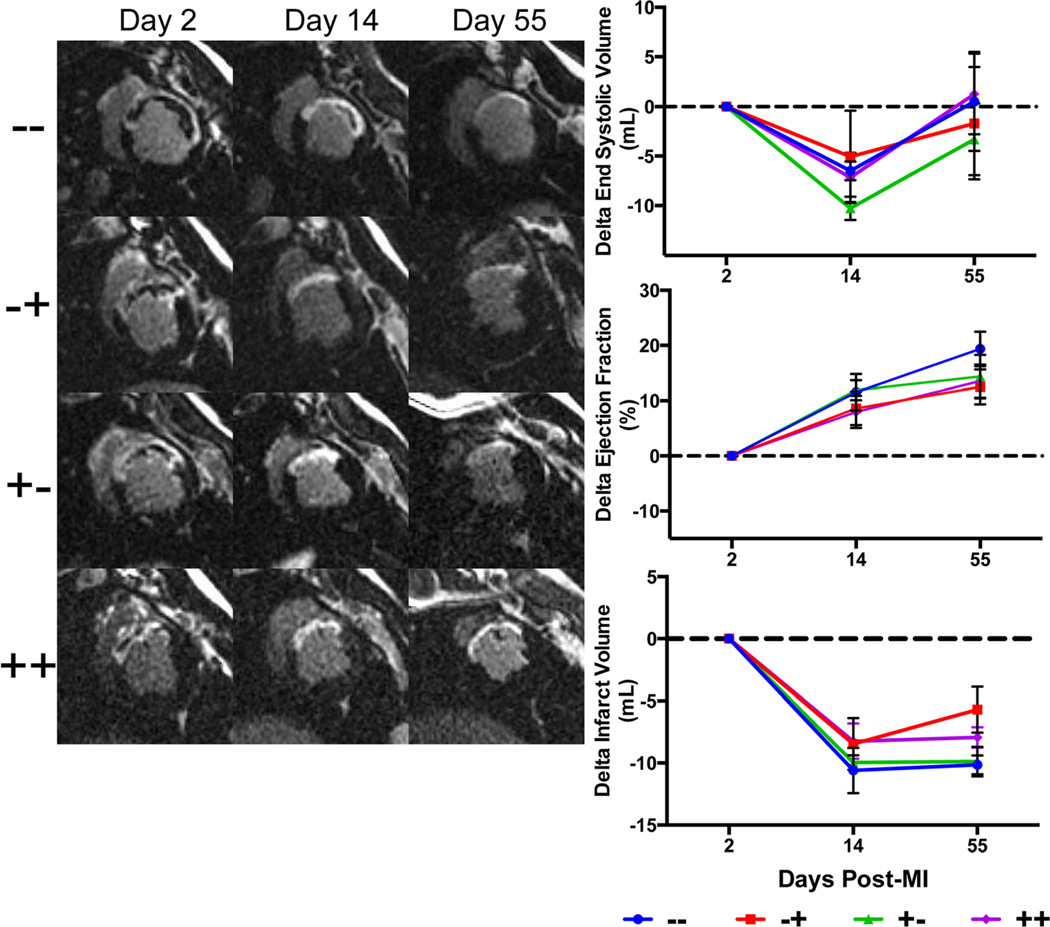
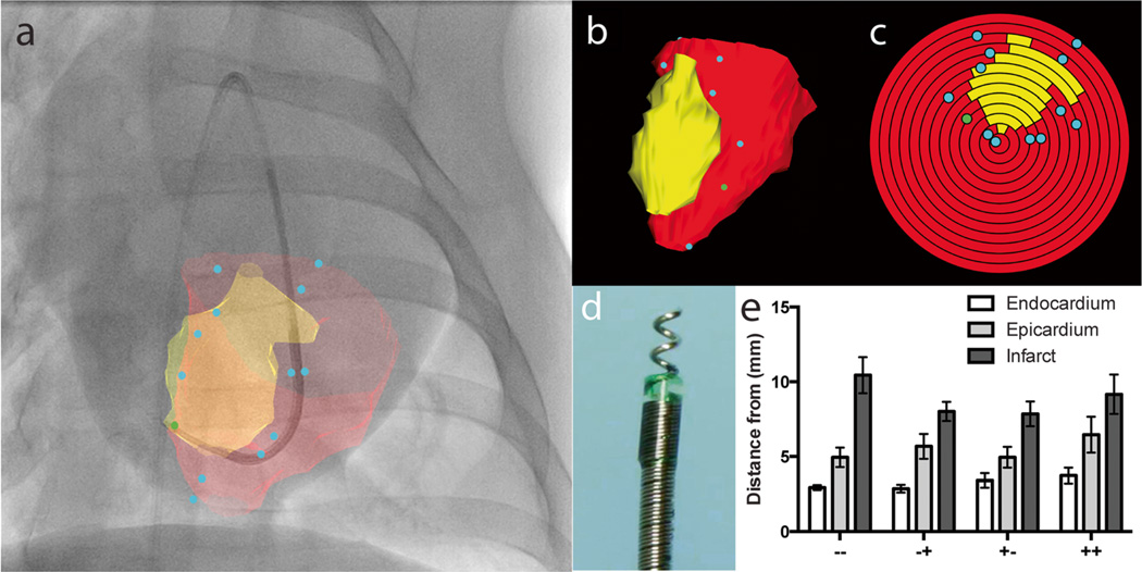
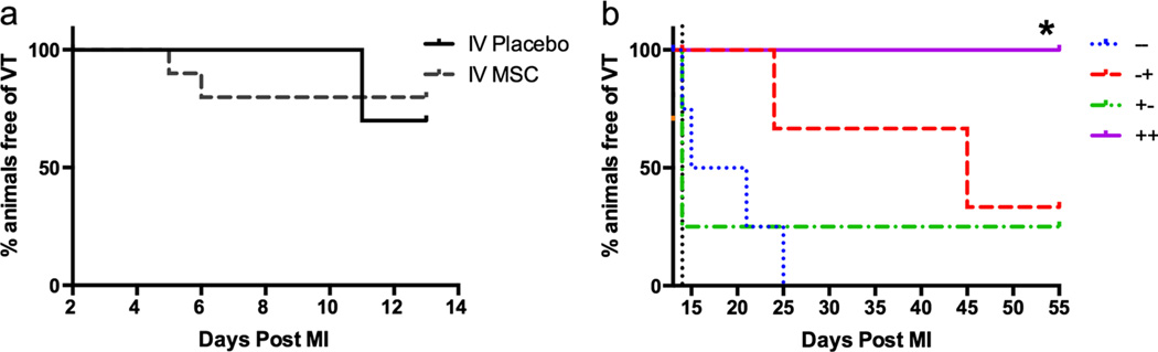
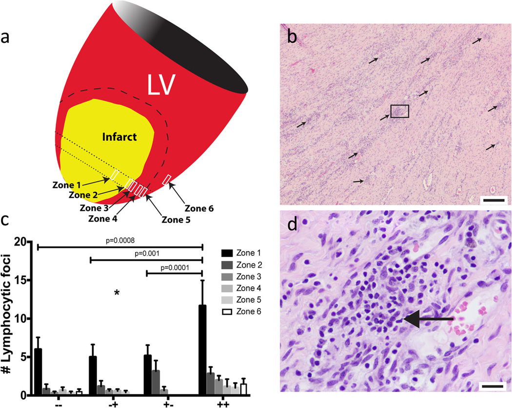
The aim of this study is to determine the effects of early intravenous (IV) infusion later followed by transendocardial (TE) injection of allogeneic mesenchymal stem cells (MSCs) following myocardial infarction (MI). Twenty-four swine underwent balloon occlusion reperfusion MI and were randomized into 4 groups: IV MSC (or placebo) infusion (post-MI day 2) and TE MSC (or placebo) injection targeting the infarct border with 2D X-ray fluoroscopy fused to 3D magnetic resonance (XFM) co-registration (post-MI day 14). Continuous ECG recording, MRI, and invasive pressure-volume analyses were performed. IV MSC plus TE MSC treated group was superior to other groups for contractility reserve (p = 0.02) and freedom from VT (p = 0.03) but had more lymphocytic foci localized to the peri-infarct region (p = 0.002). No differences were observed in post-MI remodeling parameters. IV followed by XFM targeted TE MSC therapy improves contractility reserve and suppresses VT but does not affect post-MI remodeling and may cause an immune response.

Keywords: Allogeneic; Immune response; Intravenous; Mesenchymal stem cell; Multiple dose; Myocardial infarction; Stem cell; Swine; Transendocardial.


Conflict of interest statement

The authors declare that they have no competing interests.

Figures

Fig. 1
Fig. 1
Myocardial infarction was induced by closed chest, balloon occlusion reperfusion of the LAD. Thirty-six Yorkshire swine underwent 90 min of ischemia. Twenty-four animals survived the model creation to be randomized into treatment groups. Two days post-MI swine underwent a cardiac MRI and were given either an IV infusion of porcine MSCs or a placebo. Fourteen days post-MI swine underwent a second cardiac MRI and received XFM guided transendocardial (TE) MSC injections or placebo injections. The study design resulted in four treatment groups: IV placebo plus TE placebo (−−) (n=6), IV placebo plus TE MSC (−+) (n=6), IV MSC plus TE placebo (+−) (n=6), and IV MSC plus TE MSC (++) n=6). Swine were survived out to 55 days post-MI at which time a follow-up cardiac MRI and invasive pressure-volume loop analysis was carried out prior to necropsy
Fig. 2
Fig. 2
a Contractile reserve measured was the greatest in the IV MSC plus TE MSC group (++ compared to all others (*p=0.02) and the worst in the IV MSC plus TE placebo group (+−)
Fig. 3
Fig. 3
Representative delayed enhancement cardiac MRI images at 2, 14 and 55 days post-MI for each group. Note the thinning of the infarct (white section of the LV wall) and dilation of the LV between day 2 and 55. End-systolic volumes (top) were reduced at day 14, but returned to baseline (day 2 post-MI) by day 55. Ejection fractions (middle) increased in all groups at day 14 and 55 compared to day 2 post-MI due to myocardial stunning effects. Infarct volumes reduced from day 2 to day 14 due to resolving myocardial edema. IV placebo plus TE placebo (−−), IV Placebo plus TE MSC (−+), IV MSC plus TE placebo (+−), IV MSC plus TE MSC (++)
Fig. 4
Fig. 4
Bi-plane X-ray fluoroscopy fused with MRI guided transendocardial injections. a Intraoperative display of MRI endocardial (red) and infarct (yellow) surface registered and projected on to live X-ray fluoroscopy in the anterior-posterior view. Blue dots indicate the injection locations. b Three dimensional surface rendered volume in an oblique view and c corresponding polar plot of injection locations. The apex of the ventricle represents the center of the polar plot. d Corkscrew-shaped needle tipped Helix transendocardial injection catheter. e Mean distances of injection location to the closest endocardial, epicardial, and infarct surface across all treatment groups. IV placebo plus TE placebo (−−), IV Placebo plus TE MSC (−+), IV MSC plus TE placebo (+−), and IV MSC plus TE MSC (++)
Fig. 5
Fig. 5
Time to sustained fast ventricular tachycardia. a Day 2 to day 14 analysis on the effects of IV MSC therapy on median time to first ventricular tachycardia event (IV placebo (n=12) and IV MSC (n=12)). IV MSC treatment had no significant effect on median time to sustained fast ventricular tachycardia (p=0.56). b Day 14 to day 55 analysis: When data was analyzed from day 14 (The vertical dotted black line), there was a significant reduction in VT in the ++ group compared to other groups (p=0.033)
Fig. 6
Fig. 6
a Systematic histology assessment, sampled from six zones spanning from the infarct (zone 1), radiating outwardly across the border zone (zones 2– 5), and the unaffected interventricular septum (zone 6). b Representative 4× image of diffuse lymphocytic foci (black arrows) in the ++ treated group (scale bar=200 mm). c Average number of lymphocytic foci in each group per zone. There was a significant increase in lymphocytic foci in the ++ group compared to the other groups (p= 0.002) with the greatest difference in the infarct zone (zone 1). d High power (40×) image of the lymphocytic foci in the box in panel B, the small round deeply purple nuclei are lymphocytes (scale bar=20 mm)

References

    1. Burchfield JS, Xie M, Hill JA. Pathological ventricular remodeling: mechanisms: part 1 of 2. Circulation. 2013;128(4):388–400. - PMC - PubMed
    1. Xie M, Burchfield JS, Hill JA. Pathological ventricular remodeling: therapies: part 2 of 2. Circulation. 2013;128(9):1021–1030. - PMC - PubMed
    1. Yeh RW, et al. Population trends in the incidence and outcomes of acute myocardial infarction. New England Journal of Medicine. 2010;362(23):2155–2165. - PubMed
    1. Goodrich AD, Hematti P. Mesenchymal stem cell therapies: the quest for fine-tuning. Experimental Dermatology. 2014;23(9):632–633. - PubMed
    1. Pittenger MF, et al. Multilineage potential of adult human mesenchymal stem cells. Science. 1999;284(5411):143–147. - PubMed

Publication types