

Cancer cell invasion driven by extracellular matrix remodeling is dependent on the properties of cancer-associated fibroblasts
- PMID: 26374424
- PMCID: PMC11819212
- DOI: 10.1007/s00432-015-2046-7
Cancer cell invasion driven by extracellular matrix remodeling is dependent on the properties of cancer-associated fibroblasts
Abstract
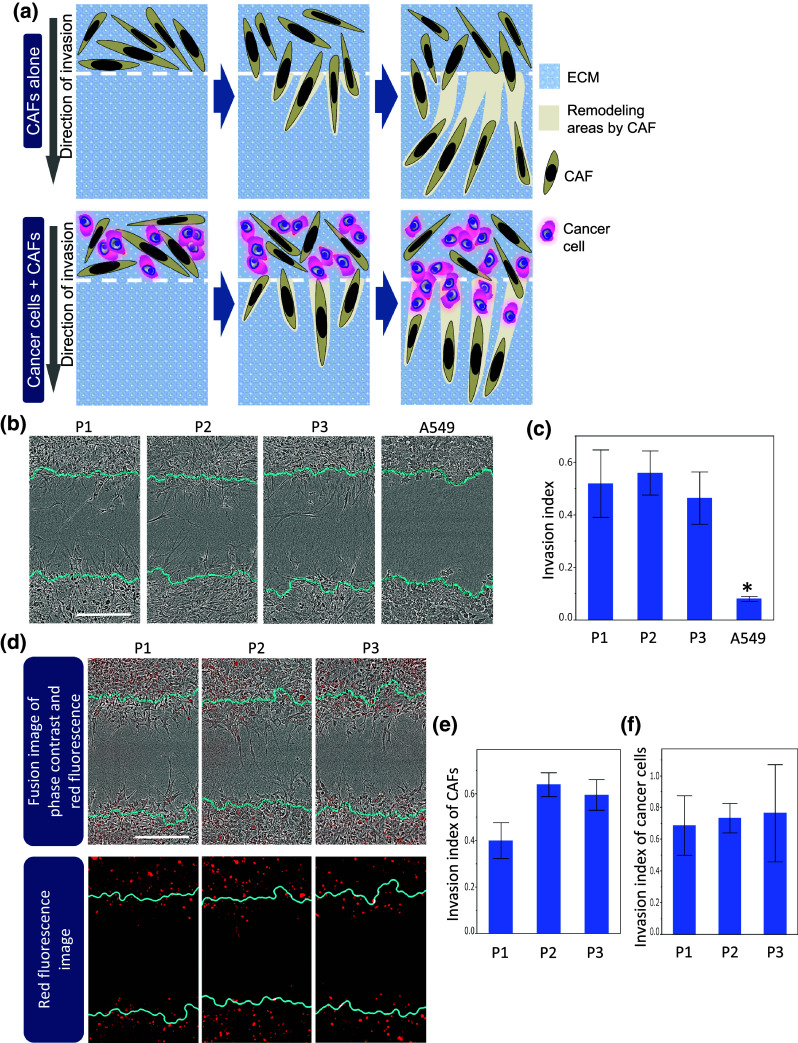
Purpose: As one form of tumor invasion, cancer cells can invade the extracellular matrix (ECM) through tracks that have been physically remodeled by cancer-associated fibroblasts (CAFs). However, CAFs are a heterogeneous population with diverse matrix-remodeling capacities. The purpose of this study was to investigate how CAFs with various matrix-remodeling capacities influence cancer cell invasion.
Methods: We established single-cell-derived clones from three primary cultures of CAFs from lung adenocarcinoma patients (Case 1, 5 clones; Case 2, 5 clones; and Case 3, 7 clones). Using a co-culture model, we evaluated the correlations between the number of invaded cancer cells and the remodeling areas generated by CAF clones in each case.
Results: When A549 lung adenocarcinoma cells and CAF clones were co-cultured, both the numbers of invaded cancer cells and the remodeling areas generated by the CAF clones varied greatly. The number of invaded cancer cells was moderately and strongly correlated with the remodeling areas generated by each CAF clone originating from Cases 1 and 2 (R(2) value = 0.53 and 0.68, respectively), suggesting that the remodeling areas in the ECM may determine the number of invaded cancer cells. In contrast, the number of invaded cancer cells was not correlated with the remodeling areas generated by CAF clones originating from Case 3, suggesting that factors other than the remodeling areas might determine the number of invading cancer cells.
Conclusions: These findings showed two types of fibroblast-dependent cancer cell invasion that are dependent on and independent of the remodeling areas generated by CAFs.
Keywords: Cancer-associated fibroblasts; Extracellular matrix remodeling; Fibroblast-dependent cancer invasion; Single-cell-derived clones.
Conflict of interest statement
The authors have no conflicts of interest to disclose.
Figures


References
-
- Allen M, Louise Jones J (2011) Jekyll and Hyde: the role of the microenvironment on the progression of cancer. J Pathol 223:162–176. doi:10.1002/path.2803 - PubMed
-
- Allinen M et al (2004) Molecular characterization of the tumor microenvironment in breast cancer. Cancer Cell 6:17–32. doi:10.1016/j.ccr.2004.06.010 - PubMed
-
- Bonaventure J, Domingues MJ, Larue L (2013) Cellular and molecular mechanisms controlling the migration of melanocytes and melanoma cells. Pigment Cell Melanoma Res 26:316–325. doi:10.1111/Pcmr.12080 - PubMed
Publication types
MeSH terms
Substances
LinkOut - more resources
Full Text Sources
Medical
Research Materials
