. 2015 Sep 16:5:14210.
doi: 10.1038/srep14210.

Luteinizing hormone induces ovulation via tumor necrosis factor α-dependent increases in prostaglandin F2α in a nonmammalian vertebrate

Affiliations

Luteinizing hormone induces ovulation via tumor necrosis factor α-dependent increases in prostaglandin F2α in a nonmammalian vertebrate

Diego Crespo et al. Sci Rep. .

Abstract

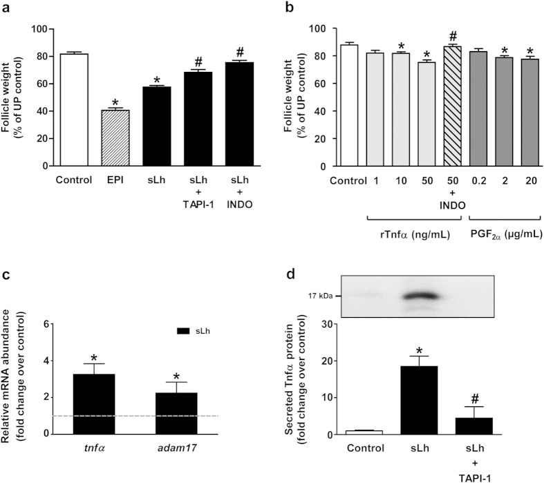
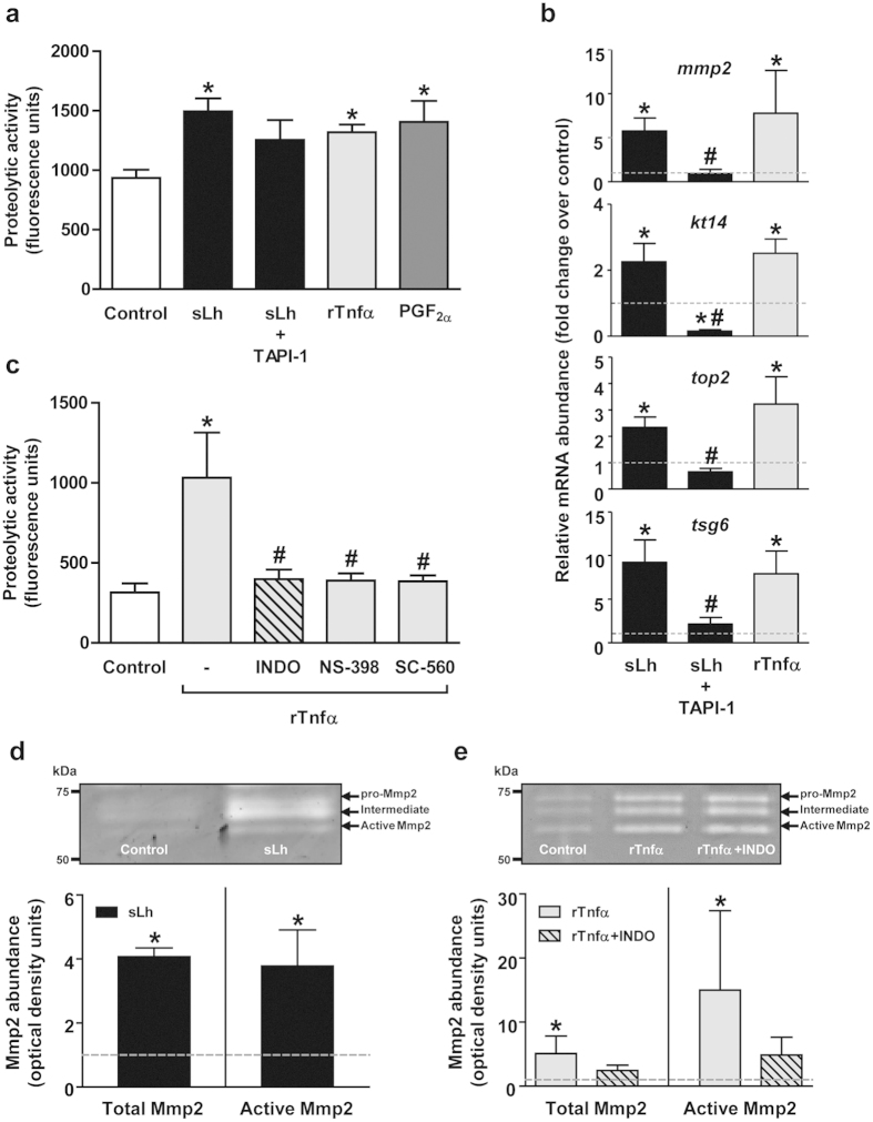
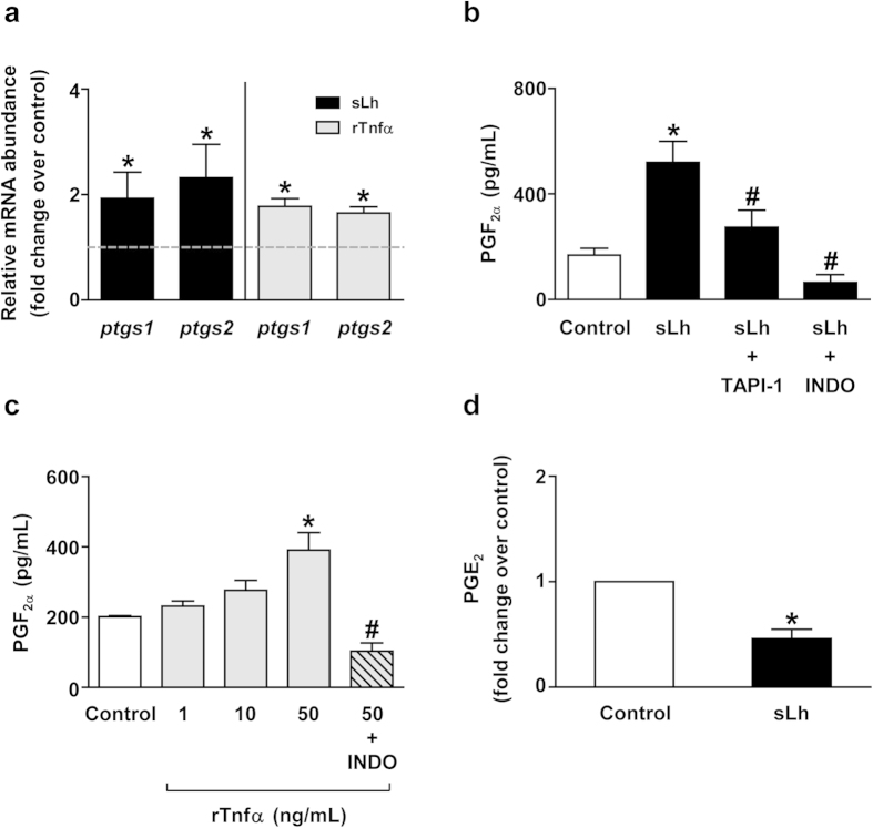
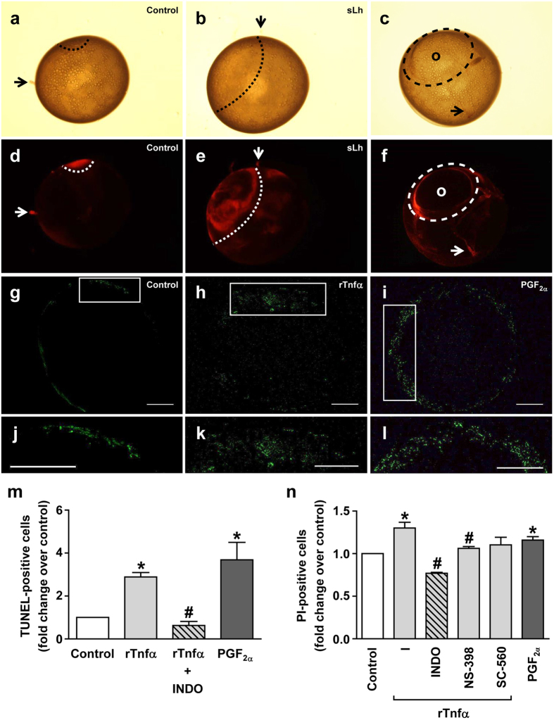
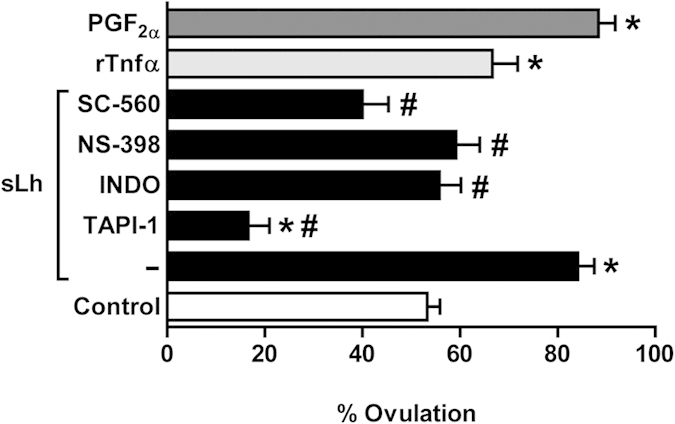
Ovulation is induced by the preovulatory surge of luteinizing hormone (LH) that acts on the ovary and triggers the rupture of the preovulatory ovarian follicle by stimulating proteolysis and apoptosis in the follicle wall, causing the release of the mature oocyte. The pro-inflammatory cytokine tumor necrosis factor α (TNFα) and prostaglandin (PG) F2α (PGF2α) are involved in the control of ovulation but their role mediating the pro-ovulatory actions of LH is not well established. Here we show that Lh induces PGF2α synthesis through its stimulation of Tnfα production in trout, a primitive teleost fish. Recombinant trout Tnfα (rTnfα) and PGF2α recapitulate the stimulatory in vitro effects of salmon Lh (sLh) on contraction, proteolysis and loss of cell viability in the preovulatory follicle wall and, finally, ovulation. Furthermore, all pro-ovulatory actions of sLh are blocked by inhibition of Tnfα secretion or PG synthesis and all actions of rTnfα are blocked by PG synthesis inhibitors. Therefore, we provide evidence that the Tnfα-dependent increase in PGF2α production is necessary for the pro-ovulatory actions of Lh. The results from this study shed light onto the mechanisms underlying the pro-ovulatory actions of LH in vertebrates and may prove important in clinical assessments of female infertility.


Figures

Figure 1
Figure 1. Mediatory effects of Tnfα and PGF on sLh-induced contraction of brown trout preovulatory follicles.
(a) Effects of sLh on follicle contraction. Punctured follicles were incubated for 16 h at 15 °C with epinephrine (EPI; 10 μM), sLh (25 ng/mL), sLh plus TAPI-1 (50 μM) and sLh plus INDO (10 μg/mL). The results are expressed as percent change with respect to the unpunctured control group that was set at 100%. (b) Effects of rTnfα and PGF on follicle contraction. Punctured preovulatory follicles were incubated for 16 h at 15 °C with rTnfα (1, 10 and 50 ng/mL), rTnfα (50 ng/mL) in the presence of INDO (10 μg/mL) and PGF (0.2, 2 and 20 μg/mL). In (a) and (b), data are expressed as percent change with respect to the unpunctured control group that was set at 100%. (c) Relative expression of tnfα and adam17 analyzed by qPCR in ovarian follicles incubated in the absence or presence of sLh (25 ng/mL). (d) Ovarian Tnfα secretion in brown trout follicle incubates by Western blot. Preovulatory follicles were incubated with sLh (25 ng/mL) in the absence or presence of TAPI-1 (50 μM) for 16 h at 15 °C. A representative Western blot is shown in the inset and the full-length blot is presented in Supplementary Figure S1. In all graphs, data are the mean ± standard error (SEM) (n = 3–5). *P < 0.05, with respect to control; #P < 0.05, with respect to sLh (a,d) and with respect to rTnfα (50 ng/mL) (b).
Figure 2
Figure 2. Induction of proteolysis by Lh, Tnfα and PGF in brown trout preovulatory follicles.
(a) In vitro effects of sLh (25 ng/mL), in the absence or presence of TAPI-1 (50 μM), rTnfα (50 ng/mL) and PGF (2 μg/mL) on trout gelatinase/collagenase activity in ovarian follicle homogenates. (b) Expression of proteolytic genes in ovarian follicles incubated with sLh (25 ng/mL), in the absence or presence of TAPI-1 (50 μM), or with rTnfα (50 ng/mL). The relative expression of mmp2, kt14, top2 and tsg6 was determined by qPCR and normalized to the abundance of 18s. (c) Inhibition of rTnfα-induced gelatinase/collagenase activity in ovarian follicle homogenates by prostaglandin synthesis inhibitors. Follicles were incubated with rTnfα (50 ng/mL) in the absence or presence of INDO (10 μg/mL), NS-398 (10 μg/mL) and SC-560 (10 μg/mL) for 16 h at 15 °C. (d,e) Determination of ovarian Mmp2 activity by gelatin zymography in follicles treated with sLh (25 ng/mL) (d) and rTnfα (50 ng/mL) in the absence or presence of INDO (10 μg/mL) (e). Clear bands in the zymogram indicated enzymatic digestion of gelatin. Densitometric analyses of Mmp2 gelatinase activity were quantified and expressed as fold increase of the total (pro-, intermediate and active Mmp2) or active Mmp2 content in control (set at 1 and represented by the dashed line) and sLh and rTnfα ± INDO-treated follicles. Representative zymograms are shown in the graph and the full-length gels are presented in Supplementary Figure S3. In all graphs, data are the mean ± SEM (n = 3–4). *P < 0.05, with respect to control; #P < 0.05, with respect to sLh (b) and with respect to rTnfα (50 ng/mL) (c).
Figure 3
Figure 3. Regulation of prostaglandin synthesis by Lh and Tnfα in brown trout preovulatory follicles.
(a) In vitro effects of sLh (25 ng/mL) and rTnfα (50 ng/mL) on the expression of enzymes involved in prostaglandin synthesis. The relative expression of ptgs1 and ptgs2 was determined by qPCR and normalized to the abundance of 18s. (b) Determination of PGF production in vitro by intact ovarian follicles treated with sLh. Trout follicles were incubated with sLh (25 ng/mL) in the absence or presence of TAPI-1 (50 μM) and INDO (10 μg/mL) for 16 h at 15 °C and PGF was measured in the incubation media. (c) PGF production in response to rTnfα treatment in vitro. Follicles were incubated with rTnfα (1, 10 and 50 ng/mL) or rTnfα (50 ng/mL) with INDO (10 μg/ml) for 16 h at 15 °C. (d) PGE2 production by brown trout preovulatory ovarian follicles. Follicles were incubated in the absence or presence of sLh (25 ng/mL) for 16 h at 15 °C and PGE2 levels were measured in the incubation media. In all graphs, data are the mean ± SEM (n = 3–7). *P < 0.05, with respect to control; #P < 0.05, with respect to sLh (b) and with respect to rTnfα (50 ng/mL) (c).
Figure 4
Figure 4. Lh, Tnfα and PGF induce cell death in trout preovulatory follicles.
(a–f) Assessment of cell death in ovarian follicles. Cell viability was assessed in follicles incubated for 18 h at 12 °C in the absence (a,d) or the presence of sLh (25 ng/mL) (b,e) and subsequently follicles were stained with propidium iodide (PI; 20 μg/mL) during 10 min at room temperature and visualized under bright light (a,b) or a fluorescent stereomicroscope (d,e). An ovulating follicle incubated under control conditions showing the point of rupture (c,f) (o, oocyte). Loss of cell viability is highlighted by a dashed line and stromal tissue debris is indicated by arrows. Representative pictures of a total of three independent experiments are shown. (g–l) Representative images of preovulatory follicles subjected to TUNEL analysis. Follicles incubated in the absence (control; g,j) or presence of rTnfα (50 ng/mL; h,k) and PGF (2 μg/mL; i,l) were analyzed by TUNEL. Green staining indicates fragmented DNA in cells undergoing apoptosis. Scale bar represents 1000 μm. (m) Quantification of TUNEL-positive cells. Quantification was performed using ImageJ software. (n) Analysis of apoptosis in isolated granulosa cells by PI staining. Granulosa cells isolated from trout preovulatory follicles incubated in the absence or presence of rTnfα (50 ng/mL), rTnfα plus PG synthesis inhibitors (INDO, NS-398 and SC-560; 10 μg/mL) and PGF (2 μg/mL) were stained with PI and analyzed by flow cytometry (FACS analysis). Data are the mean ± SEM (n = 3). *P < 0.05, with respect to control; #P < 0.05, with respect to rTnfα.
Figure 5
Figure 5. Effects of Lh, Tnfα and PGF on in vitro ovulation in brook trout preovulatory follicles.
Trout follicles were incubated for 36 h at 12 °C in the presence or absence of sLh (25 ng/mL), sLh plus different inhibitors (TAPI-1, 50 μM; INDO, 10 μg/mL; NS-398, 10 μg/mL; and SC-560, 10 μg/mL), rTnfα (50 ng/mL) and PGF (2 μg/mL). Data are the mean ± SEM (n = 4). *P < 0.05, with respect to control; #P < 0.05, with respect to sLh.

Similar articles

Cited by

References

    1. Espey L. L. & Richards J. S. Ovulation in The Physiology of Reproduction (ed. Neil J. D.) 425–474 (Academic Press, 2006).
    1. Rao C. V. & Lei Z. M. Consequences of targeted inactivation of LH receptors. Mol Cell Endocrinol 187, 57–67 (2002). - PubMed
    1. Curry T. E. J. & Smith M. F. Impact of extracellular matrix remodeling on ovulation and the folliculo-luteal transition. Semin Reprod Med 24, 228–241 (2006). - PubMed
    1. Reich R. et al. Preovulatory changes in ovarian expression of collagenases and tissue metalloproteinase inhibitor messenger ribonucleic acid: role of eicosanoids. Endocrinology 129, 1869–1875 (1991). - PubMed
    1. Gottsch M. L., Van Kirk E. A. & Murdoch W. J. Tumour necrosis factor alpha up-regulates matrix metalloproteinase-2 activity in periovulatory ovine follicles: metamorphic and endocrine implications. Reprod Fertil Dev 12, 75–80 (2000). - PubMed

Publication types