. 2015 Sep 29;112(39):E5391-400.
doi: 10.1073/pnas.1515737112. Epub 2015 Sep 15.

Crystal structure reveals specific recognition of a G-quadruplex RNA by a β-turn in the RGG motif of FMRP

Affiliations

Crystal structure reveals specific recognition of a G-quadruplex RNA by a β-turn in the RGG motif of FMRP

Nikita Vasilyev et al. Proc Natl Acad Sci U S A. .

Abstract

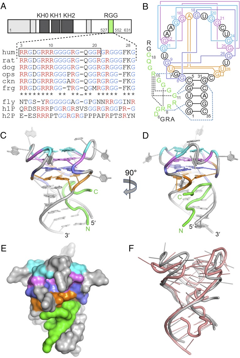
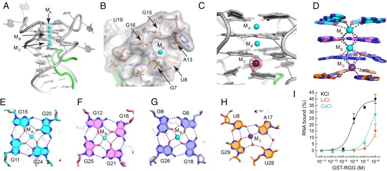
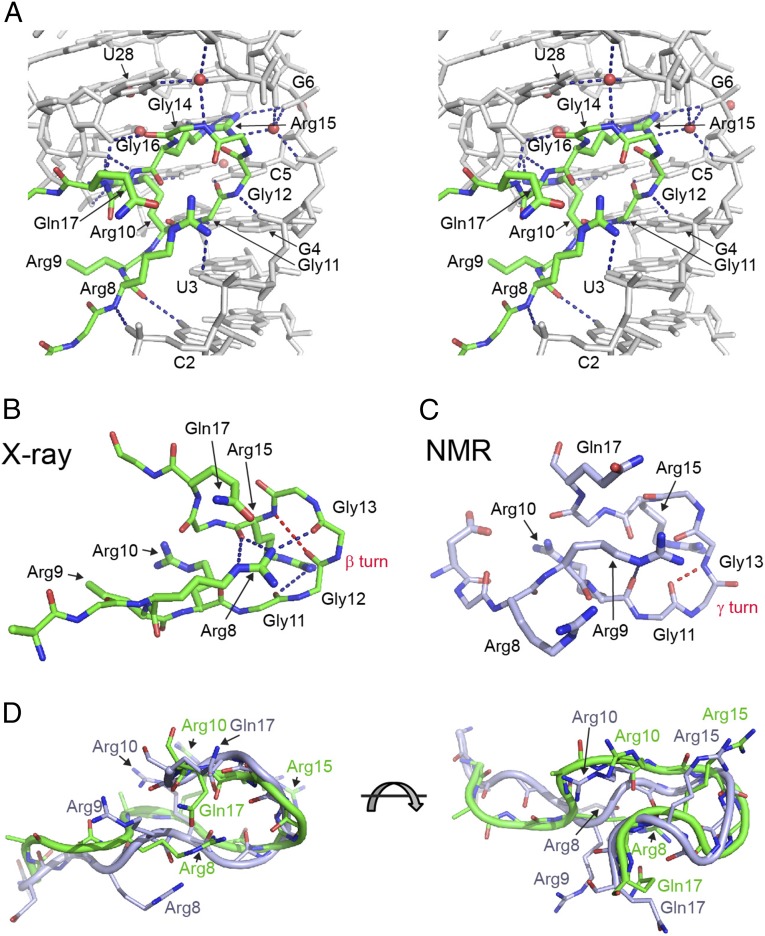
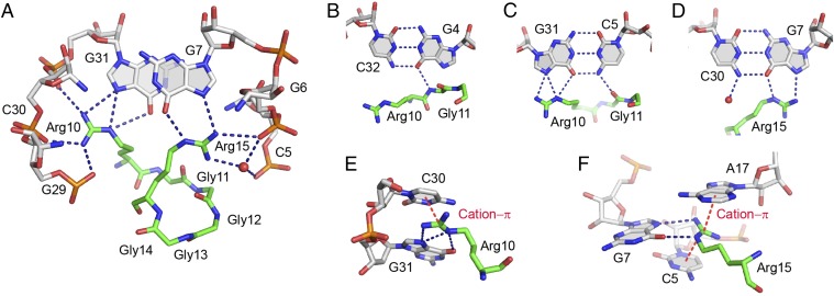
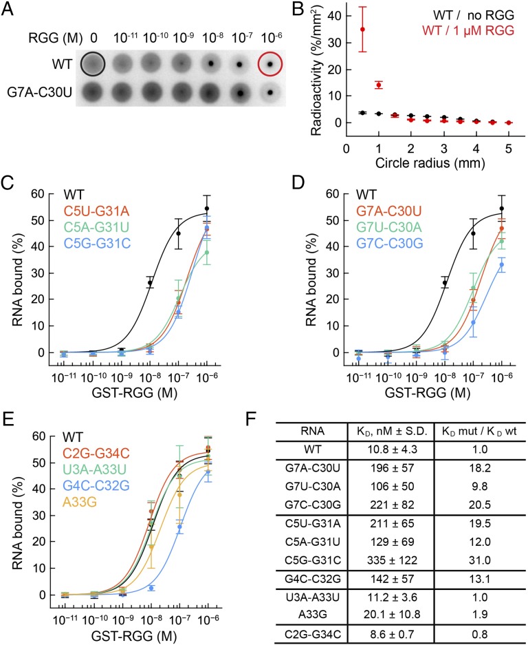
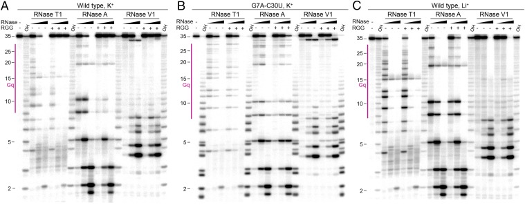
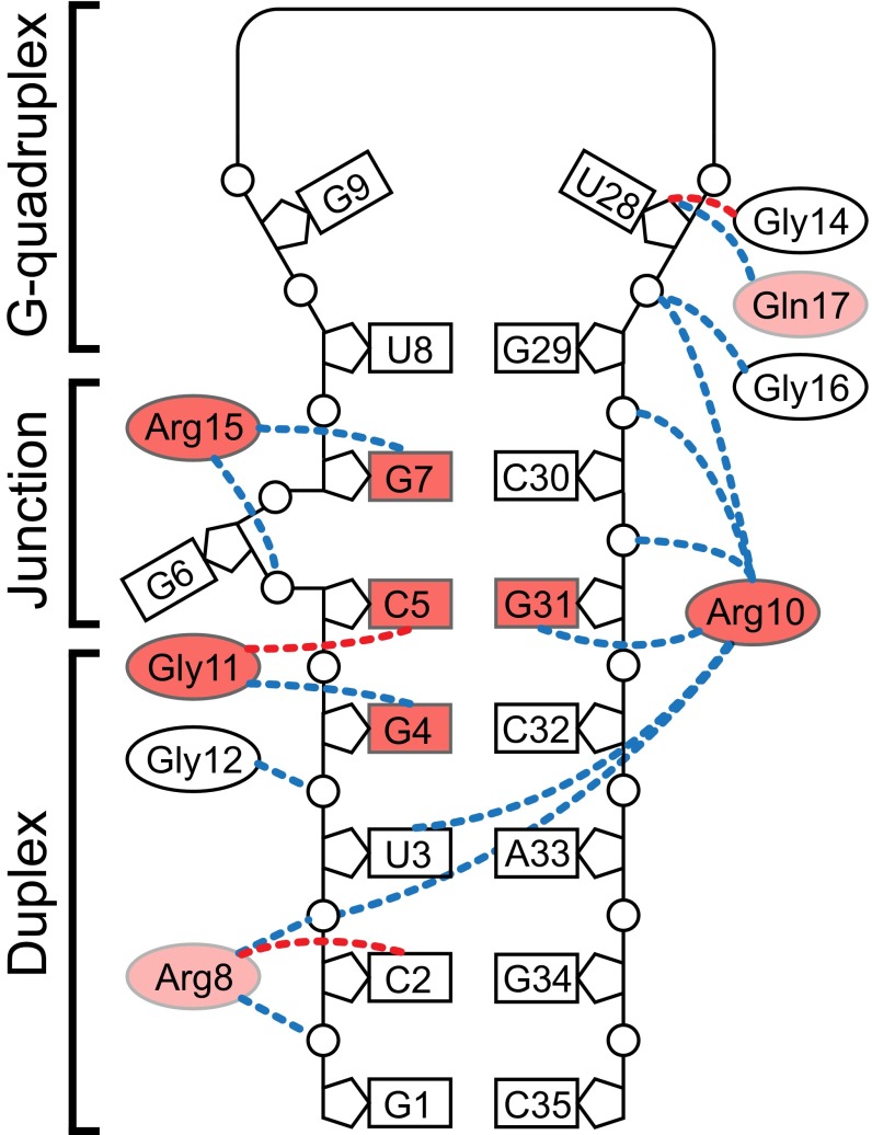
Fragile X Mental Retardation Protein (FMRP) is a regulatory RNA binding protein that plays a central role in the development of several human disorders including Fragile X Syndrome (FXS) and autism. FMRP uses an arginine-glycine-rich (RGG) motif for specific interactions with guanine (G)-quadruplexes, mRNA elements implicated in the disease-associated regulation of specific mRNAs. Here we report the 2.8-Å crystal structure of the complex between the human FMRP RGG peptide bound to the in vitro selected G-rich RNA. In this model system, the RNA adopts an intramolecular K(+)-stabilized G-quadruplex structure composed of three G-quartets and a mixed tetrad connected to an RNA duplex. The RGG peptide specifically binds to the duplex-quadruplex junction, the mixed tetrad, and the duplex region of the RNA through shape complementarity, cation-π interactions, and multiple hydrogen bonds. Many of these interactions critically depend on a type I β-turn, a secondary structure element whose formation was not previously recognized in the RGG motif of FMRP. RNA mutagenesis and footprinting experiments indicate that interactions of the peptide with the duplex-quadruplex junction and the duplex of RNA are equally important for affinity and specificity of the RGG-RNA complex formation. These results suggest that specific binding of cellular RNAs by FMRP may involve hydrogen bonding with RNA duplexes and that RNA duplex recognition can be a characteristic RNA binding feature for RGG motifs in other proteins.

Keywords: FMRP; G-quadruplex; RGG box; RNA structure; fragile X syndrome.


Conflict of interest statement

The authors declare no conflict of interest.

Figures

Fig. 1.
Fig. 1.
Crystal structure of the complex between the FMRP RGG box and sc1 RNA. (A) FMRP schematic and the RGG motif sequence alignment for FMRP and its autosomal paralogs. FMRPs were from opossum (ops), chicken (ckn), Xenopus (frg), and Drosophila (fly). h1P, human FXR1P; h2P, human FXR2P. The boxed sequence was used for the design of the crystallization construct. (B) Secondary structure schematic and interactions in the RGG–sc1 complex. RNA is in gray color with guanine quartets in cyan, violet and blue, and the mixed tetrad in orange. Pairing alignments in RNA are shown by solid lines. The well defined region of the RGG peptide is in green and intramolecular hydrogen bonds are depicted by black dashed lines. Blue dashed lines show base-specific intermolecular interactions. (CD) Overall view of the complex in front (C) and side (D) views. (E) A surface view of the complex. (F) Superposition of the X-ray (current study, gray color) and NMR (salmon color) (37) structures.
Fig. 2.
Fig. 2.
Formation of the G-quadruplex. (A) The RGG–sc1 structure shown with K+ cations (cyan spheres) and the FoFc electron density map contoured at the 5 σ level (cyan mesh). The map was generated before addition of metal cations to the refined RNA model and therefore is unbiased for cations. (B) General cation-binding site shown in surface representation with bound K+ cation in the MD position. (C) Anomalous difference Fourier map contoured at the 6 σ level (magenta) indicates replacement of K+ cation by Cs+ (purple sphere). (D) Coordination of K+ cations by base tetrads. Coordination bonds are depicted with dashed lines. (EH) Nucleotide tetrad alignments and cation binding. Dashed lines show coordination and hydrogen bonds. Note that each cation binds two layers of the quadruplex and this figure does not depict all interactions. (I) Binding curves of the RGG peptide and sc1 RNA in the presence of various cations.
Fig. 3.
Fig. 3.
Protein–RNA interactions in the RGG–sc1 complex. Putative hydrogen bonds are shown as blue dashed lines. (A) Stereoview of hydrogen bonding between the RGG peptide (green) and sc1 RNA (gray). (B and C) Comparison of the RGG peptides from the X-ray (B, current study) and NMR (C) (37) structures. Hydrogen bonds characteristic for peptide turns are in red dashed lines. (D) Superposition of the RGG peptide from the X-ray (green) and NMR (light blue) structures.
Fig. 4.
Fig. 4.
Details of protein–RNA interactions in the RGG–sc1 complex. (A) Specific recognition of RNA by Arg10 and Arg15 of the RGG peptide. (BD) Base-specific recognition of the RNA base pairs in the upper part of the RNA duplex. (E and F) Cation–π interactions (red dashed lines) involving guanidinium groups of arginines and nucleotide bases.
Fig. 5.
Fig. 5.
DRaCALA binding experiments with the GST-RGG protein and sc1 RNA mutants. KDs are average values from at least three independent experiments. Error bars are SDs. (A) Diffusion of radioactively labeled wild type (Upper) and G7A-C30U mutant (Lower) RNAs on nitrocellulose filters in the DRaCALA assay. Black and red circles show maximal diffusion limits used for quantification of binding affinity. (B) Quantification of the wild type RNA diffusion in the absence and presence of the protein. (CE) Binding curves of the RNA variants. Mutated base pairs are shown in insets. (F) Dissociation constants and effects of mutations expressed as a ratio between KDs for wild-type and mutant RNAs.
Fig. 6.
Fig. 6.
Representative gels with ribonuclease T1, A, and V1 footprinting of the RGG–sc1 complex. Enzyme concentrations were: RNase T1, 0.01, 0.1, and 1.0 U/µL; RNase A, 0.01, 0.1 and 1.0 ng/µL; RNase V1, 0.001, 0.01, and 0.1 U/µL. OH, alkaline ladder. Gq, G-quadruplex region. (A) Wild-type RNA in K+. (B) G7A-C30U mutant RNA in K+. (C) Wild-type RNA in Li+.
Fig. 7.
Fig. 7.
Molecular determinants of RGG–sc1 recognition. Effects of selected mutations (current study and ref. 37) are color-coded: red and light red are >10 and 2- to 10-fold reduction of binding affinity, respectively. Putative intermolecular hydrogen bonds involving C-O and N-H main chain functionalities of the peptide are depicted by dashed lines of red and blue colors, respectively.

References

    1. Baltz AG, et al. The mRNA-bound proteome and its global occupancy profile on protein-coding transcripts. Mol Cell. 2012;46(5):674–690. - PubMed
    1. Castello A, et al. Insights into RNA biology from an atlas of mammalian mRNA-binding proteins. Cell. 2012;149(6):1393–1406. - PubMed
    1. Ashley CT, Jr, Wilkinson KD, Reines D, Warren ST. FMR1 protein: Conserved RNP family domains and selective RNA binding. Science. 1993;262(5133):563–566. - PubMed
    1. De Boulle K, et al. A point mutation in the FMR-1 gene associated with fragile X mental retardation. Nat Genet. 1993;3(1):31–35. - PubMed
    1. Myrick LK, et al. Fragile X syndrome due to a missense mutation. Eur J Hum Genet. 2014;22(10):1185–1189. - PMC - PubMed

Publication types

Substances