. 2015 Nov 15;24(22):6540-51.
doi: 10.1093/hmg/ddv364. Epub 2015 Sep 15.

Genetic dissection of the Down syndrome critical region

Affiliations

Genetic dissection of the Down syndrome critical region

Xiaoling Jiang et al. Hum Mol Genet. .

Abstract

Down syndrome (DS), caused by trisomy 21, is the most common chromosomal disorder associated with developmental cognitive deficits. Despite intensive efforts, the genetic mechanisms underlying developmental cognitive deficits remain poorly understood, and no treatment has been proven effective. The previous mouse-based experiments suggest that the so-called Down syndrome critical region of human chromosome 21 is an important region for this phenotype, which is demarcated by Setd4/Cbr1 and Fam3b/Mx2. We first confirmed the importance of the Cbr1-Fam3b region using compound mutant mice, which carry a duplication spanning the entire human chromosome 21 orthologous region on mouse chromosome 16 [Dp(16)1Yey] and Ms1Rhr. By dividing the Setd4-Mx2 region into complementary Setd4-Kcnj6 and Kcnj15-Mx2 intervals, we started an unbiased dissection through generating and analyzing Dp(16)1Yey/Df(16Setd4-Kcnj6)Yey and Dp(16)1Yey/Df(16Kcnj15-Mx2)Yey mice. Surprisingly, the Dp(16)1Yey-associated cognitive phenotypes were not rescued by either deletion in the compound mutants, suggesting the possible presence of at least one causative gene in each of the two regions. The partial rescue by a Dyrk1a mutation in a compound mutant carrying Dp(16)1Yey and the Dyrk1a mutation confirmed the causative role of Dyrk1a, whereas the absence of a similar rescue by Df(16Dyrk1a-Kcnj6)Yey in Dp(16)1Yey/Df(16Dyrk1a-Kcnj6)Yey mice demonstrated the importance of Kcnj6. Our results revealed the high levels of complexities of gene actions and interactions associated with the Setd4/Cbr1-Fam3b/Mx2 region as well as their relationship with developmental cognitive deficits in DS.


Figures

Figure 1.
Figure 1.
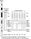
Mouse mutants and their cognitive phenotypes. Hsa21 and the orthologous region on Mmu16 are shown. Dashed lines indicate the locations of specific genes. PR, partial rescue observed; NR, no rescue observed.
Figure 2.
Figure 2.
Analysis of Dp(16)1;Ms1Rhr mice. (A) Dp(16)1;Ms1Rhr (n = 15), Dp(16)/+ (n = 13) and +/+ (n = 13) mice were examined in the T-maze. (B) The three groups of mice in (A) were examined in a contextual fear-conditioning test. The percentages of time frozen before the foot shock and during the 24 h contextual tests are shown. *P < 0.05 and **P < 0.01.
Figure 3.
Figure 3.
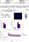
Generation and analysis of Dp(16)1/Df(16)5 mice. (A) Strategy to generate Df(16)5. 5′, 5′ half of a HPRT minigene; 3′, 3′ half of a HPRT minigene; N, neomycin resistance gene; P, puromycin resistance gene; Ty, tyrosinase transgene; Ag, K14-Agouti transgene; ►, loxP; A, AflII; K, KpnI. (B) Genomic locations of BAC probes for FISH analysis. (C) FISH analysis of metaphase chromosomes prepared from Df(16)5/+ ES cells. (D) Southern blot analysis, using Probe 5C, of AflII-digested tail DNA from a Df(16)5/+ mouse. (E) Dp(16)1/Df(16)5 (n = 13), Dp(16)/+ (n = 13) and +/+ (n = 16) mice were examined in the T-maze. (F) The three groups of mice in (E) were examined in a contextual fear-conditioning test. The percentages of time frozen before the foot shock and during the 24 h contextual tests are shown. (G) Analysis of hippocampal LTP. Brain slices from Dp(16)1/Df(16)5 (n = 9), Dp(16)1/+ (n = 9) and +/+ (n = 9) mice were analyzed using electrophysiological recordings in the CA1 region of the hippocampus. Recordings of fEPSPs were carried out before and after TBS inductions. Evoked potentials were normalized to the fEPSPs recorded prior to TBS induction. *P < 0.05 and **P < 0.01.
Figure 4.
Figure 4.
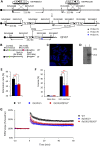
Generation and analysis of Dp(16)1/Df(16)6 mice. (A) Strategy to generate Df(16)6. For the definitions of the abbreviations, see the legend in Figure 3A. A, AflII; K, KpnI; X, XbaI. (B) Genomic locations of BAC probes for FISH analysis. (C) FISH analysis of metaphase chromosomes prepared from Df(16)6/+ ES cells. (D) Southern blot analysis, using Probe 6C, of AflII-digested tail DNA from a Df(16)6/+ mouse. (E) Dp(16)1/Df(16)6 (n = 10), Dp(16)1/+ (n = 12) and +/+ (n = 11) mice were examined in the T-maze. (F) The three groups of mice in (E) were examined in a contextual fear-conditioning test. The percentages of time frozen before the foot shock and during the 24 h contextual tests are shown. (G) Analysis of hippocampal LTP. Brain slices from Dp(16)1/Df(16)6 (n = 8), Dp(16)1/+ (n = 9) and +/+ (n = 9) mice were analyzed using electrophysiological recordings in the CA1 region of the hippocampus. Recordings of fEPSPs were carried out before and after TBS inductions. Evoked potentials were normalized to the fEPSPs recorded prior to TBS induction. *P < 0.05 and **P < 0.01.
Figure 5.
Figure 5.
Generation and analysis of Dp(16)1/Dyrk1am1mice. (A) The gene trap insertion site in Dyrk1a in the ES cell clone QX0369. Arrow heads indicate the primer locations for RT–PCR. (B) Confirmation of the gene trap event by RT–PCR. RNAs were purified from the QX0369 ES cells and the brains of a Dyrk1am1/+ mouse and a wild-type littermate. RT–PCR was performed using the forward primer located in exon 4 of Dyrk1a and the reverse primer in the beta-Geo cassette at the annealing temperature of 70°C. The resulting product was 296 bp. Lane 1, Dyrk1am1/+ mouse; lane 2, wild-type littermate; lane 3, Dyrk1a gene trap cell clone QX0369; MW, 100 bp DNA ladder. Sequencing of the PCR products confirms the presence of a fusion mRNA (Supplementary Material, Fig. S1). (C) Dp(16)1/Dyrk1am1 (n = 16), Dp(16)1/+ (n = 16) and +/+ (n = 16) mice were examined in the T-maze. (D) The three groups of mice in (C) were examined in a contextual fear-conditioning test. The percentages of time frozen before the foot shock and during the 24 h contextual tests are shown. (E) Analysis of LTP. Brain slices from Dp(16)1/Dyrk1am1 (n = 13), Dp(16)1/+ (n = 13) and +/+ (n = 13) mice were analyzed using electrophysiological recordings in the CA1 region of the hippocampus. Recordings of fEPSPs were carried out before and after TBS inductions. Evoked potentials were normalized to the fEPSPs recorded prior to TBS induction. *P < 0.05 and **P < 0.01.
Figure 6.
Figure 6.
Generation and analysis of Dp(16)1/Df(16)7 mice. (A) Strategy to generate Df(16)7. For the definitions of the abbreviations, see the legend in Figure 3A. H, HpaI; K, KpnI; X, XbaI. (B) Genomic locations of BAC probes for FISH analysis. (C) FISH analysis of metaphase chromosomes prepared from Df(16)7/+ ES cells. (D) Southern blot analysis, using Probe 7C, of HpaI-digested tail DNA from a Df(16)7/+ mouse. (E) Dp(16)1/Df(16)7 (n = 14), Dp(16)1/+ (n = 14) and +/+ (n = 14) mice were examined in the T-maze. (F) The three groups of mice in (E) were examined in a contextual fear-conditioning test. The percentages of time frozen before the foot shock and during the 24 h contextual tests are shown. (G) Analysis of hippocampal LTP. Brain slices from Dp(16)1/Df(16)7 (n = 8), Dp(16)1/+ (n = 9) and +/+ (n = 9) mice were analyzed using electrophysiological recordings in the CA1 region of the hippocampus. Recordings of fEPSPs were carried out before and after TBS inductions. Evoked potentials were normalized to the fEPSPs recorded prior to TBS induction. *P < 0.05 and **P < 0.01.
Figure 7.
Figure 7.
Actions and interactions of the genes in the Setd4/Cbr1-Fam3b/Mx2 region in relationship to DS-associated cognitive deficits. D, Dyrk1a; K, Kcnj6; X, a gene located in the deletion interval of Df(16)6. PR, partial rescue observed; NR, no rescue observed.

References

    1. Antonarakis S.E., Lyle R., Dermitzakis E.T., Reymond A., Deutsch S. (2004) Chromosome 21 and down syndrome: from genomics to pathophysiology. Nat. Rev. Genet., 5, 725–738. - PubMed
    1. Roizen N.J., Patterson D. (2003) Down's syndrome. Lancet, 361, 1281–1289. - PubMed
    1. Parker S.E., Mai C.T., Canfield M.A., Rickard R., Wang Y., Meyer R.E., Anderson P., Mason C.A., Collins J.S., Kirby R.S. et al. (2010) Updated National Birth Prevalence estimates for selected birth defects in the United States, 2004–2006. Birth Defects Res. A Clin. Mol. Teratol., 88, 1008–1016. - PubMed
    1. Khoshnood B., Greenlees R., Loane M., Dolk H., Committee E.P.M., Group E.W. (2011) Paper 2: EUROCAT public health indicators for congenital anomalies in Europe. Birth Defects Res. A Clin. Mol. Teratol., 91(Suppl. 1), S16–S22. - PMC - PubMed
    1. Natoli J.L., Ackerman D.L., McDermott S., Edwards J.G. (2012) Prenatal diagnosis of Down syndrome: a systematic review of termination rates (1995–2011). Prenat. Diagn., 32, 142–153. - PubMed

Publication types

Supplementary concepts