. 2015 Nov:88:14-28.
doi: 10.1016/j.yjmcc.2015.09.001. Epub 2015 Sep 12.

Overexpressing superoxide dismutase 2 induces a supernormal cardiac function by enhancing redox-dependent mitochondrial function and metabolic dilation

Affiliations

Overexpressing superoxide dismutase 2 induces a supernormal cardiac function by enhancing redox-dependent mitochondrial function and metabolic dilation

Patrick T Kang et al. J Mol Cell Cardiol. 2015 Nov.

Abstract

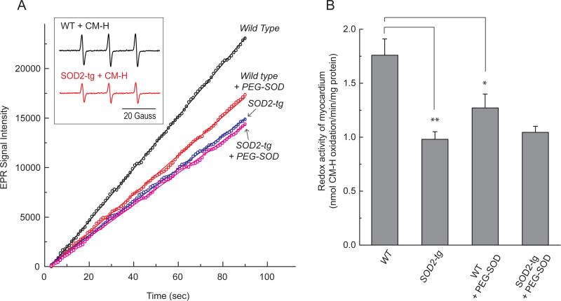
During heightened cardiac work, O2 consumption by the heart benefits energy production via mitochondria. However, some electrons leak from the respiratory chain and yield superoxide, which is rapidly metabolized into H2O2 by SOD2. To understand the systemic effects of the metabolic dilator, H2O2, we studied mice with cardiac-specific SOD2 overexpression (SOD2-tg), which increases the H2O2 produced by cardiac mitochondria. Contrast echocardiography was employed to evaluate cardiac function, indicating that SOD2-tg had a significantly greater ejection fraction and a lower mean arterial pressure (MAP) that was partially normalized by intravenous injection of catalase. Norepinephrine-mediated myocardial blood flow (MBF) was significantly enhanced in SOD2-tg mice. Coupling of MBF to the double product (Heart Rate×MAP) was increased in SOD2-tg mice, indicating that the metabolic dilator, "spilled" over, inducing systemic vasodilation. The hypothesis that SOD2 overexpression effectively enhances mitochondrial function was further evaluated. Mitochondria of SOD2-tg mice had a decreased state 3 oxygen consumption rate, but maintained the same ATP production flux under the basal and L-NAME treatment conditions, indicating a higher bioenergetic efficiency. SOD2-tg mitochondria produced less superoxide, and had lower redox activity in converting cyclic hydroxylamine to stable nitroxide, and a lower GSSG concentration. EPR analysis of the isolated mitochondria showed a significant decrease in semiquinones at the SOD2-tg Qi site. These results support a more reductive physiological setting in the SOD2-tg murine heart. Cardiac mitochondria exhibited no significant differences in the respiratory control index between WT and SOD2-tg. We conclude that SOD2 overexpression in myocytes enhances mitochondrial function and metabolic vasodilation, leading to a phenotype of supernormal cardiac function.

Keywords: Bioenergetics; Cardiac function; Metabolic dilation; Mitochondria; Redox regulation; Superoxide dismutase 2 (SOD2); Transgenic mice.


Figures

Fig. 1
Fig. 1. Cardiac-specific SOD2 overexpression diminished O2 mediated by mitochondria
Hearts were removed from cardiac-specific SOD2-tg and wild type mice, and subjected to mitochondrial and cytosolic preparations. In the top panel, mitochondria (mito., 100 μg loaded) and cytosol (100 μg loaded) were immunoblotted with anti-GAPDH monoclonal antibody (1:500). In the middle panel, mitochondria were immunoblotted using anti-SOD2 Ab (1:2000), an antibody against the 70 kDa subunit of complex II (1:30,000), and anti-Cox I Ab (1:3000). Lower panel, b and e: O2 generation mediated by mitochondria isolated from murine hearts of wild type and SOD2-tg mice, respectively; c and f: the same as b and e, except that antimycin A (10 μM) was included in the system.
Fig. 2
Fig. 2. Cardiac structure and enhanced function are detected in SOD2-tg mice by echocardiography
A and B, Representative images taken using 12-week-old mice. The images demonstrate intact left ventricular (LV) wall integrity, greater wall thickness, and reduced chamber size in SOD2-tg hearts (n=6). Echocardiographic assessment of wild type and SOD2-tg mice (12 weeks old, n=6) were summarized in the Table I.
Fig. 3
Fig. 3
Cardiac-specific SOD2 overexpression decreases heart rate in A, mean arterial pressure (MAP) in B,, increases norepinephrine (NE)-mediated myocardial blood flow (MBF) in C, and enhances the efficacy of coupling RPP to MBF in D. *p<0.05, **p<0.01 (n=6)
Fig. 4
Fig. 4
Mitochondria were prepared from the myocardia of wild type, SOD2-tg (in A-D) and L-NAME-treated wild type mice (in D), and then subjected to measurement of oxygen consumption rate (OCR) by oxygen polarography at 30 °C and NADH-linked ATP production as described under “Experimental Procedures”. A-B, state 3, state 4, and FCCP-mediated oxygen consumption rates by the isolated mitochondria. C, Respiratory control index obtained from the ratio of state 3 OCR to state 4 OCR. D, the ATP/O ratio is calculated from the ratio of ATP production rate to state 3 OCR. The effect of AK2 inhibitor was assessed by pre-incubation of as-isolated mitochondria with Ap5A (20 μM) prior to ATP production assay. *p<0.05, **p<0.01, ***p<0.001, assessed by student's t-test between WT control and SOD2-tg (in B-C, n=12) or WT control and L-NAME-treated WT (n=6) or L-NAME-treated WT and L-NAME-treated SOD2-tg (n=5) or WT+Ap5A and SOD2-tg+Ap5A (n=6) or L-NAME-treated WT+Ap5A and L-NAME-treated SOD2-tg+Ap5A (n=3) (in D,). Comparison among the three groups (WT control, SOD2-tg, and L-NAME-treated WT) in D was analyzed by one-way ANOVA followed by Tukey's post hoc test, indicating that there is a significant difference among the three groups (p < 0.001). There is no significant difference between SOD2-tg and L-NAME-treated SOD2-tg or between SOD2-tg+Ap5A and L-NAME-treated SOD2-tg+Ap5A.
Fig. 5
Fig. 5
A, Upper panel, increased SOD2 expression in the HL-1 by pBI-eGFP-SOD2 transfection was measured by Western blotting (80 μg of cell lysates for each sample was loaded) using a polyclonal antibody against SOD2 (400 ×), and the 70 kDa FAD-binding subunit of Complex II (20,000 ×) was used as a loading control. Lower panel, the effect of SOD2 overexpression in HL-1 on the antimycin A-stimulated O2 production measured by EPR spin-trapping with DMPO. The peGFP was employed as a control scramble plasmid. 1.2 × 10⁷ of HL-1 myocytes (~ 6.5 mg/ml) were used for EPR assay. B, Upper panel, the chart of extracellular flux analysis showing representative measurements of the percentage decrease in oxygen consumption rates (OCR) and relative rates in control, peGFP (scramble plasmid) transfected, and pBI-eGFP-SOD2 transfected HL-1 myocytes in response to glucose (10 mM) and pyruvate (0.2 mM) as the substrates. Lower panel, the effect of overexpressing SOD2 in HL-1 through pBI-eGFP-SOD2 transfection on the OCR of HL-1 myocytes was measured by extracellular flux analyzer.
Fig. 6
Fig. 6
Hearts were removed from wild type and SOD2-tg mice, and subjected to mitochondrial preparation. A, the activities of electron transport chain components in the as-isolated mitochondria were assayed by the methods described in Experimental Procedure (n= 6, *p<0.05, **p<0.01, ***p<0.001).” Inset in gray box, the mitochondrial preparation (5 mg/ml) was incubated with Q2 (20 μM) in ice for 30 min. An aliquots of Q2-reconstituted SOD2-tg mitochondria (SOD2-tg mito. + Q2) was then subjected to assay for SCR activity. B, Protein expression of complex I in the mitochondria and SMP was assessed by Western blot using antibodies against the 51 kDa subunit (nuclear DNA-encoded, Ab51, 10,000 × and ND1 (mitochondrial DNA-encoded, 1,000 ×) of complex I [48]. The protein expression levels of subunit I (70 kDa flavoprotein, nuclear DNA-encoded, 20,000×) of complex II in mitochondria and subunit I (COXI, mitochondrial DNA-encoded, 4,000×) of complex IV in SMP were used as loading controls. The density ratio of the blotting signals was quantitated by Image J software (n=6 for 51 kDa/70 kDa in mitochondria, p<0.001; n=4 for ND1/COXI in SMP, p<0.05). C, A mitochondrial preparation (15 mg/ml in M-buffer) was frozen in liquid N2, and then transferred to an HS cavity equipped with a cryostat operated at 100° K. Left: the spectra of antimycin A-sensitive semiquinone were obtained from the differential spectra of mitochondria with or without antimycin A treatment (50 μM). Right: the spin quantitation of ubisemiquinone was obtained by double integration of simulation spectrum (n=3, *p<0.05). EPR setting: center field, 3350 G, sweep width: 100 G, power: 20 mW, receiver gain: 5 × 10⁴, modulation amplitude: 0.8 G, time constant: 81.92 ms, conversion time: 838.8 ms/G, number of scan: 20.
Fig. 7
Fig. 7
A and B, The kinetics of H2O2 generation mediated by mitochondria in the presence of glutamate and malate (NADH-linked) under state 3 and state 4 conditions was assessed by an HRP/Amplex Red assay (n=5). C, Rate of H2O2 generation mediated by the mitochondria of WT and SOD2-tg murine hearts (n=5, ***p<0.001).
Fig. 8
Fig. 8
A-C, Overexpressing SOD2 in myocytes enhanced the catalase activity in mitochondria (A and B, n=4, **p < 0.01), and decreased the activity of cytosolic catalase (C, n=7, ***p < 0.001). The enzymatic activity of the catalase were measured at 25 ° C as described in “Experimental Procedures”. D, The effect of H2O2 on the mean arterial pressures of wild type and SOD2-tg mice was elevated by intravenous injection of catalase (15.000 and 35,000 units/kg). Mouse MAP was accessed by the approach of jugular and femoral characterization as described in the “Experimental Procedures”. The MAP of SOD2-tg was significantly elevated and normalized to the level of wild type by infusion of catalase at a dosage of 35,000 units/kg (n=4, *p< 0.05).
Fig. 9
Fig. 9. The redox activities of the myocardium from wild type and SOD2-tg mice were measured by EPR with the cyclic hydroxylamine spin probe CM-H
A, The tissue homogenates (0.5 mg/ml) of wild type or SOD2-tg myocardium in SHE buffer (250 mM sucrose, 10 mM HEPES, and 1 mM EDTA, pH 7.4) containing 1 mM DTPA and 1 mM CM-H were subjected to EPR measurement after converting CM-H to a stable nitroxide at 298° K. . The O2–dependent CM-H oxidation was assessed by pre-treatment of tissue homogenates with PEG-SOD (0.04 unit/μl) prior to EPR measurement. B, The effect of PEG-SOD on the redox activity of converting CM-H to stable nitroxide is presented in bar graph (n= 3-6, **p<0.01 and *p<0.05). The instrumental settings used for detecting the three-line spectrum of the nitroxide are: center field, 3360.3 G; sweep width, 60 G; microwave frequency, 9.43 GHz; power, 20 mW; receiver gain, 5.02 × 10³; modulation frequency, 100kHz; modulation amplitude, 1 G; time constant, 163.84 ms; conversion time, 41 ms; sweep time, 42 sec; number of X-scans, 1. The parameters for the kinetic mode are: static field, 3360.3 G; receiver gain, 2 × 10³; time constant, 2624.44 ms; conversion time, 1,000 ms; and sweep time, 300s; number of scan, 1.
Fig. 10
Fig. 10. Diagram showing the mechanism by which transgenic overexpression of SOD2 in vivo mediates supernormal cardiac function by enhancing metabolic dilation and mitochondrial function
In comparison to basal conditions, overexpression of SOD2 increases conversion of O2 to H2O2 (brick red coarse arrows). Accumulation of H2O2 in mitochondria is further facilitated by down-regulation of GPx2. Excess mitochondrial H2O2 diffusing to the vasculature serves as a metabolic dilator, mediating smooth muscle cell (SMC) relaxation, enhancing myocardial blood flow (MBF) and cardiac function. The process is also mediated by down-regulation of the catalase activity in the cytosol of myocytes. Oxidant stress induced by excess H2O2 is presumably neutralized by a more reductive physiological setting and up-regulation of catalase activity in mitochondria. SOD2 overexpression in vivo further down-regulates the function of complex I, complex III, and the SCR supercomplex to minimize the electron leakage, preserve membrane integrity, and increase ATP production in response to oxygen consumption (i.e. increasing ATP/O ratio, indicated by blue fine and coarse arrows), thus enhancing mitochondrial function.

References

    1. Liu Y, Zhao H, Li H, Kalyanaraman B, Nicolosi AC, Gutterman DD. Mitochondrial sources of H2O2 generation play a key role in flow-mediated dilation in human coronary resistance arteries. Circ Res. 2003;93:573–80. Epub 2003 Aug 14. - PubMed
    1. Zhang DX, Gutterman DD. Mitochondrial reactive oxygen species-mediated signaling in endothelial cells. Am J Physiol Heart Circ Physiol. 2007;292:H2023–31. Epub 07 Jan 19. - PubMed
    1. Zhang DX, Borbouse L, Gebremedhin D, Mendoza SA, Zinkevich NS, Li R, et al. H2O2-induced dilation in human coronary arterioles: role of protein kinase G dimerization and large-conductance Ca2+-activated K+ channel activation. Circ Res. 2012;110:471–80. doi: 10.1161/CIRCRESAHA.111.258871. Epub 2011 Dec 8. - PMC - PubMed
    1. Saitoh S, Zhang C, Tune JD, Potter B, Kiyooka T, Rogers PA, et al. Hydrogen peroxide: a feed-forward dilator that couples myocardial metabolism to coronary blood flow. Arterioscler Thromb Vasc Biol. 2006;26:2614–21. Epub 006 Oct 5. - PubMed
    1. Rogers PA, Chilian WM, Bratz IN, Bryan RM, Jr., Dick GM. H2O2 activates redox- and 4-aminopyridine-sensitive Kv channels in coronary vascular smooth muscle. Am J Physiol Heart Circ Physiol. 2007;292:H1404–11. Epub 2006 Oct 27. - PubMed

Publication types

MeSH terms

LinkOut - more resources