Review
. 2015 Jul-Aug;90(4):529-43.
doi: 10.1590/abd1806-4841.20153370.

Female Pattern Hair Loss: a clinical and pathophysiological review

Affiliations
Review

Female Pattern Hair Loss: a clinical and pathophysiological review

Paulo Müller Ramos et al. An Bras Dermatol. 2015 Jul-Aug.

Abstract

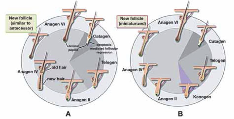
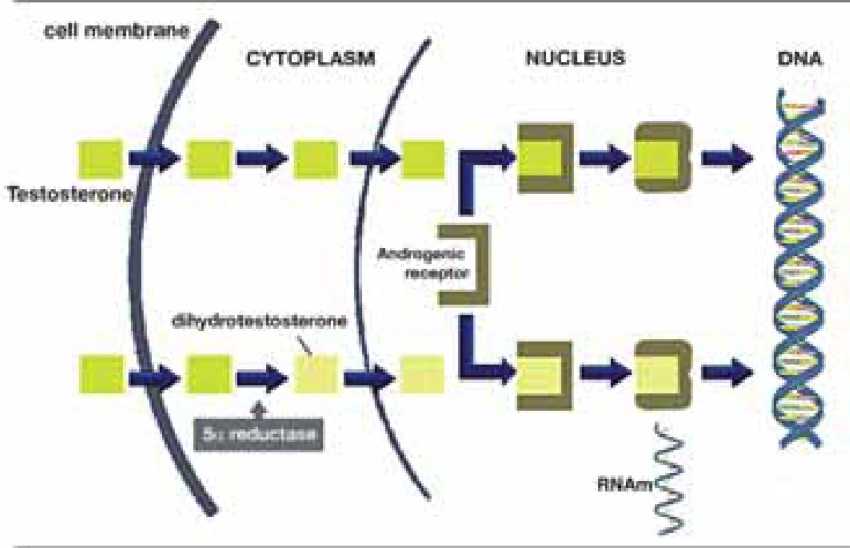
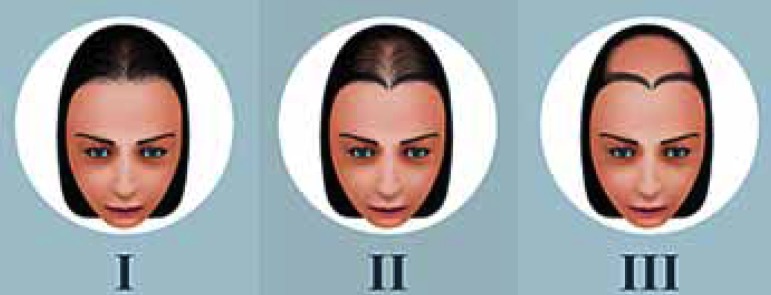
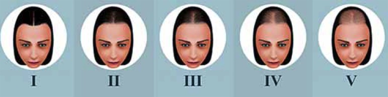
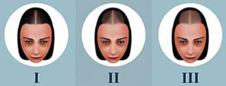
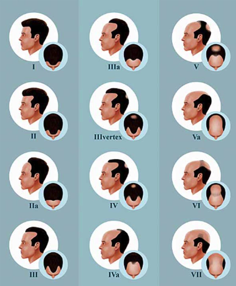
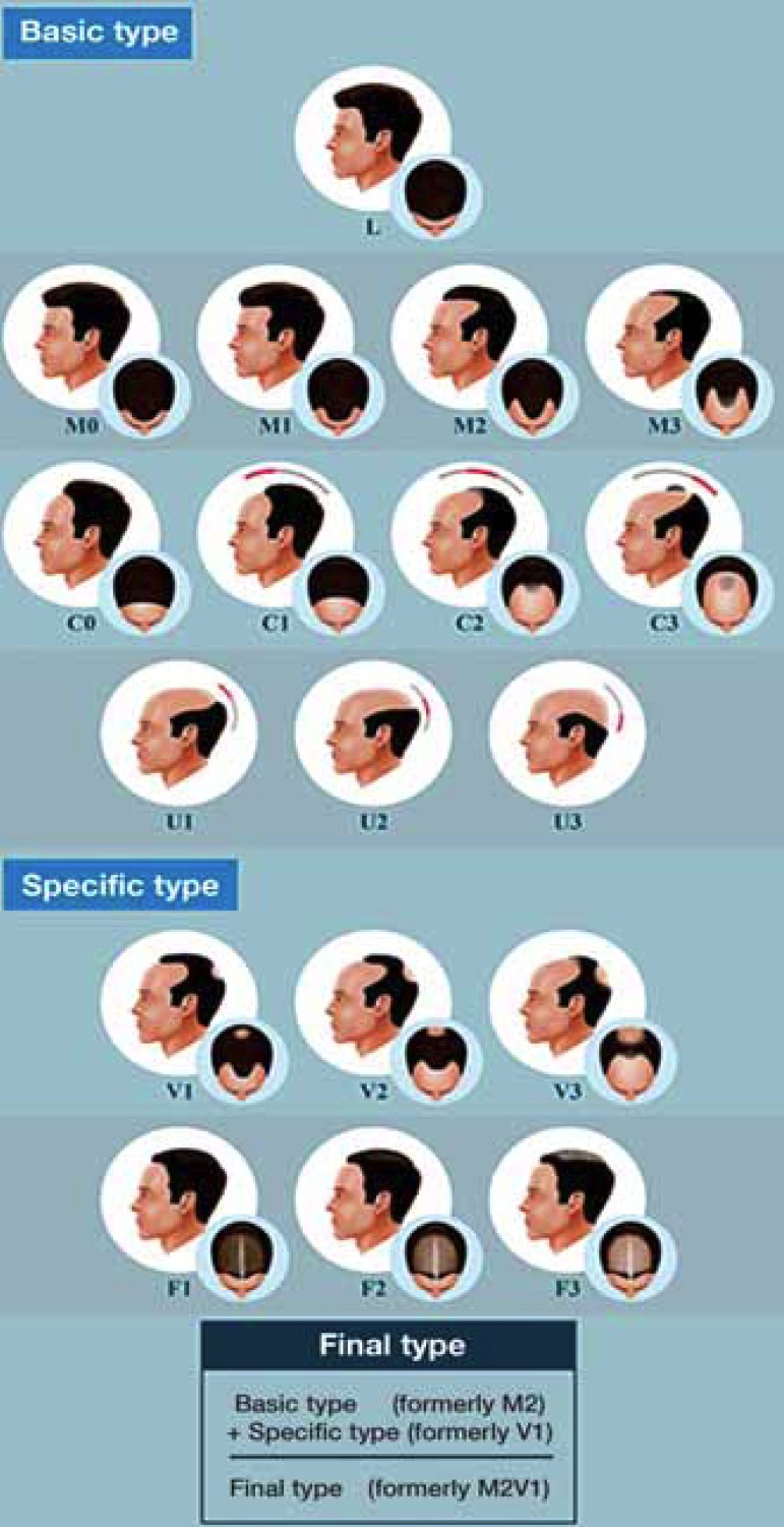
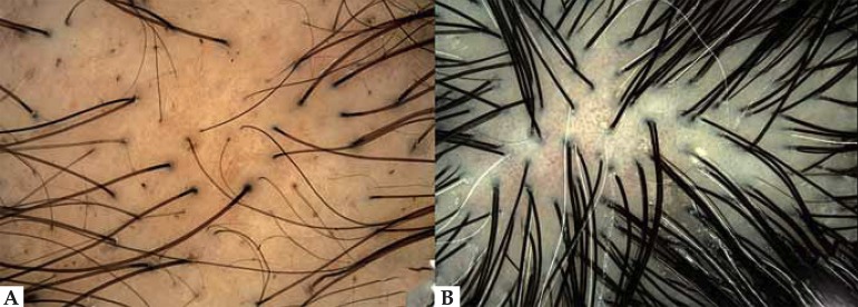
Female Pattern Hair Loss or female androgenetic alopecia is the main cause of hair loss in adult women and has a major impact on patients' quality of life. It evolves from the progressive miniaturization of follicles that lead to a subsequent decrease of the hair density, leading to a non-scarring diffuse alopecia, with characteristic clinical, dermoscopic and histological patterns. In spite of the high frequency of the disease and the relevance of its psychological impact, its pathogenesis is not yet fully understood, being influenced by genetic, hormonal and environmental factors. In addition, response to treatment is variable. In this article, authors discuss the main clinical, epidemiological and pathophysiological aspects of female pattern hair loss.


Conflict of interest statement

Conflicts of Interest: None

Figures

FIGURE 1
FIGURE 1
Female Pattern Hair Loss. Diffuse thinning of the hairs in the frontal and parietal regions, preserving the anterior hair implantation line
FIGURE 2
FIGURE 2
Representative schemes of the hair cycle. A - Normal cycle of the follicle. B - Alterations occurring in baldness: shortening of the anagen phase, increase in the latency period (kenogen phase) and hair follicle minituarization. These alterations may occur together or individually both in FPHL and MPA.
FIGURE 3
FIGURE 3
Representative scheme of the cellular action of androgens. Testosterone (T) enters the cell and is converted by the 5α-reductase enzyme into dihydrotestosterone (DHT). Both T and DHT link themselves to the androgen receptor to promote alterations in cell DNA transcription
FIGURE 4
FIGURE 4
Ludwig´s classification. First proposed classification of MPA. MPA was divided into three intensity degrees.
FIGURE 5
FIGURE 5
Sinclair’s classification. MPA is divided into four levels of intensity based on the normal scalp to the left.
FIGURE 6
FIGURE 6
Olsen´s classification. Other than diffuse thinning, there is also a frontal accentuation opening towards the anterior hair implantation line, creating a “Christmas tree” pattern.
FIGURE 7
FIGURE 7
Hamilton-Nordwood´s classification. Created for the classification of MPA. This pattern rarely affects women
FIGURE 8
FIGURE 8
The basic type (BA) is defined by the shape of the anterior hair implantation line. There are 4 types and they are designated by letters. Type L, there is no hair loss in the frontal hair implantation line, linear pattern Type M, the bilateral frontotemporal recess is more prominent than the central recess. It has the shape of the letter M. Type C, the central recess is more prominent than the frontotemporal recess. It has the shape od the letter C. Types M and C are subdivided into 4 groups, according to the intensity. Type U, the frontal line is behind the vertex. It has the shape of a horseshoe or of the letter U. Type U is subdivided into 3 groups, according to the position of the hair implantation line between the vertex and the occipital protuberance. The specific type (SP) represents the capillary density in certain areas. Type F (frontal), decreased hair density across the top area of the scalp, except the anterior line. Type V is the rarefaction in the vertex region. Specific types are subdivided into 3 groups, according to the intensity. When the patient presents both types (F and V), both types should be described. The fi nal type is decided by the combination of the assigned basic and specific types.
FIGURE 9
FIGURE 9
Dermoscopy of the scalp. A) FPHL. There is great variability in the thickness of the hair shaft, hairs emerging individually from the follicular ostium, reduced follicle density (late fi nding) and incipient pigment network between follicles. B) Normal. Uniformity in the thickness of the hairs and several hairs emerging from the same ostium
FIGURE 10
FIGURE 10
Histopathological examination of FPHL. a) Transverse section evidencing wide variability in diameter of the follicles. b) terminal follicle in detail. c) miniaturized follicle, perifollicular fibrosis and sparse mononuclear inflammatory infiltrate in detail. (HE 40x)

Similar articles

Cited by

References

    1. Rezende JM. Linguagem médica: Alopécia, alopecia. 2011. [11 nov 2013]. Disponível em: Jmrezende.com.br. Internet. a Disponível em: http://www.jmrezende.com.br/alopecia.htm.
    1. Busca no vocabulário. Academia Brasileira de Letras; 2009. [11 nov 2013]. Disponível em: Academia.org.br. Internet. Disponível em: http://www.academia.org.br/abl/cgi/cgilua.exe/sys/start.htm?sid=23.
    1. Descritores em ciências da saúde. Biblioteca Virtual em Saúde; 2013. [11 nov 2013]. Disponível em: Decs.bvs.br. Internet. Disponível em: http://decs.bvs.br/cgi-bin/wxis1660.exe/decsserver/
    1. Plenck JJ. Doctrina de Morbis Cutaneis. Qua hi morbi in suas classes, genera et species redingtur. Vienna: Van Overbeke; 1776.
    1. Sinclair R. Winding the clock back on female androgenetic alopecia. Br J Dermatol. 2012;166:1157–1158. - PubMed

Publication types