

Case Reports
doi: 10.1590/abd1806-4841.20153732.
Scanning electron microscopy of the collodion membrane from a self-healing collodion baby
Affiliations
- PMID: 26375232
- PMCID: PMC4560552
- DOI: 10.1590/abd1806-4841.20153732
Item in Clipboard
Case Reports
Scanning electron microscopy of the collodion membrane from a self-healing collodion baby
An Bras Dermatol.
2015 Jul-Aug.
Abstract
Self-healing collodion baby is a well-established subtype of this condition. We examined a male newborn, who was covered by a collodion membrane. The shed membrane was examined with scanning electron microscopy. The outer surface showed a very compact keratin without the normal elimination of corneocytes. The lateral view of the specimen revealed a very thick, horny layer. The inner surface showed the structure of lower corneocytes with polygonal contour. With higher magnifications villous projections were seen in the cell membrane.
Conflict of interest statement
Conflict of Interest: None.
Figures

Clinical aspects with covering membrane on the trunk and face, with light
ectropion and no eclabium.
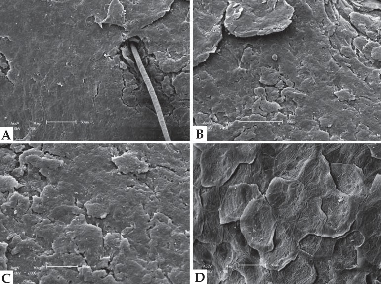
Scanning electron microscopy of outer surface- a. compact membrane
with a perforating vellous hair (x300). b. compact membrane without
released corneocytes (x450). c. detail of the membrane with stony
aspect (x800). d. comparison with normal control releasing
corneocytes (x700).
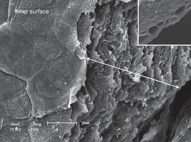
Scanning electron microscopy - lateral view of the collodion membrane showing
multilayered corneocytes (x1.500). Inset with normal skin showing the “basket
weave “aspect of the horny layer (x1.200).

Scanning electron microscopy - general view of the inner surface showing irregular
lower corneocytes (x700).
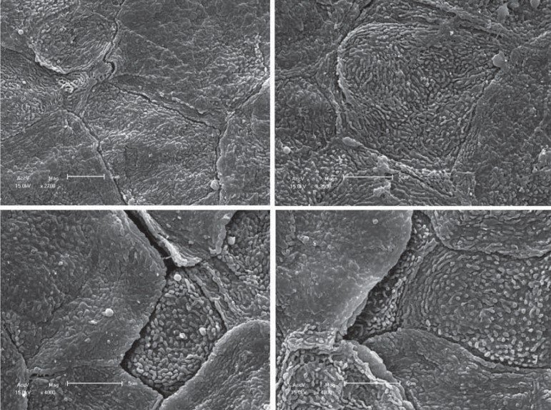
Scanning electron microscopy - detail of the lower corneocytes showing irregular
cell outline, intercellular clefts and villous projections (x2.700- 4.000).
Similar articles
-
Collodion baby and lamellar ichthyosis.J Cutan Pathol. 1998 Feb;25(2):116-21. doi: 10.1111/j.1600-0560.1998.tb01699.x. J Cutan Pathol. 1998. PMID: 9521501
-
Genotype-phenotype correlations with TGM1: clustering of mutations in the bathing suit ichthyosis and self-healing collodion baby variants of lamellar ichthyosis.Br J Dermatol. 2010 Feb 1;162(2):448-51. doi: 10.1111/j.1365-2133.2009.09537.x. Epub 2009 Oct 26. Br J Dermatol. 2010. PMID: 19863506 No abstract available.
-
Collodion baby: ultrastructure and distribution of cornified cell envelope proteins and keratins.Dermatology. 1997;195(2):164-8. doi: 10.1159/000245724. Dermatology. 1997. PMID: 9310728
-
[Cutaneous fissures in collodion babies: incidence and treatment].Ann Dermatol Venereol. 2008 Apr;135(4):279-85. doi: 10.1016/j.annder.2008.01.008. Epub 2008 Mar 28. Ann Dermatol Venereol. 2008. PMID: 18420074 Review. French.
-
Collodion baby concomitant with congenital hypothyroidism: a patient report and review of the literature.J Pediatr Endocrinol Metab. 1998 Jul-Aug;11(4):569-73. doi: 10.1515/jpem.1998.11.4.569. J Pediatr Endocrinol Metab. 1998. PMID: 9777579 Review.
Cited by
-
Collagen XVII deficiency alters epidermal patterning.Lab Invest. 2022 Jun;102(6):581-588. doi: 10.1038/s41374-022-00738-2. Epub 2022 Feb 10. Lab Invest. 2022. PMID: 35145203
References
-
- Hallopeu H, Watelet R. Sur une forme atténuée de la maladie dite ichtyose fetale. Ann Dermatol Syphilol. 1884;3:149–152.
-
- Goldsmith L, Katz S, Gilchrest B, Paller A, Leffel D, Wolff K. Fitzpatrick's Dermatology in Gereral Medicine. 8th Ed. New York: MacGraw Hill; 2012.
-
- Tüzün Y, Isçimen A, Pehlivan O. Collodion Baby. J Turk Acad Dermatol. 2008;2:82201r–82201r.
Publication types
MeSH terms
Supplementary concepts
LinkOut - more resources
Full Text Sources
Other Literature Sources