Review
. 2016 Mar;1858(3):457-466.
doi: 10.1016/j.bbamem.2015.09.013. Epub 2015 Sep 12.

Assembling the puzzle: Oligomerization of α-pore forming proteins in membranes

Affiliations
Review

Assembling the puzzle: Oligomerization of α-pore forming proteins in membranes

Katia Cosentino et al. Biochim Biophys Acta. 2016 Mar.

Abstract

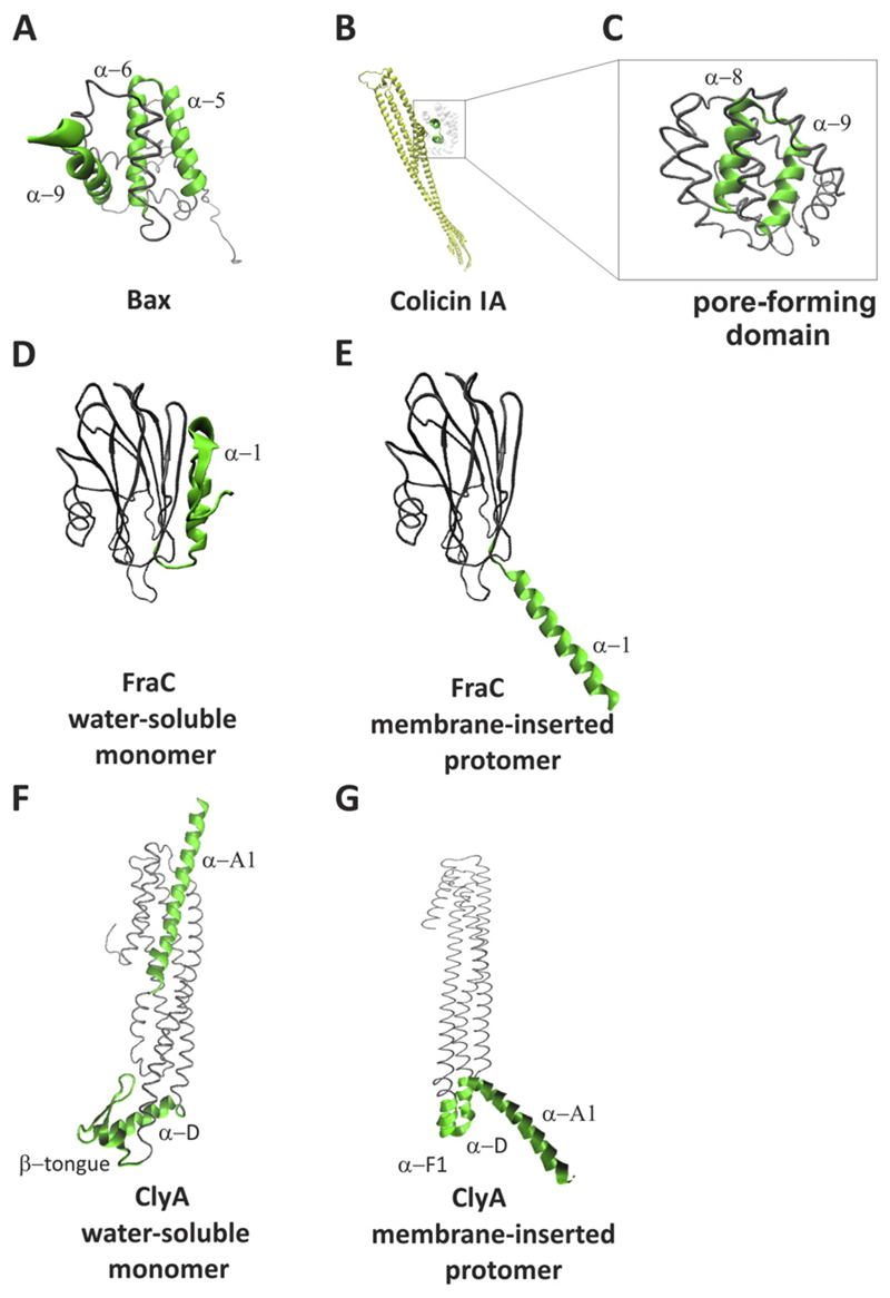
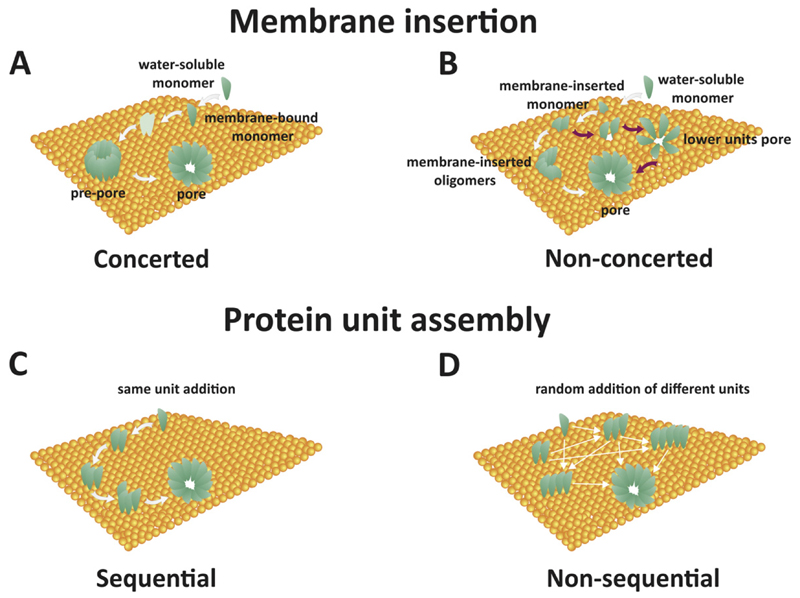
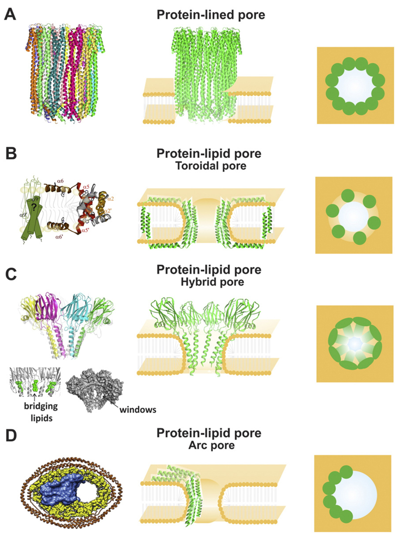
Pore forming proteins (PFPs) share the ability of creating pores that allow the passage of ions, proteins or other constituents through a wide variety of target membranes, ranging from bacteria to humans. They often cause cell death, as pore formation disrupts the membrane permeability barrier required for maintaining cell homeostasis. The organization into supramolecular complexes or oligomers that pierce the membrane is a common feature of PFPs. However, the molecular pathway of self-assembly and pore opening remains unclear. Here, we review the most recent discoveries in the mechanism of membrane oligomerization and pore formation of a subset of PFPs, the α-PFPs, whose pore-forming domains are formed by helical segments. Only now we are starting to grasp the molecular details of their function, mainly thanks to the introduction of single molecule microscopy and nanoscopy techniques. This article is part of a Special Issue entitled: Pore-forming toxins edited by Mauro Dalla Serra and Franco Gambale.

Keywords: Membrane; Pore forming proteins (PFPs); Pore forming toxins (PFTs); Pore structure; Protein oligomerization.


Conflict of interest statement

The authors declare no competing financial interest.

Figures

Fig. 1
Fig. 1. 3D structure of some representative α-PFPs.
A) water-soluble structure of Bax (PDB: 1F16), B) water-soluble structure of full-length colicin IA (PDB: 1CII) and C) its pore-forming domain, D) water-soluble structure of FraC (PDB: 3ZWG) and E) its protomer conformation (PDB: 4TSY), F) water-soluble structure of ClyA (PDB: 1QOY) and E) its protomer conformation (PDB: 2WCD). The pore-forming domains are shown in green and highlighted as thicker structures.
Fig. 2
Fig. 2. Mechanisms of PFPs membrane insertion and protein assembly.
A) Concerted mechanism of membrane insertion. Water-soluble monomers bind to the membrane. Oligomerization and pre-pore formation take place in a fast step. Low-stoichiometry oligomers (shown in light green) are commonly not detected. Membrane insertion occurs after pre-pore formation. B) Non-concerted mechanism of membrane insertion. Water-soluble monomers bind to the membrane in a first fast step. Membrane insertion may take place before or concomitantly with oligomerization. Intermediate pore stages with lower stoichiometry can be detected (magenta arrows pathway). C) Sequential model of protein assembly in the membrane. Addition of units of fixed molecularity (i.e. monomers or dimers) takes place. D) Non-sequential mechanism of protein assembly in the membrane. Random addition of units of different molecularities takes place.
Fig. 3
Fig. 3. Different models of pore-formation by α-PFPs.
A) Protein-lined pores. Left panel: 3D structure of the pore formed by ClyA, determined in detergents (PDB: 2WCD). Central panel: side view graphical representation. Right panel: top view graphical representation. B) Protein–lipid pores: toroidal pores. Left panel: clamp model proposed for Bax pore (adapted from [60]). Central panel: side view graphical representation. Right panel: top view graphical representation. C) Protein–lipid pores: hybrid pores. Left panel: 3D structure of the pore formed by FraC and liposomes (PDB: 4TSY). The bridging lipids connecting two adjacent molecules of FraC are shown in the bottom left side (green). Windows in the pore are also shown in the bottom right side. Central panel: side view graphical representation. Right panel: top view graphical representation. B) Protein–lipid pores: arc pores. Left panel: pore formed by Bax in nanodisc (adapted from [117]). Central panel: side view graphical representation. Right panel: top view graphical representation. In all the graphical representations protein molecules are shown in green and lipids are shown in orange.

References

    1. Iacovache I, Bischofberger M, van der Goot FG. Structure and assembly of pore-forming proteins. Curr Opin Struct Biol. 2010;20:241–246. - PubMed
    1. Bischofberger M, Iacovache I, Gisou van der Goot F. Pathogenic pore-forming proteins: function and host response. Cell Host Microbe. 2012;12:266–275. - PubMed
    1. Pipkin ME, Lieberman J. Delivering the kiss of death: progress on understanding how perforin works. Curr Opin Immunol. 2007;19(3):301–308. - PMC - PubMed
    1. García-Sáez AJ. The secrets of the Bcl-2 family. Cell Death Differ. 2012;19:1733–1740. - PMC - PubMed
    1. Cosentino K, García-Sáez AJ. Mitochondrial alterations in apoptosis. Chem Phys Lipids. 2014;181:62–75. - PubMed

Publication types

LinkOut - more resources