. 2015 Sep 16;10(9):e0138222.
doi: 10.1371/journal.pone.0138222. eCollection 2015.

Cyclosporine A Treatment Inhibits Abcc6-Dependent Cardiac Necrosis and Calcification following Coxsackievirus B3 Infection in Mice

Affiliations

Cyclosporine A Treatment Inhibits Abcc6-Dependent Cardiac Necrosis and Calcification following Coxsackievirus B3 Infection in Mice

Jennifer Marton et al. PLoS One. .

Abstract

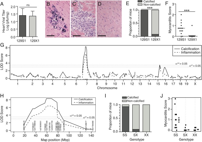
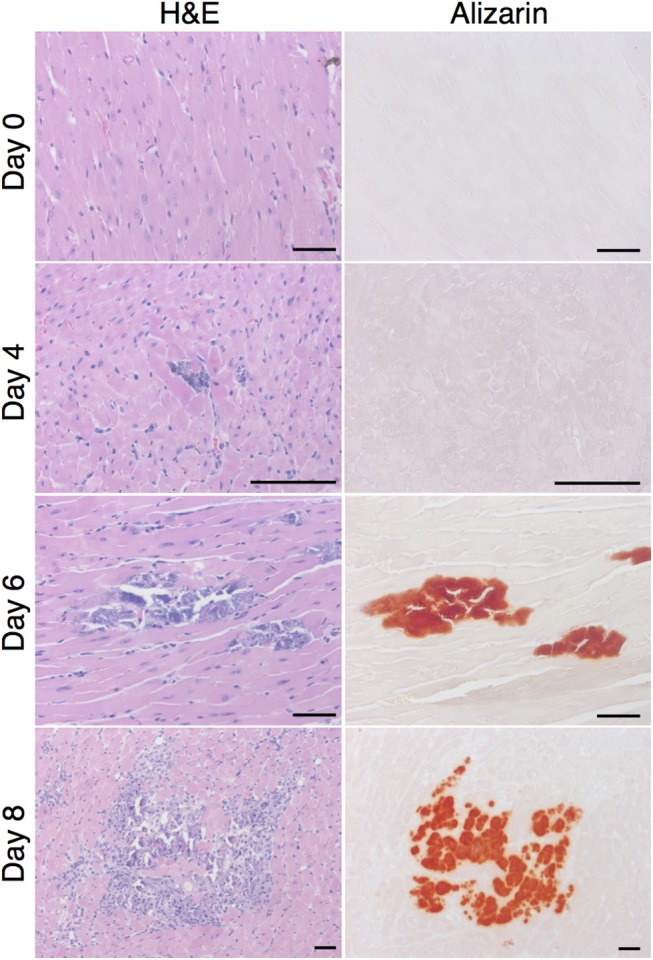
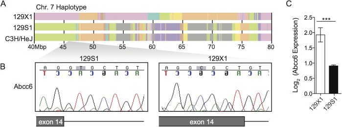
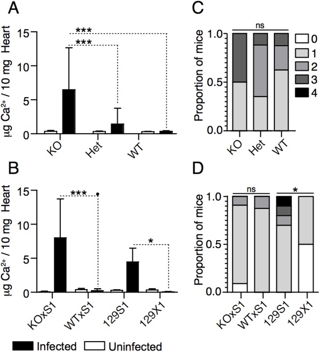
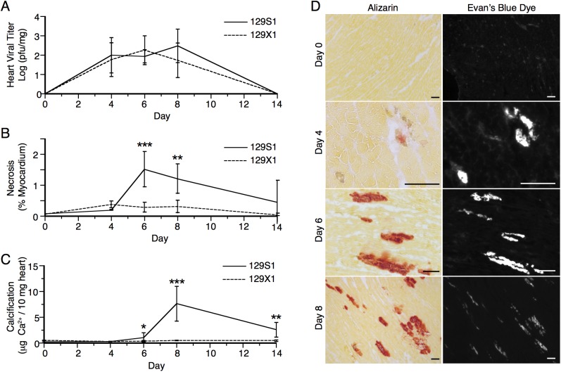
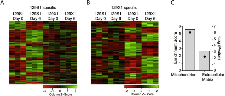
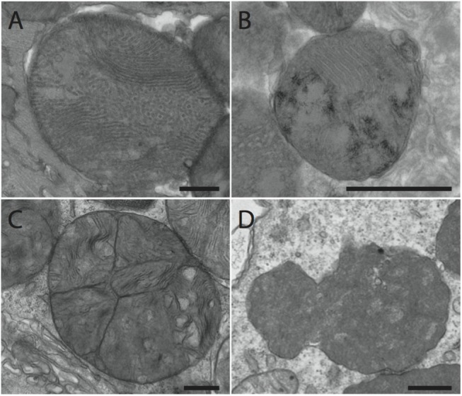
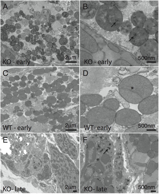
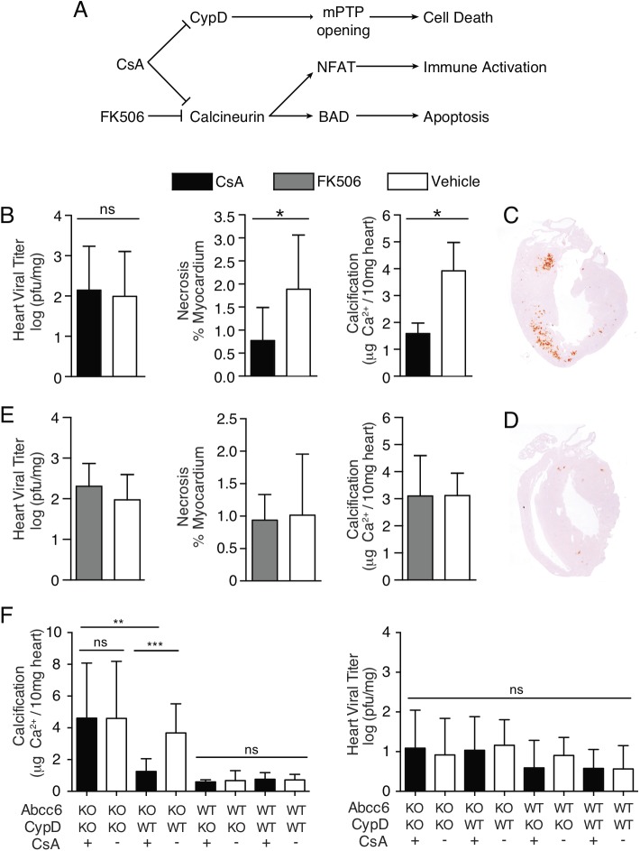
Coxsackievirus type B3 (CVB3) is a cardiotropic enterovirus. Infection causes cardiomyocyte necrosis and myocardial inflammation. The damaged tissue that results is replaced with fibrotic or calcified tissue, which can lead to permanently altered cardiac function. The extent of pathogenesis among individuals exposed to CVB3 is dictated by a combination of host genetics, viral virulence, and the environment. Here, we aimed to identify genes that modulate cardiopathology following CVB3 infection. 129S1 mice infected with CVB3 developed increased cardiac pathology compared to 129X1 substrain mice despite no difference in viral burden. Linkage analysis identified a major locus on chromosome 7 (LOD: 8.307, P<0.0001) that controlled the severity of cardiac calcification and necrosis following infection. Sub-phenotyping and genetic complementation assays identified Abcc6 as the underlying gene. Microarray expression profiling identified genotype-dependent regulation of genes associated with mitochondria. Electron microscopy examination showed elevated deposition of hydroxyapatite-like material in the mitochondrial matrices of infected Abcc6 knockout (Abcc6-/-) mice but not in wildtype littermates. Cyclosporine A (CsA) inhibits mitochondrial permeability transition pore opening by inhibiting cyclophilin D (CypD). Treatment of Abcc6 -/- mice with CsA reduced cardiac necrosis and calcification by more than half. Furthermore, CsA had no effect on the CVB3-induced phenotype of doubly deficient CypD-/-Abcc6-/- mice. Altogether, our work demonstrates that mutations in Abcc6 render mice more susceptible to cardiac calcification following CVB3 infection. Moreover, we implicate CypD in the control of cardiac necrosis and calcification in Abcc6-deficient mice, whereby CypD inhibition is required for cardioprotection.


Conflict of interest statement

Competing Interests: The authors have declared that no competing interests exist.

Figures

Fig 1
Fig 1. Differential susceptibility to viral myocarditis is controlled by a chr. 7 locus.
Cardiac viral titer and histopathology were examined in 129S1 and 129X1 mice infected with 500pfu/g CVB3. Viral titer was equal between both strains (A). 129S1 mice developed calcified lesions (B, E) and increased inflammation (C, F) in response to CVB3 infection. 129X1 mice very rarely presented with either (D, E, F). Analysis of phenotypic segregation and linkage mapping in an (129S1 x 129X1) F2 population identified a common, highly significant locus for each phenotype (Calcification: LOD = 8.307, P<0.0001; Inflammation: LOD = 6.370, P<0.0001) (G). Peak linkage for both traits occurred on Chr. 7 between markers D7Mit69 (56.3 Mbp) and rs32401703 (68.2 Mbp) (H). Recessive inheritance of homozygous S1 alleles conferred susceptibility to both phenotypes (I,J). Statistical tests were chi-squared test and student’s t test for panels E and F respectively. *** P<0.001. Bar = 50m.
Fig 2
Fig 2. Spatio-temporal relationship between calcification and inflammation following CVB3 infection.
Presence and location of calcification and inflammation were assessed at days 0, 4, 6, and 8 post infection in 129S1 mice using alizarin and H&E staining respectively. Normal histology was observed at day 0. Non-calcified lesions were observed 4 days post infection. These expanded in size and number by day 6 post infection at which time calcification became apparent. By day 8 post infection, inflammation was observed and colocalized completely with calcified lesions. Bar = 100μm.
Fig 3
Fig 3. Abcc6 is a promising candidate gene.
(A) 129S1 mice share a common haplotype with C3H/HeJ in the chr.7 QTL interval. (B) Both C3H/HeJ [32] and 129S1 mice possess a mutation in exon 14 of Abcc6 that creates a new splice donor site and causes a 5 bp deletion. This mutation is absent in 129X1 mice and causes a twofold decrease in Abcc6 expression in 129S1 livers (C). The statistical test used was a student’s t test. ***P<0.001.
Fig 4
Fig 4. Causative gene identification by allelic complementation tests.
Cardiac calcification was quantified in Abcc6 KO, Het, WT, (Abcc6 KO x 129S1)F1, (Abcc6 WTxS1)F1, 129S1, and 129X1 mice before infection and at day 8 post-infection. Abcc6 KO, Het, and WT mice were infected with 50pfu/g CVB3; the rest were infected with 500pfu/g CVB3. The concentration of calcium in uninfected mice was equal between all strains (A, B). At day 8 post-infection, increased calcification was consistently observed only in Abcc6 KO, (Abcc6 KO x 129S1)F1, and 129S1 mice (A,B). The severity of inflammation was evaluated in the same mice. Although differences between 129S1 and 129X1 were apparent, inflammation severity did not differ significantly between the other groups (C, D). The statistical test for panels A and B was a two-way ANOVA followed by Bonferonni post tests. The statistical test for panels C and D was a one-way ANOVA followed by a Dunn’s post-test. *: P<0.05, ***: P<0.001, ns: not significant.
Fig 5
Fig 5. Impact of Abcc6-deficiency on the kinetics of viral titer, necrosis, and calcification.
The temporal relationship between viral replication (A), necrosis (B), and calcification (C) was evaluated at days 0, 4, 6, 8, and 14 post-infection with 500pfu/g CVB3 in 129S1 and 129X1 mice. Viral replication did not differ significantly between strains at any time point post-infection (A). Quantitatively significant differences in necrosis and calcification were observed by day 6 post-infection (B,C). The spatial relationship between necrosis and calcification was assessed at days 0, 4, 6, and 8 post-infection using Evan’s blue incorporation and alizarin staining respectively (D). Non-calcified necrotic lesions were apparent by day 4 post-infection. Complete co-localization of necrotic and calcified lesions was observed at days 6 and 8 post-infection. The statistical test for panels A-C was a two-way ANOVA followed by Bonferonni post tests. *: P<0.05, **: P<0.01, ***: P<0.001. Bar = 100mm.
Fig 6
Fig 6. Identification of transcriptional signatures of calcification using microarrays.
Gene expression data was assessed before infection and at day 6 post-infection with 500pfu/g CVB3 in 129S1 and 129X1 mice using microarrays. Genotype-specific expression changes were determined. The heatmaps represent 129S1 and 129X1-specific transcriptomic signatures of infection (A, B). Functional clustering of these genes using DAVID identified significant enrichment of two clusters: mitochondrion and extracellular matrix (C). Bars represent the enrichment score and points represent the–log (Bonferonni pvalue).
Fig 7
Fig 7. Representative images of abnormal mitochondria used to categorized TEM images from Abcc6 KO and WT CVB3-infected hearts.
Representative images were taken from heart sections from Abcc6 KO mice infected with 50pfu/g CVB3. (A) Disrupted cristae. (B) Fusion/fission events. (C) Hydroxyapatite deposition. (D) Abnormal mitochondria characterized by dense staining and irregular shape. Bar: 500nm
Fig 8
Fig 8. Genotype-dependent differences in mitochondrial morphology following infection.
Abcc6 KO (A,B,E,F) and WT (C,D) mice were infected with 50pfu/g CVB3 or mock infected with PBS. Mitochondria were characterized using transmission electron microscopy. At the early stage of necrosis, disruption of mitochondrial cristae was evident in both the Abcc6 KO and WT mice (A-D). Fission and fusion abnormalities were also evident in both strains (D, asterisk). Electron dense deposits consistent with hydroxyapatite were only observed in Abcc6 KO mice (A, B, arrow). At later stages of necrosis, the electron dense deposits became visible in extra-mitochondrial areas only in Abcc6 KO mice (E, F, arrow).
Fig 9
Fig 9. Inhibition of CypD in Abcc6-deficient mice prevents cardiac necrosis and calcification.
(A) Schematic representing the molecular targets of CsA and FK506 and their downstream cellular effects. (B) Daily CsA (10mg/kg/day) treatment of Abcc6 KO mice infected with 50pfu/g CVB3 attenuates CVB3-induced calcification and necrosis despite no effect on viral replication. (C, D) Representative images of vehicle treated (C) and CsA treated (D) hearts stained with alizarin, a calcium specific stain (red areas). Daily FK506 (1mg/kg/day) treatment of Abcc6 KO mice infected with 50pfu/g CVB3 has no effect on cardiac viral titer, necrosis, or calcification (E). CsA treatment of Abcc6 / CypD double knockout mice infected with 50pfu/g CVB3 has no effect on calcification or viral titer (F). The statistical test used for panels B and C was a student’s t test. The statistical test used for panel D was a two way ANOVA followed by Bonferonni post tests *: P<0.05, **: P<0.01, ***P<0.001.

Similar articles

Cited by

  • Drug Repositioning for Hand, Foot, and Mouth Disease.
    Yan R, He J, Liu G, Zhong J, Xu J, Zheng K, Ren Z, He Z, Zhu Q. Yan R, et al. Viruses. 2022 Dec 27;15(1):75. doi: 10.3390/v15010075. Viruses. 2022. PMID: 36680115 Free PMC article. Review.
  • Mitochondrial damage activates the NLRP10 inflammasome.
    Próchnicki T, Vasconcelos MB, Robinson KS, Mangan MSJ, De Graaf D, Shkarina K, Lovotti M, Standke L, Kaiser R, Stahl R, Duthie FG, Rothe M, Antonova K, Jenster LM, Lau ZH, Rösing S, Mirza N, Gottschild C, Wachten D, Günther C, Kufer TA, Schmidt FI, Zhong FL, Latz E. Próchnicki T, et al. Nat Immunol. 2023 Apr;24(4):595-603. doi: 10.1038/s41590-023-01451-y. Epub 2023 Mar 20. Nat Immunol. 2023. PMID: 36941400
  • Clinical and molecular characterization of COVID-19 hospitalized patients.
    Benetti E, Giliberti A, Emiliozzi A, Valentino F, Bergantini L, Fallerini C, Anedda F, Amitrano S, Conticini E, Tita R, d'Alessandro M, Fava F, Marcantonio S, Baldassarri M, Bruttini M, Mazzei MA, Montagnani F, Mandalà M, Bargagli E, Furini S; GEN-COVID Multicenter Study; Renieri A, Mari F. Benetti E, et al. PLoS One. 2020 Nov 18;15(11):e0242534. doi: 10.1371/journal.pone.0242534. eCollection 2020. PLoS One. 2020. PMID: 33206719 Free PMC article.
  • Mt10 Vaccine Protects Diversity Outbred Mice from CVB3 Infection by Producing Virus-Specific Neutralizing Antibodies and Diverse Antibody Isotypes.
    Rasquinha MT, Mone K, Sur M, Lasrado N, Massilamany C, Kachman SD, Steffen D, Reddy J. Rasquinha MT, et al. Vaccines (Basel). 2024 Mar 4;12(3):266. doi: 10.3390/vaccines12030266. Vaccines (Basel). 2024. PMID: 38543901 Free PMC article.
  • Guidelines for evaluating myocardial cell death.
    Mishra PK, Adameova A, Hill JA, Baines CP, Kang PM, Downey JM, Narula J, Takahashi M, Abbate A, Piristine HC, Kar S, Su S, Higa JK, Kawasaki NK, Matsui T. Mishra PK, et al. Am J Physiol Heart Circ Physiol. 2019 Nov 1;317(5):H891-H922. doi: 10.1152/ajpheart.00259.2019. Epub 2019 Aug 16. Am J Physiol Heart Circ Physiol. 2019. PMID: 31418596 Free PMC article. Review.

References

    1. World Health Organization. Global atlas on cardiovascular disease prevention and control. 2011.
    1. Yajima T, Knowlton KU. Viral Myocarditis From the Perspective of the Virus. Circulation. 2009;119(19):2615–24. 10.1161/Circulationaha.108.766022 . - DOI - PubMed
    1. Feldman AM, McNamara D. Myocarditis. N Engl J Med. 2000;343(19):1388–98. Epub 2000/11/09. 10.1056/NEJM200011093431908 . - DOI - PubMed
    1. McCaffrey FM, Braden DS, Strong WB. Sudden cardiac death in young athletes. A review. Am J Dis Child. 1991;145(2):177–83. Epub 1991/02/01. . - PubMed
    1. Whitton JL, Cornell CT, Feuer R. Host and virus determinants of picornavirus pathogenesis and tropism. Nat Rev Microbiol. 2005;3(10):765–76. Epub 2005/10/06. doi: nrmicro1284 [pii] 10.1038/nrmicro1284 . - DOI - PubMed

Publication types

MeSH terms

Substances