Review
. 2015 Dec;88(6):1240-1249.
doi: 10.1038/ki.2015.254. Epub 2015 Sep 16.

Do kidney stone formers have a kidney disease?

Affiliations
Review

Do kidney stone formers have a kidney disease?

Anna L Zisman et al. Kidney Int. 2015 Dec.

Abstract

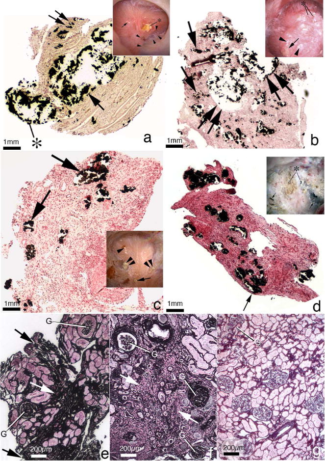
Nephrolithiasis is a highly prevalent disorder affecting approximately one in eleven people and is associated with multiple complications including hypertension, cardiovascular disease, and chronic kidney disease. Significant epidemiologic associations with chronic kidney disease and ESRD have been noted and are reviewed herein, but debate persists in the literature as to whether kidney stone formation is a pathogenic process contributing to kidney disease. Corroborating evidence supporting the presence of kidney disease in stone formers includes the variability of renal function by stone type, the positive association of stone size with renal dysfunction, the presence of markers of renal injury in the urine of even asymptomatic stone formers, and direct evidence of renal tissue injury on histopathology. Proposed pathogenic mechanisms include recurrent obstruction and comorbid conditions such as recurrent urinary tract infections and structural abnormalities. Recent work evaluating the renal histopathology of different groups of stone formers adds further granularity, suggesting variability in mechanisms of renal injury by stone type and confirming the pathogenic effects of crystal formation. Genetic abnormalities leading to stone formation including cystinuria and primary hyperoxaluria, among others, contribute to the burden of disease in the stone-forming population.


Figures

Figure 1
Figure 1
Histopathology of renal papilla and cortex from CaOx stone formers. Papillary tissue of ICSF patients (panel A) shows initial sites of individual calcium phosphate deposits (arrowheads) by Yasue stain (brown-black deposits) in the basement membranes of thin loops of Henle when viewed by light microscopy. These individual deposits coalesce in the interstitial space to form islands of mineral encased in matrix material (arrows) that can extend to the basal side of the urothelium. No evidence of cell injury or inflammation has been noted in the collecting ducts (CD) or interstitium of these papilla. Endoscopic examination of the papilla of ICSF patients reveals small stones (insert panel A, asterisk) attached to the papilla by way of white or interstitial plaque (arrow). In addition to sites of interstitial plaque (panel B, double arrowhead), the papilla of patients with small bowel resection show intraluminal plugs of varying size in the IMCD (arrows). These deposits contained a mixture of CaOx, apatite and ammonium acid urate. Note the extensive interstitial fibrosis surrounding the IMCD plug. These patients also have attached stones (insert panel B, double arrow) at sites of white plaque (arrows). Areas of yellow plaque are also seen (arrowheads). Papilla of intestinal bypass patients for obesity possesses little to no interstitial plaque (panel C) but have a few intraluminal plugs in IMCD (arrow) and ducts of Bellini (double arrow) composed of apatite and CaOx. The insert in panel C shows a dilated opening to a BD (arrowhead). The initial layer of mineral to coat the apical surface of the lining cells of IMCD of these patients is Yasue positive (panel D, arrows) and particularly clear at the arrowhead. Panel E shows a cortical biopsy from an ICSF patient revealing only minimal interstitial fibrosis and glomerular (G) changes. A moderate level of change is seen panel F from a patient with small bowel resection. Panels G and H show sites of hyaluronan staining (arrows) in papillary tissue from intestinal bypass patients revealing areas of cell surface changes at sites of crystal deposits (panel H, arrowheads) or away from deposits (panel G).
Figure 2
Figure 2
Histopathology of renal papilla and cortex from apatite and brushite stone formers. Panel A is a low magnification light microscopic image of a renal papilla from a brushite stone former showing an enormous amount of Yasue-positive deposits (arrow) filling a grossly dilated IMCD and BD. The crystalline material is protruding (asterisk) from the dilated opening of a BD. Sites of interstitial plaque are noted through out papilla (double arrow). Insert to panel A shows endoscopic view of papillum from brushite patients characterized by areas of white plaque (arrows), yellowish crystalline material protruding from dilated BD (asterisk), pitting (arrowheads) and sites of yellow plaque. Panel B is a histologic section from an apatite stone former with large fields of interstitial plaque termed “novel interstitial plaque structures” (NIPS, arrows) located near greatly dilated IMCD and BD filled with Yasue-positive deposits (double arrows) which are hydroxyapatite. Extensive regions of interstitial fibrosis surround the dilated IMCD and NIPS. The insert in panel B shows endoscopic view of a papilla from an apatite stone former characterized by sites of white plaque (arrowheads), protruding plugs for dilated opening of BD (asterisk) and an occasional site of yellow plaque (arrow). Panel C is a histologic section from a patient with primary hyperparathyroidism with stones. These patients show numerous IMCD with apatite plugs (arrows), extensive interstitial fibrosis at sites of intraluminal plugs and areas of interstitial plaque. The insert in panel C shows an endoscopic view of a papilla from a patient with primary hyperparathyroidism characterized by attached stone to white plaque, sites of yellow plaque (double arrows), interstitial plaque (arrowhead), and dilated BD with protruding plug (double arrowhead). Panel D is a histologic section from a patient with distal renal tubular acidosis presenting with diffuse IMCD plugging (arrows), severe and diffuse interstitial fibrosis and little to no interstitial plaque. The insert to panel D shows endoscopic view of a papilla from a patient with distal renal tubular acidosis characterized extensive papillary damage with retraction, loss of normal architecture, multiple dilated BD that produce a pitted appearance (arrows), some with protruding mineral deposits (asterisks) and a whitish thickened membrane appearance. Panel E shows a cortical biopsy from an apatite stone former revealing a moderate level of interstitial fibrosis (arrow) and glomerular (G) changes while panel F shows a severe level of interstitial fibrosis (arrow) and glomerular (G) change in a brushite stone former. Panel H shows a cortical biopsy from a patient with distal renal tubular acidosis revealing only a minimal level of change.
Figure 3
Figure 3
Histopathology of renal papilla from primary hyperoxaluria, cystine and DHA stone formers. Panel A is a histologic section from a primary hyperoxaluric stone former with normal renal function showing isolated dilated IMCD filled with CaOx mineral (arrow) encased in regions of extensive interstitial fibrosis (arrowhead). Sites of interstitial plaque were rare. Insert to panel A shows endoscopic view of papillum from a primary hyperoxaluric stone former characterized by a few dilated ducts of Bellini with and without (asterisk) protruding plugs were seen scattered between sites of yellow plaque (arrowheads). Panel B is a higher magnification Yasue-stained tissue section showing regions of extensive interstitial fibrosis adjacent to and away from (arrowheads) sites of intraluminal IMCD deposits. Panel C is a Yasue-stained histologic section from a patient with cystine stones revealing numerous dilated IMCD (double arrows) and BD plugged primarily apatite with a few BD containing cystine deposits. Occasionally a thin loop of Henle was noted plugged with apatite deposits (arrow). Panel D is a higher magnification image of a region from panel C showing extensive interstitial fibrosis (double arrow) adjacent to sites of intraluminal IMCD deposits (asterisk) and an occasional site of interstitial plaque (arrow). Panels E and F show the morphology of 2,8-dihydroxyadenine (DHA) crystal in kidneys from adenine phosphoribosyltranserase (Aprt)-deficient mice that resembles human Aprt deficiency. In panel E two proximal (PT, arrows) and a distal tubule (DT, arrow) is seen, each with large brownish-red crystals in the tubular lumens. Interstitial fibrosis is noted around glomeruli and various tubular segments. The insert in panel E shows by scanning electron microscopy an individual DHA crystal in the lumen of a proximal tubule. Panel F shows numerous DHA crystals (arrows) in the cortex of an Aprt-deficient mouse with advanced pathology (score of 4) characterized by interstitial fibrosis and glomerulosclerosis (G).

References

    1. Ferraro PM, Taylor EN, Eisner BH, et al. History of kidney stones and the risk of coronary heart disease. JAMA. 2013;310(4):408–415. - PMC - PubMed
    1. Liu Y, Li S, Zeng Z, et al. Kidney stones and cardiovascular risk: a meta-analysis of cohort studies. Am J Kidney Dis. 2014;64(3):402–410. - PubMed
    1. Domingos F, Serra A. Metabolic syndrome: a multifaceted risk factor for kidney stones. Scand J Urol. 2014;48(5):414–419. - PubMed
    1. Sarnak MJ, Levey AS, Schoolwerth AC, et al. Kidney disease as a risk factor for development of cardiovascular disease: a statement from the American Heart Association Councils on Kidney in Cardiovascular Disease, High Blood Pressure Research, Clinical Cardiology, and Epidemiology and Prevention. Hypertension. 2003;42(5):1050–1065. - PubMed
    1. Schiffrin EL, Lipman ML, Mann JF. Chronic kidney disease: effects on the cardiovascular system. Circulation. 2007;116(1):85–97. - PubMed