Review
. 2015 Nov;88(5):974-89.
doi: 10.1038/ki.2015.252. Epub 2015 Sep 16.

New developments in the genetics, pathogenesis, and therapy of IgA nephropathy

Affiliations
Review

New developments in the genetics, pathogenesis, and therapy of IgA nephropathy

Riccardo Magistroni et al. Kidney Int. 2015 Nov.

Abstract

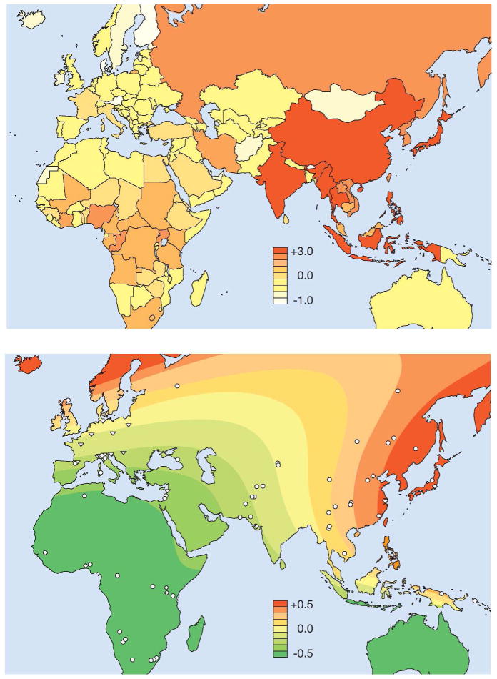
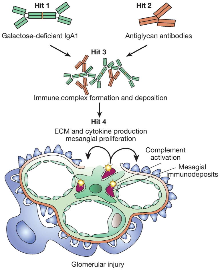
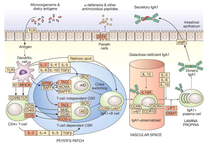
Recent years have brought notable progress in the field of IgA nephropathy. Here, we highlight important new directions and latest developments, including successful discovery of several genetic susceptibility loci, formulation of the multihit pathogenesis model, introduction of the Oxford pathology scoring system, and formalization of the Kidney Disease Improving Global Outcomes (KDIGO) consensus treatment guidelines. We focus on the latest genetic findings that confirm a strong contribution of inherited factors and explain some of the geoethnic disparities in disease susceptibility. Most IgA nephropathy susceptibility loci discovered to date encode genes involved in the maintenance of the intestinal epithelial barrier and response to mucosal pathogens. The concerted pattern of interpopulation allelic differentiation across all genetic loci parallels the disease prevalence and correlates with variation in local pathogens, suggesting that multilocus adaptation might have shaped the present-day landscape of IgA nephropathy. Importantly, the 'Intestinal Immune Network for IgA Production' emerged as one of the new targets for potential therapeutic intervention. We place these findings in the context of the multihit pathogenesis model and existing knowledge of IgA immunobiology. Lastly, we provide our perspective on the existing treatment options, discuss areas of clinical uncertainty, and outline ongoing clinical trials and translational studies.


Figures

Figure 1
Figure 1. Pathologic features of IgA nephropathy by light microscopy, immunofluorescence and electron microscopy
(a) The glomerulus has global mesangial proliferation with at least 4 cells per mesangial area. When >50% of glomeruli exhibit mesangial hypercellularity, the biopsy receives a score of M1 according to the Oxford/IgA MEST system (H&E, x600). (b) Segmental endocapillary proliferation obliterates capillary lumina (score E1 when a biopsy contains one or more such lesions). The adjacent glomerular segments have mild mesangial hypercellularity (H&E, x600). (c) The stain for IgA is intense and globally outlines the mesangial framework of the glomerulus (immunofluorecence, x600). (d) Segmental glomerular scarring develops as postinflammatory sclerosis, mimicking the changes in focal segmental glomerulosclerosis. (score S1 when a biopsy contains one or more such lesions), (Jones methenamine silver, x600). (e) A case with high chronicity contains globally sclerotic glomeruli and exhibits more than 50% tubular atrophy/interstitial fibrosis (score T2), (Masson trichrome, x200). (f) The immunofluorescence staining for C3 is similar in distribution as the mesangial staining for IgA (shown from the same glomerulus as in 1C) but exhibits weaker intensity and a more punctate, granular texture (immunofluorescence, x600). (g) A severe example has a cellular crescent that compresses the glomerular tuft. Global mesangial expansion is present (Jones methenamine silver, x400). (h) One or more red blood cell casts are commonly encountered at biopsy and may be numerous, especially in cases with gross hematuria and acute tubular injury (H&E, x600). (i) By electron microscopy, large mesangial deposits elevate the glomerular basement membrane reflection over the mesangium, bulging towards the urinary space. This deposit involves the entire mesangium but is most prominent in the paramesangial region, beneath the GBM reflection. The mesangial cellularity is increased but the capillary lumen is patent (electron micrograph, x5000).
Figure 2
Figure 2. Geospatial Pattern of Genetic Risk for IgA Nephropathy and Worldwide Map of Helminth Diversity
Top Panel: Surface interpolation of the standardized genetic risk over Africa and Euroasia. Symbols represent the locations of sampled populations: Human Genome Diversity Panel (HGDP; 1,050 individuals representative of 52 worldwide populations), HapMap III (1,184 individuals representative of 11 populations), other population samples (4,547 individuals representative of 25 populations); from Kiryluk et al. PLoS Genetics 2012;8(6):e1002765. Bottom Panel: Standardized values for the diversity of helminth species infecting humans per country; data from the Global Infectious Disease and Epidemiology Online Network (GIDEON), see URL.
Figure 3
Figure 3
The Multi-hit Pathogenesis Model of IgA Nephropathy.
Figure 4
Figure 4. Genetic Hits to the “Intestinal Immune Network for IgA Production”
IgA is the most abundant antibody isotype in the body, with the majority of IgA found in mucosal secretions. Mucosal IgA production is induced by T cell-dependent and T cell-independent mechanisms. T cell-independent production of IgA is primarily stimulated by IL-6, IL-10, TGF-β, BAFF, and APRIL produced by intestinal epithelial, dendritic, and stromal cells. In this environment, intestinal B cells undergo class switching from IgM to IgA1. IgA-secreting plasma cells migrate to lamina propria, where they release dimeric IgA1. The dimers are formed through an interaction of two IgA1 molecules with a joining chain (J-chain), which is synthesized by plasma cells. IgA1 dimers can bind to the polymeric Ig receptor (pIgR) on the basolateral surface of the mucosal epithelium and undergo transcytosis to the apical surface, where they dissociate from pIgR and are secreted into the lumen carrying the secretory component of the receptor. The secretory component protects IgA molecules from proteolytic enzymes in the gut lumen. The bacteriostatic effects of secretory IgA1 are accompanied by antimicrobial peptides, such as defensins, secreted into the gut lumen by Paneth cells. The key molecules involved in the intestinal immune network for IgA production are indicated in orange; molecules implicated by GWAS are marked in red. The risk alleles generally lead to increased IgA1 responsiveness stimulating IgA1 production; increased levels of polymeric IgA1 in the circulation may represent a consequence of “spill-over” from mucosal sites and/or “mis-trafficking” of stimulated plasma cell to bone marrow sites.

References

    1. Berger J, Hinglais N. Intercapillary deposits of IgA-IgG. Journal d’urologie et de nephrologie. 1968;74:694–5. - PubMed
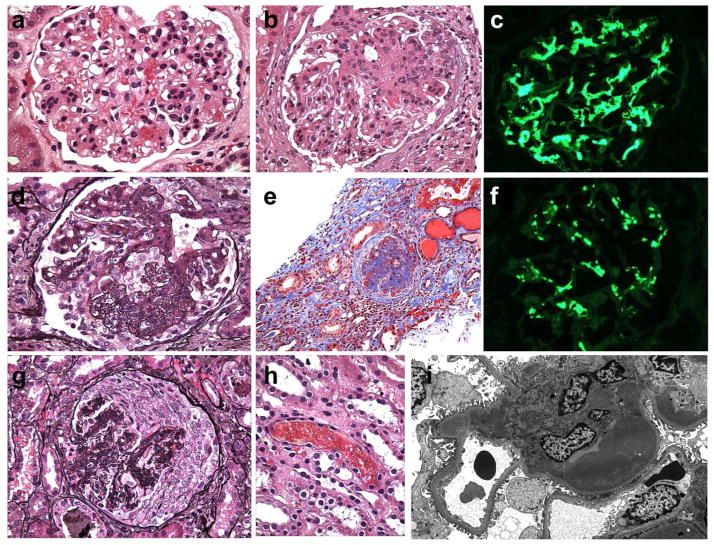
    1. Roberts IS. Pathology of IgA nephropathy. Nature reviews Nephrology. 2014;10:445–54. - PubMed
    1. Working Group of the International Ig ANN, the Renal Pathology S. Roberts IS, et al. The Oxford classification of IgA nephropathy: pathology definitions, correlations, and reproducibility. Kidney international. 2009;76:546–56. - PubMed
    1. Jennette JC. The immunohistology of IgA nephropathy. American journal of kidney diseases: the official journal of the National Kidney Foundation. 1988;12:348–52. - PubMed
    1. Valentijn RM, Radl J, Haaijman JJ, et al. Circulating and mesangial secretory component-binding IgA-1 in primary IgA nephropathy. Kidney international. 1984;26:760–6. - PubMed

Publication types

MeSH terms