A murine model of targeted infusion for intracranial tumors
- PMID: 26376657
- PMCID: PMC5489113
- DOI: 10.1007/s11060-015-1942-7
A murine model of targeted infusion for intracranial tumors
Abstract
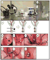
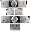
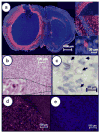
Historically, intra-arterial (IA) drug administration for malignant brain tumors including glioblastoma multiforme (GBM) was performed as an attempt to improve drug delivery. With the advent of percutaneous neuorovascular techniques and modern microcatheters, intracranial drug delivery is readily feasible; however, the question remains whether IA administration is safe and more effective compared to other delivery modalities such as intravenous (IV) or oral administrations. Preclinical large animal models allow for comparisons between treatment routes and to test novel agents, but can be expensive and difficult to generate large numbers and rapid results. Accordingly, we developed a murine model of IA drug delivery for GBM that is reproducible with clear readouts of tumor response and neurotoxicities. Herein, we describe a novel mouse model of IA drug delivery accessing the internal carotid artery to treat ipsilateral implanted GBM tumors that is consistent and reproducible with minimal experience. The intent of establishing this unique platform is to efficiently interrogate targeted anti-tumor agents that may be designed to take advantage of a directed, regional therapy approach for brain tumors.
Keywords: GBM; Glioblastoma multiforme; Infusion; Intra-arterial; Mouse; Regional cancer therapy.
Conflict of interest statement
The authors have no financial interests to disclose regarding this manuscript.
Figures


Similar articles
-
Intratumoral delivery of bortezomib: impact on survival in an intracranial glioma tumor model.J Neurosurg. 2018 Mar;128(3):695-700. doi: 10.3171/2016.11.JNS161212. Epub 2017 Apr 14. J Neurosurg. 2018. PMID: 28409734
-
Distribution kinetics of targeted cytotoxin in glioma by bolus or convection-enhanced delivery in a murine model.J Neurosurg. 2004 Dec;101(6):1004-11. doi: 10.3171/jns.2004.101.6.1004. J Neurosurg. 2004. PMID: 15597761
-
Orthotopic glioblastoma stem-like cell xenograft model in mice to evaluate intra-arterial delivery of bevacizumab: from bedside to bench.J Clin Neurosci. 2012 Nov;19(11):1568-72. doi: 10.1016/j.jocn.2012.03.012. Epub 2012 Sep 15. J Clin Neurosci. 2012. PMID: 22985932
-
Intraarterial drug delivery for glioblastoma mutiforme: Will the phoenix rise again?J Neurooncol. 2015 Sep;124(3):333-43. doi: 10.1007/s11060-015-1846-6. Epub 2015 Jun 25. J Neurooncol. 2015. PMID: 26108656 Review.
-
Intra-arterial chemotherapy for malignant gliomas: a critical analysis.Interv Neuroradiol. 2011 Sep;17(3):286-95. doi: 10.1177/159101991101700302. Epub 2011 Oct 17. Interv Neuroradiol. 2011. PMID: 22005689 Free PMC article. Review.
Cited by
-
Advances in drug delivery to high grade gliomas.Brain Pathol. 2016 Nov;26(6):689-700. doi: 10.1111/bpa.12423. Epub 2016 Aug 24. Brain Pathol. 2016. PMID: 27488680 Free PMC article. Review.
-
The first endovascular rat glioma model for pre-clinical evaluation of intra-arterial therapeutics.Interv Neuroradiol. 2025 Aug;31(4):447-456. doi: 10.1177/15910199231169597. Epub 2023 May 8. Interv Neuroradiol. 2025. PMID: 37157800 Free PMC article.
References
-
- Andratschke N, Grosu AL, Molls M, Nieder C. Perspectives in the treatment of malignant gliomas in adults. Anticancer research. 2001;21:3541–3550. - PubMed
-
- Lefranc F, Rynkowski M, DeWitte O, Kiss R. Present and potential future adjuvant issues in high-grade astrocytic glioma treatment. Advances and technical standards in neurosurgery. 2009;34:3–35. - PubMed
-
- Phillips HS, Kharbanda S, Chen R, Forrest WF, Soriano RH, Wu TD, Misra A, Nigro JM, Colman H, Soroceanu L, Williams PM, Modrusan Z, Feuerstein BG, Aldape K. Molecular subclasses of high-grade glioma predict prognosis, delineate a pattern of disease progression, and resemble stages in neurogenesis. Cancer cell. 2006;9:157–173. doi: 10.1016/j.ccr.2006.02.019. - DOI - PubMed
-
- Zustovich F, Cartei G, Ceravolo R, Zovato S, Della Puppa A, Pastorelli D, Mattiazzi M, Bertorelle R, Gardiman MP. A phase I study of cisplatin, temozolomide and thalidomide in patients with malignant brain tumors. Anticancer research. 2007;27:1019–1024. - PubMed
Publication types
MeSH terms
Substances
Grants and funding
LinkOut - more resources
Full Text Sources
Other Literature Sources
Medical

