. 2016 Jan 10;24(2):99-112.
doi: 10.1089/ars.2015.6347. Epub 2015 Dec 14.

Heme Attenuation Ameliorates Irritant Gas Inhalation-Induced Acute Lung Injury

Affiliations

Heme Attenuation Ameliorates Irritant Gas Inhalation-Induced Acute Lung Injury

Saurabh Aggarwal et al. Antioxid Redox Signal. .

Abstract

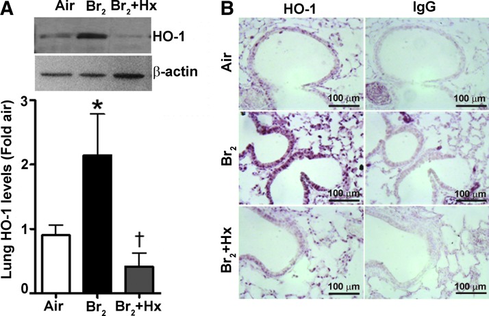
Aims: Exposure to irritant gases, such as bromine (Br2), poses an environmental and occupational hazard that results in severe lung and systemic injury. However, the mechanism(s) of Br2 toxicity and the therapeutic responses required to mitigate lung damage are not known. Previously, it was demonstrated that Br2 upregulates the heme degrading enzyme, heme oxygenase-1 (HO-1). Since heme is a major inducer of HO-1, we determined whether an increase in heme and heme-dependent oxidative injury underlies the pathogenesis of Br2 toxicity.

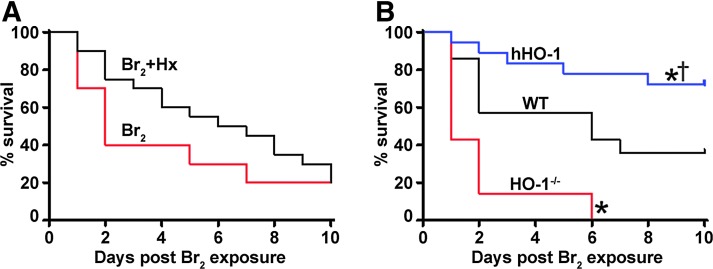
Results: C57BL/6 mice were exposed to Br2 gas (600 ppm, 30 min) and returned to room air. Thirty minutes postexposure, mice were injected intraperitoneally with a single dose of the heme scavenging protein, hemopexin (Hx) (3 μg/gm body weight), or saline. Twenty-four hours postexposure, saline-treated mice had elevated total heme in bronchoalveolar lavage fluid (BALF) and plasma and acute lung injury (ALI) culminating in 80% mortality after 10 days. Hx treatment significantly lowered heme, decreased evidence of ALI (lower protein and inflammatory cells in BALF, lower lung wet-to-dry weight ratios, and decreased airway hyperreactivity to methacholine), and reduced mortality. In addition, Br2 caused more severe ALI and mortality in mice with HO-1 gene deletion (HO-1-/-) compared to wild-type controls, while transgenic mice overexpressing the human HO-1 gene (hHO-1) showed significant protection.

Innovation: This is the first study delineating the role of heme in ALI caused by Br2.

Conclusion: The data suggest that attenuating heme may prove to be a useful adjuvant therapy to treat patients with ALI.


Figures

<b>FIG. 1.</b>
FIG. 1.
Lung injury and mortality in mice exposed to Br2 gas inhalation. Male and female C57BL/6 mice were exposed for different durations and various doses of Br2 gas and assessed for mortality. Exposure to 600 ppm of Br2 for 30 min caused mortality in 50% of mice within 6–7 days (A). There was no significant difference in the mortality rate between male and female mice (n = 12 for male [400 ppm, 30 min], n = 8 for male [600 ppm, 30 min], n = 4 for male [600 ppm, 45 min], and n = 12 for female [600 ppm, 45 min]) (A). Analysis of the BALF after 600 ppm Br2 gas inhalation for 30 min showed a significant increase in BALF protein levels (n = 4–5) (B) and total cell count (n = 4–5) (C) within 24 h postexposure. Hematoxylin and eosin staining and the staining for myeloperoxidase (n = 3–4) (E) of peripheral lung tissue 24 and 48 h post-Br2 gas (600 ppm, 30 min) exposure demonstrated an increase in the lung injury score (n = 3–4) (D), as depicted by the disruption of the airway parenchyma (black arrow), an increase in cellularity, proteinacious debris accumulation, an increase in the alveolar and interstitial accumulation of neutrophils, and alveolar septal thickening. Values are mean ± SEM. All animals were males except where indicated. *p < 0.05 versus air exposed C57BL/6 mice. BALF, bronchoalveolar lavage fluid; Br2, bromine.
<b>FIG. 2.</b>
FIG. 2.
Br2 gas inhalation increases the susceptibility to hemolysis and the apoptosis/necrosis of lung inflammatory cells. C57BL/6 mice were exposed to Br2 gas (600 ppm) for 30 min and then brought to room air. The mice were harvested 24 h postexposure and their blood and BALF was obtained. An ex vivo hemolytic assay performed on the blood of these mice demonstrated an increased susceptibility to hemolysis at lower concentrations of the detergent, SDS, compared to the blood obtained from air exposed mice (n = 10) (A). Flow cytometry analysis of Annexin V FITC-stained cells collected from the BALF showed a significant increase in late apoptotic/necrotic (Quadrant [Q] 2) and necrotic (Quadrant [Q] 1) cells in the Br2 exposed mice (n = 3–6) (B). Quadrants (Q) 3 and 4 show alive and early apoptotic cells, respectively. In addition, Br2 inhalation also increased LDH levels in the BALF of exposed mice (n = 8–10) (C). Values are mean ± SEM. All animals were males. *p < 0.05 versus air exposed C57BL/6 mice. LDH, lactate dehydrogenase; SDS, sodium dodecyl sulfate.
<b>FIG. 3.</b>
FIG. 3.
Br2 gas inhalation increases lung HO-1 protein expression. C57BL/6 mice were exposed to Br2 gas (600 ppm) for 30 min and then brought to room air. Mice were injected intraperitoneally with Hx (3 μg/g body weight) 30 min after the exposure. After 24 h, peripheral lung tissue was harvested. Immunoblot analysis demonstrated that Br2 inhalation significantly increased lung HO-1 protein levels (n = 6) (A). Hx treatment prevented the increase in HO-1 protein levels. Similarly, immunohistological staining indicated higher HO-1 expression in the Br2 exposed mice compared to the air exposed mice or Hx-treated mice (n = 4) (B). Values are mean ± SEM. All animals were males. *p < 0.05 versus air exposed C57BL/6 mice, p < 0.05 versus Br2+Hx-treated C57BL/6 mice. HO-1, heme oxygenase-1; Hx, hemopexin.
<b>FIG. 4.</b>
FIG. 4.
Heme is increased in C57BL/6 mice exposed to Br2 gas inhalation. C57BL/6 mice were exposed to Br2 gas (600 ppm) for 30 min and then brought to room air. Mice were given an intraperitoneal injection of Hx (3 μg/g body weight) 30 min after the exposure. After 24 h, Br2 inhalation significantly increased total heme levels in the BALF (A), the plasma (B), and the peripheral lung tissue (C) of the exposed C57BL/6 mice. Hx treatment reduced heme in these animals. Lung fractionation demonstrated that Br2 mainly increased cytosolic heme (D), while the heme content in the mitochondria (E) and the microsomes (F) did not change after Br2. Values are mean ± SEM (n = 5–7). All animals were males. *p < 0.05 versus air exposed C57BL/6 mice, p < 0.05 versus Br2+Hx-treated C57BL/6 mice.
<b>FIG. 5.</b>
FIG. 5.
HO-1 attenuates Br2-induced heme. Immunoblot analysis demonstrated the expression of HO-1 protein in the peripheral lung tissue of the WT mice, the mice overexpressing the human heme oxygenase-1 enzyme (hHO-1), and the mice lacking endogenous HO-1 (HO-1−/−) (n = 5–6) (A). These mice were exposed to Br2 gas (600 ppm) for 30 min and then brought to room air. After 24 h, Br2 inhalation significantly increased total heme levels in the BALF of the WT and the HO-1−/− mice compared to their air exposed controls (B). Br2 exposure also increased total heme levels in the plasma (C) and the peripheral lung tissue (D) of the HO-1−/− mice in comparison to the air exposed mice. In addition, the BALF, plasma, and whole lung heme levels were higher in the Br2 exposed HO-1−/− mice compared to the Br2 exposed WT mice. Br2 did not alter heme in the hHO-1 mice. Values are mean ± SEM (n = 3 for air exposed mice and 5–6 for Br2 exposed mice). All animals were males. *p < 0.05 versus respective air exposed control mice, p < 0.05 versus Br2 exposed WT mice, and p < 0.05 versus Br2 exposed HO-1−/− mice. WT, wild type.
<b>FIG. 6.</b>
FIG. 6.
Heme induces lung oxidative stress in Br2 gas exposed mice. C57BL/6 mice, WT mice, the mice overexpressing the human heme oxygenase 1 enzyme (hHO-1), and the mice lacking endogenous HO-1 (HO-1−/−) were exposed to Br2 gas (600 ppm) for 30 min and then brought to room air. C57BL/6 mice were given an intraperitoneal injection of Hx (3 μg/g body weight) 30 min after the exposure. After 24 h, carbonyl (aldehydes and ketones) adducts, a hallmark of the oxidation status of proteins, were measured by gel electrophoresis and Western blotting using the protein obtained from the whole lung tissue. Hx prevented the Br2-dependent increase in protein carbonylation in C57BL/6 mice (A). Similarly, after Br2 gas exposure, the lung protein carbonylation levels were significantly higher in the HO-1−/− mice compared to the WT mice or the hHO-1 mice (B). Values are mean ± SEM (n = 4). All animals were males. *p < 0.05 versus air exposed C57BL/6 mice or Br2 exposed WT mice, p < 0.05 versus Br2+Hx-treated C57BL/6 mice or Br2 exposed HO-1−/− mice.
<b>FIG. 7.</b>
FIG. 7.
Br2-induced lung injury is mediated by heme. C57BL/6 mice, WT mice, the mice overexpressing the human heme oxygenase 1 enzyme (hHO-1), and the mice lacking endogenous HO-1 (HO-1−/−) were exposed to Br2 gas (600 ppm) for 30 min and then brought to room air. C57BL/6 mice were given an intraperitoneal injection of Hx (3 μg/g body weight) 30 min after the exposure. After 24 h, the BALF protein levels, total and differential cell counts, were measured. Hx significantly attenuated the Br2-dependent increase in BALF protein levels (n = 6) (A) and BALF total cell count (n = 5–6) (B) and prevented the extravasation of neutrophils in C57BL/6 mice (n = 5–6) (C). Furthermore, BALF protein levels (n = 5–6) (D), but not the total number of cells (n = 5–6) (E), were higher in the HO-1−/− mice compared to the WT mice or the hHO-1 mice. Yet, the neutrophil count in the BALF was significantly higher in the HO-1−/− mice compared to the WT mice and the hHO-1 mice (n = 5–6) (F). Values are mean ± SEM. All animals were males except in (D–F), where two animals were females in each of the three groups: WT, HO-1−/−, and hHO-1. *p < 0.05 versus air exposed C57BL/6 mice or Br2 exposed WT mice, p < 0.05 versus Br2+Hx-treated C57BL/6 mice or Br2 exposed HO-1−/− mice.
<b>FIG. 8.</b>
FIG. 8.
Heme reduction improves airway function post-Br2 gas exposure. Lung wet-to-dry ratio, methacholine-dependent peripheral lung resistance (R), and central (Newtonian) airway resistance (Rn) were measured in mice exposed to Br2 gas (600 ppm) for 30 min. Br2 increased the lung wet-to-dry weight ratio in C57BL/6 mice, which was attenuated by treatment (30 min postexposure) with Hx (3 μg/g body weight) (n = 6) (A). The methacholine-dependent increase in R (n = 4–10) (B) and Rn (n = 6) (C) was also higher in the Br2 exposed C57BL/6 mice compared to the air exposed mice. Hx prevented the increase in both R and Rn (B, C). Similarly, the mice lacking the endogenous heme oxygenase-1 gene (HO-1−/−) had a higher lung wet-to-dry ratio (n = 5–7) (D) and an elevated methacholine-dependent increase in R (n = 3–7) (E) and Rn (n = 4–7) (F) compared to the WT mice and the mice overexpressing the human HO-1 gene (hHO-1). Values are mean ± SEM. All animals were males. *p < 0.05 versus air exposed C57BL/6 mice or Br2 exposed WT mice, p < 0.05 versus Br2+Hx-treated mice or Br2 exposed HO-1−/− mice.
<b>FIG. 9.</b>
FIG. 9.
Heme attenuation improves survival after Br2 gas exposure. Mice were exposed to Br2 gas (600 ppm) for 30 min and then immediately brought to room air. After 30 min of exposure, C57BL/6 mice were given an intraperitoneal injection of Hx (3 μg/g body weight). Heme scavenging by Hx reduced the early increase in mortality within the first 2 days from 60% in the vehicle-treated to 25% in the Hx-treated mice after Br2 exposure. However, the overall survival according to the log-rank test was not significant between the two groups in the C57BL/6 mice (n = 20) (A). The Kaplan–Meier curve also demonstrated that the mice overexpressing the human heme oxygenase 1 enzyme (hHO-1) had lower mortality in comparison to the WT mice and the mice lacking endogenous HO-1 (HO-1−/−) postexposure to Br2 gas (n = 7 for HO-1−/−, 14 for WT, and 17 for hHO-1) (B). All animals were males. *p < 0.05 versus Br2 exposed WT mice, p < 0.05 versus Br2 exposed HO-1−/− mice.

References

    1. Alam J. and Smith A. Heme-hemopexin-mediated induction of metallothionein gene expression. J Biol Chem 267: 16379–16384, 1992 - PubMed
    1. Alam J. and Smith A. Receptor-mediated transport of heme by hemopexin regulates gene expression in mammalian cells. J Biol Chem 264: 17637–17640, 1989 - PubMed
    1. Balakrishna S, Song W, Achanta S, Doran SF, Liu B, Kaelberer MM, Yu Z, Sui A, Cheung M, Leishman E, Eidam HS, Ye G, Willette RN, Thorneloe KS, Bradshaw HB, Matalon S, and Jordt SE. TRPV4 inhibition counteracts edema and inflammation and improves pulmonary function and oxygen saturation in chemically induced acute lung injury. Am J Physiol Lung Cell Mol Physiol 307: L158–L172, 2014 - PMC - PubMed
    1. Bitron MD. and Aharonson EF. Delayed mortality of mice following inhalation of acute doses of CH2O, SO2Cl2, and Br2. Am Ind Hyg Assoc J 39: 129–138, 1978 - PubMed
    1. Brass CA, Immenschuh S, Song DX, Liem HH, and Eberhard UM. Hemopexin decreases spontaneous chemiluminescence of cold preserved liver after reperfusion. Biochem Biophys Res Commun 248: 574–577, 1998 - PubMed

Publication types