

Impaired survival of regulatory T cells in pulmonary sarcoidosis
- PMID: 26376720
- PMCID: PMC4574219
- DOI: 10.1186/s12931-015-0265-8
Impaired survival of regulatory T cells in pulmonary sarcoidosis
Abstract
Background: Impaired regulatory T cell (Treg) function is thought to contribute to ongoing inflammatory responses in sarcoidosis, but underlying mechanisms remain unclear. Moreover, it is not known if increased apoptotic susceptibility of Tregs may contribute to an impaired immunosuppressive function in sarcoidosis. Therefore, the aim of this study is to analyze proportions, phenotype, survival, and apoptotic susceptibility of Tregs in sarcoidosis.
Methods: Patients with pulmonary sarcoidosis (n = 58) were included at time of diagnosis. Tregs were analyzed in broncho-alveolar lavage fluid and peripheral blood of patients and healthy controls (HC).
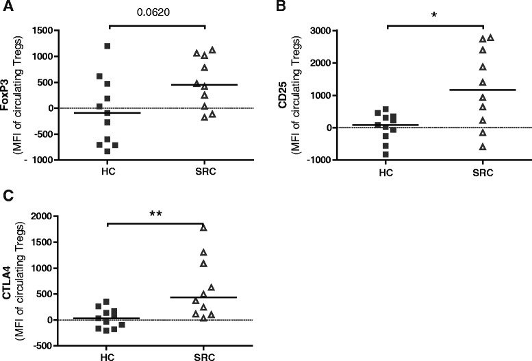
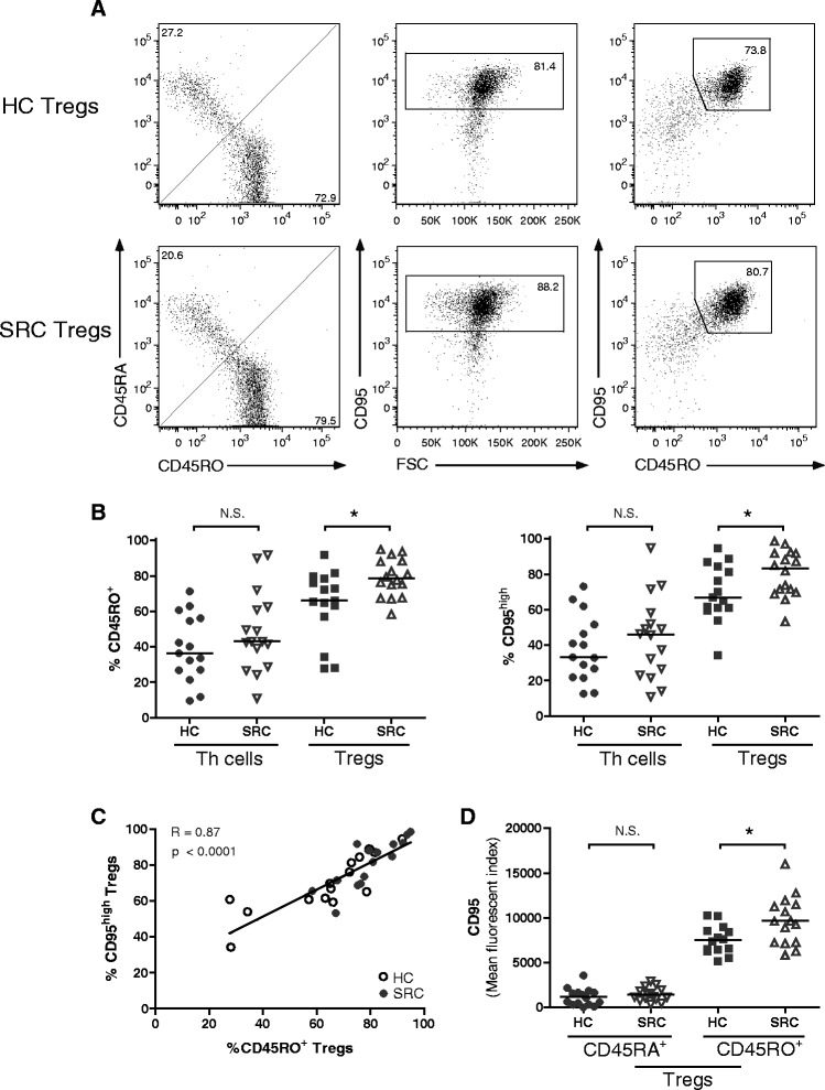
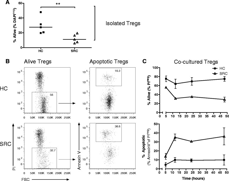
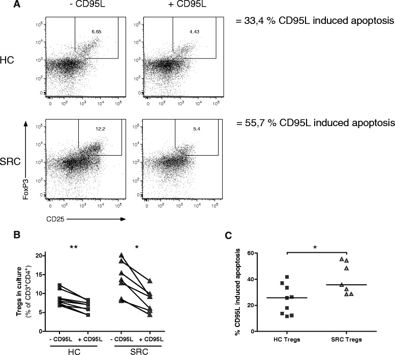
Results: In sarcoidosis patients no evidence was found for a relative deficit of Tregs, neither locally nor systemically. Rather, increased proportions of circulating Tregs were observed, most prominently in patients developing chronic disease. Sarcoidosis circulating Tregs displayed adequate expression of FoxP3, CD25 and CTLA4. Remarkably, in sarcoidosis enhanced CD95 expression on circulating activated CD45RO(+) Tregs was observed compared with HC, and proportions of these cells were significantly increased. Specifically sarcoidosis Tregs--but not Th cells--showed impaired survival compared with HC. Finally, CD95L-mediated apoptosis was enhanced in sarcoidosis Tregs.
Conclusion: In untreated patients with active pulmonary sarcoidosis, Tregs show impaired survival and enhanced apoptotic susceptibility towards CD95L. Increased apoptosis likely contributes to the insufficient immunosuppressive function of sarcoidosis Tregs. Further research into this field will help determine whether improvement of Treg survival holds a promising new therapeutic approach for chronic sarcoidosis patients.
Figures

Similar articles
-
Molecular profiling of regulatory T cells in pulmonary sarcoidosis.J Autoimmun. 2018 Nov;94:56-69. doi: 10.1016/j.jaut.2018.07.012. Epub 2018 Jul 23. J Autoimmun. 2018. PMID: 30049532
-
Global impairment of CD4+CD25+FOXP3+ regulatory T cells in idiopathic pulmonary fibrosis.Am J Respir Crit Care Med. 2009 Jun 15;179(12):1121-30. doi: 10.1164/rccm.200812-1936OC. Epub 2009 Apr 2. Am J Respir Crit Care Med. 2009. PMID: 19342412
-
The immune paradox of sarcoidosis and regulatory T cells.J Exp Med. 2006 Feb 20;203(2):359-70. doi: 10.1084/jem.20050648. Epub 2006 Jan 23. J Exp Med. 2006. PMID: 16432251 Free PMC article.
-
FOXP3+ regulatory T-cells in chronic kidney disease: molecular pathways and clinical implications.Adv Exp Med Biol. 2009;665:163-70. doi: 10.1007/978-1-4419-1599-3_12. Adv Exp Med Biol. 2009. PMID: 20429423 Review.
-
T-cell immunology in sarcoidosis: Disruption of a delicate balance between helper and regulatory T-cells.Curr Opin Pulm Med. 2016 Sep;22(5):476-83. doi: 10.1097/MCP.0000000000000303. Curr Opin Pulm Med. 2016. PMID: 27379969 Review.
Cited by
-
Infliximab therapy balances regulatory T cells, tumour necrosis factor receptor 2 (TNFR2) expression and soluble TNFR2 in sarcoidosis.Clin Exp Immunol. 2016 Aug;185(2):263-70. doi: 10.1111/cei.12808. Epub 2016 Jul 12. Clin Exp Immunol. 2016. PMID: 27158798 Free PMC article.
-
Imbalanced distribution of regulatory T cells and Th17.1 cells in the peripheral blood and BALF of sarcoidosis patients: relationship to disease activity and the fibrotic radiographic phenotype.Front Immunol. 2023 Jul 13;14:1185443. doi: 10.3389/fimmu.2023.1185443. eCollection 2023. Front Immunol. 2023. PMID: 37520566 Free PMC article.
-
Increased proportions of circulating PD-1+ CD4+ memory T cells and PD-1+ regulatory T cells associate with good response to prednisone in pulmonary sarcoidosis.Respir Res. 2024 May 7;25(1):196. doi: 10.1186/s12931-024-02833-y. Respir Res. 2024. PMID: 38715030 Free PMC article.
-
Key Players and Biomarkers of the Adaptive Immune System in the Pathogenesis of Sarcoidosis.Int J Mol Sci. 2020 Oct 7;21(19):7398. doi: 10.3390/ijms21197398. Int J Mol Sci. 2020. PMID: 33036432 Free PMC article. Review.
-
The Role of Diverse Immune Cells in Sarcoidosis.Front Immunol. 2021 Nov 19;12:788502. doi: 10.3389/fimmu.2021.788502. eCollection 2021. Front Immunol. 2021. PMID: 34868074 Free PMC article. Review.
References
-
- Statement on sarcoidosis. Joint Statement of the American Thoracic Society (ATS), the European Respiratory Society (ERS) and the World Association of Sarcoidosis and Other Granulomatous Disorders (WASOG) adopted by the ATS Board of Directors and by the ERS Executive Committee, February 1999. Am J Respir Crit Care Med. 1999;160(2):736–55. doi:10.1164/ajrccm.160.2.ats4-99. - PubMed
Publication types
MeSH terms
Substances
LinkOut - more resources
Full Text Sources
Other Literature Sources
Research Materials
Miscellaneous
