

Pirt contributes to uterine contraction-induced pain in mice
- PMID: 26376721
- PMCID: PMC4574137
- DOI: 10.1186/s12990-015-0054-x
Pirt contributes to uterine contraction-induced pain in mice
Abstract
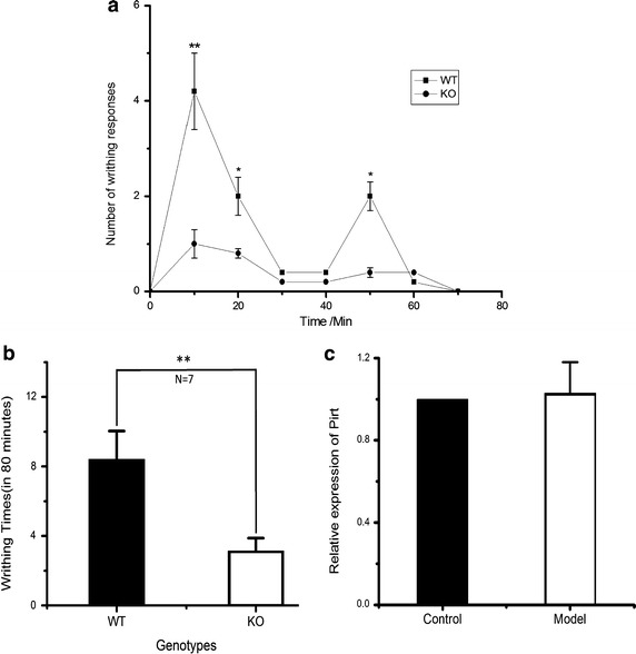
Uterine contraction-induced pain (UCP) represents a common and severe form of visceral pain. Nerve fibers that innervate uterine tissue express the transient receptor potential vanilloid channel 1 (TRPV1), which has been shown to be involved in the perception of UCP. The phosphoinositide-interacting regulator of TRP (Pirt) may act as a regulatory subunit of TRPV1. The intraperitoneal injection of oxytocin into female mice after a 6-day priming treatment with estradiol benzoate induces writhing responses, which reflect the presence of UCP. Here, we first compared writhing response between Pirt (+/+) and Pirt (-/-) mice. Second, we examined the innervation of Pirt-expressing nerves in the uterus of Pirt (-/-) mice by immunofluorescence and two-photon microscopy. Third, we identified the soma of dorsal root ganglion (DRG) neurons that innerve the uterus using retrograde tracing and further characterized the neurochemical properties of these DRG neurons. Finally, we compared the calcium response of capsaicin between DRG neurons from Pirt (+/+) and Pirt (-/-) mice. We found that the writhing responses were less intensive in Pirt (-/-) mice than in Pirt (+/+) mice. We also observed Pirt-expressing nerve fibers in the myometrium of the uterus, and that retrograde-labeled cells were small-diameter, unmyelinated, and Pirt-positive DRG neurons. Additionally, we found that the number of capsaicin-responding neurons and the magnitude of evoked calcium response were markedly reduced in DRG neurons from Pirt (-/-) mice. Taken together, we speculate that Pirt plays an important role in mice uterine contraction-induced pain.
Figures


Similar articles
-
Pirt Together with TRPV1 Is Involved in the Regulation of Neuropathic Pain.Neural Plast. 2018 Apr 2;2018:4861491. doi: 10.1155/2018/4861491. eCollection 2018. Neural Plast. 2018. PMID: 29808083 Free PMC article.
-
Pirt, a phosphoinositide-binding protein, functions as a regulatory subunit of TRPV1.Cell. 2008 May 2;133(3):475-85. doi: 10.1016/j.cell.2008.02.053. Cell. 2008. PMID: 18455988 Free PMC article.
-
Pirt functions as an endogenous regulator of TRPM8.Nat Commun. 2013;4:2179. doi: 10.1038/ncomms3179. Nat Commun. 2013. PMID: 23863968 Free PMC article.
-
Pirt reduces bladder overactivity by inhibiting purinergic receptor P2X3.Nat Commun. 2015 Jul 7;6:7650. doi: 10.1038/ncomms8650. Nat Commun. 2015. PMID: 26151598
-
Distinct chemical classes of medium-sized transient receptor potential channel vanilloid 1-immunoreactive dorsal root ganglion neurons innervate the adult mouse jejunum and colon.Neuroscience. 2008 Oct 2;156(2):334-43. doi: 10.1016/j.neuroscience.2008.06.071. Epub 2008 Jul 25. Neuroscience. 2008. PMID: 18706490
Cited by
-
Facilitation of MrgprD by TRP-A1 promotes neuropathic pain.FASEB J. 2019 Jan;33(1):1360-1373. doi: 10.1096/fj.201800615RR. Epub 2018 Aug 27. FASEB J. 2019. PMID: 30148678 Free PMC article.
-
Pirt Together with TRPV1 Is Involved in the Regulation of Neuropathic Pain.Neural Plast. 2018 Apr 2;2018:4861491. doi: 10.1155/2018/4861491. eCollection 2018. Neural Plast. 2018. PMID: 29808083 Free PMC article.
-
Ultrasound Guided Surgery as a Refinement Tool in Oncology Research.Animals (Basel). 2022 Dec 6;12(23):3445. doi: 10.3390/ani12233445. Animals (Basel). 2022. PMID: 36496966 Free PMC article. Review.
-
Phosphoinositide-interacting regulator of TRP (PIRT) has opposing effects on human and mouse TRPM8 ion channels.J Biol Chem. 2018 Jun 15;293(24):9423-9434. doi: 10.1074/jbc.RA118.003563. Epub 2018 May 3. J Biol Chem. 2018. PMID: 29724821 Free PMC article.
-
TRPV1 in Pain and Itch.Adv Exp Med Biol. 2021;1349:249-273. doi: 10.1007/978-981-16-4254-8_12. Adv Exp Med Biol. 2021. PMID: 35138618
References
-
- Berkley KJ, Hotta H, Robbins A, Sato Y. Functional properties of afferent fibers supplying reproductive and other pelvic organs in pelvic nerve of female rat. J Neurophysiol. 1990;63:256–272. - PubMed
-
- Jones RC, III, Xu L, Gebhart GF. The mechanosensitivity of mouse colon afferent fibers and their sensitization by inflammatory mediators require transient receptor potential vanilloid 1 and acid-sensing ion channel 3. J Neurosci. 2005;25:10981–10989. doi: 10.1523/JNEUROSCI.0703-05.2005. - DOI - PMC - PubMed
Publication types
MeSH terms
Substances
LinkOut - more resources
Full Text Sources
Other Literature Sources
Medical
Molecular Biology Databases
