. 2015 Dec 1;24(23):6736-55.
doi: 10.1093/hmg/ddv380. Epub 2015 Sep 16.

Conditional depletion of intellectual disability and Parkinsonism candidate gene ATP6AP2 in fly and mouse induces cognitive impairment and neurodegeneration

Affiliations

Conditional depletion of intellectual disability and Parkinsonism candidate gene ATP6AP2 in fly and mouse induces cognitive impairment and neurodegeneration

Aline Dubos et al. Hum Mol Genet. .

Abstract

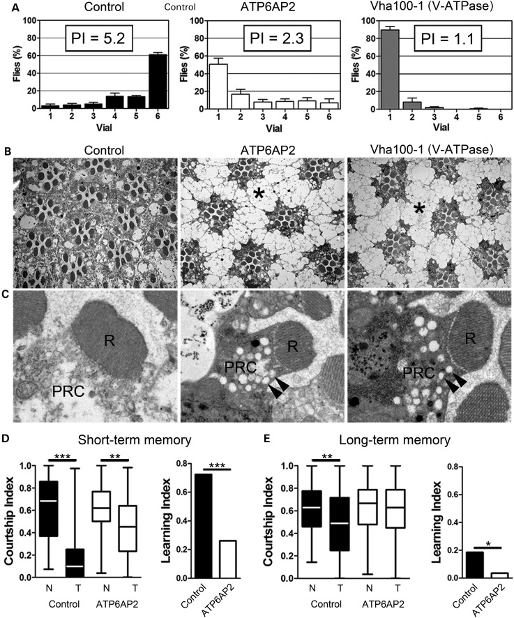
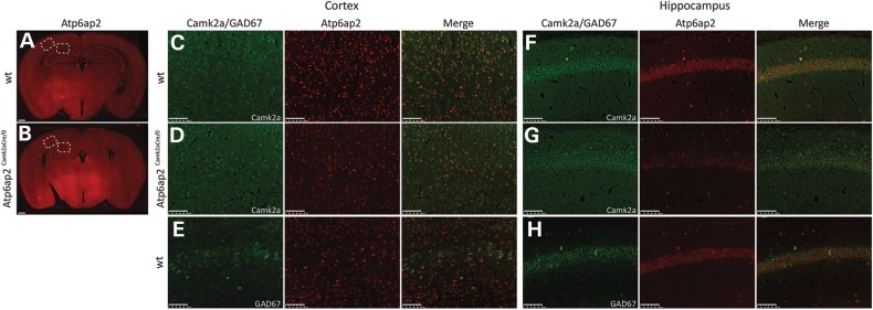
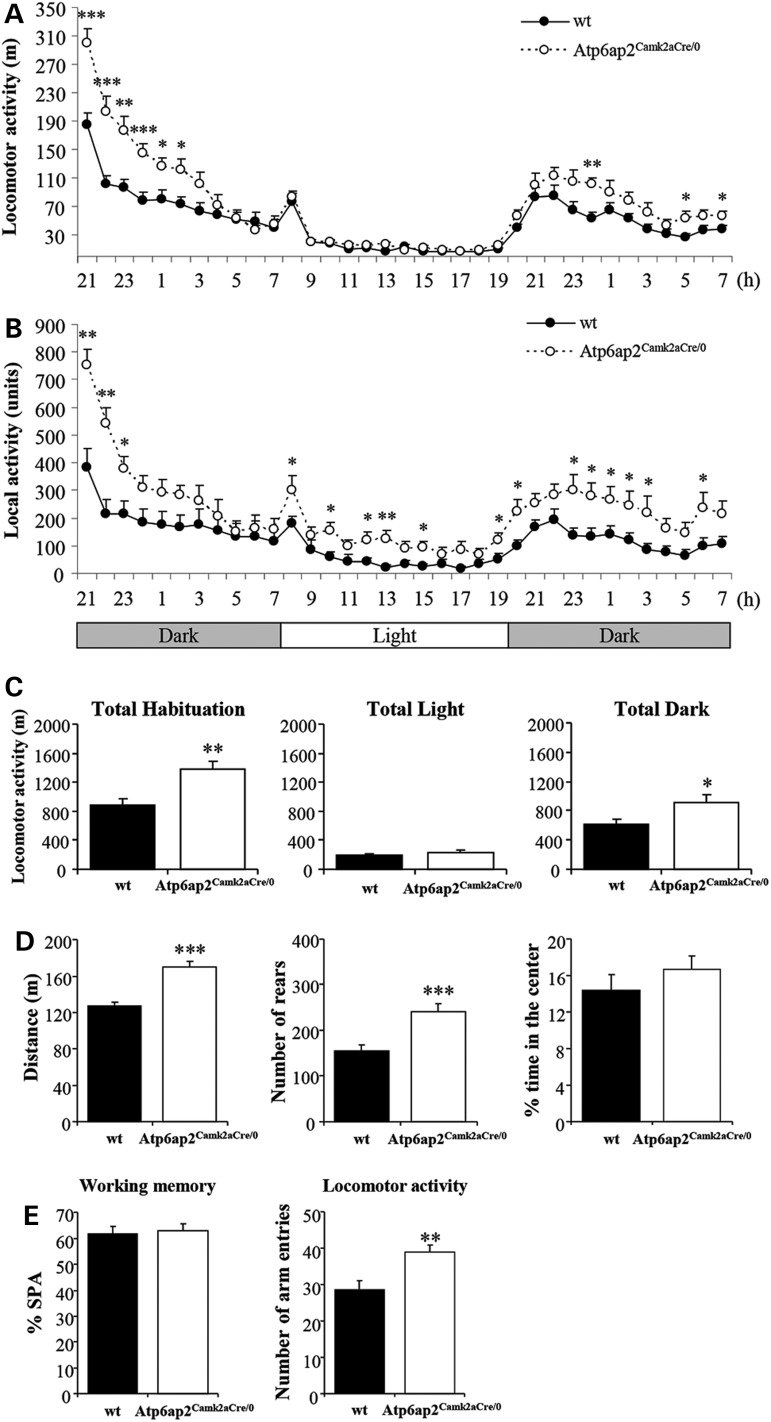
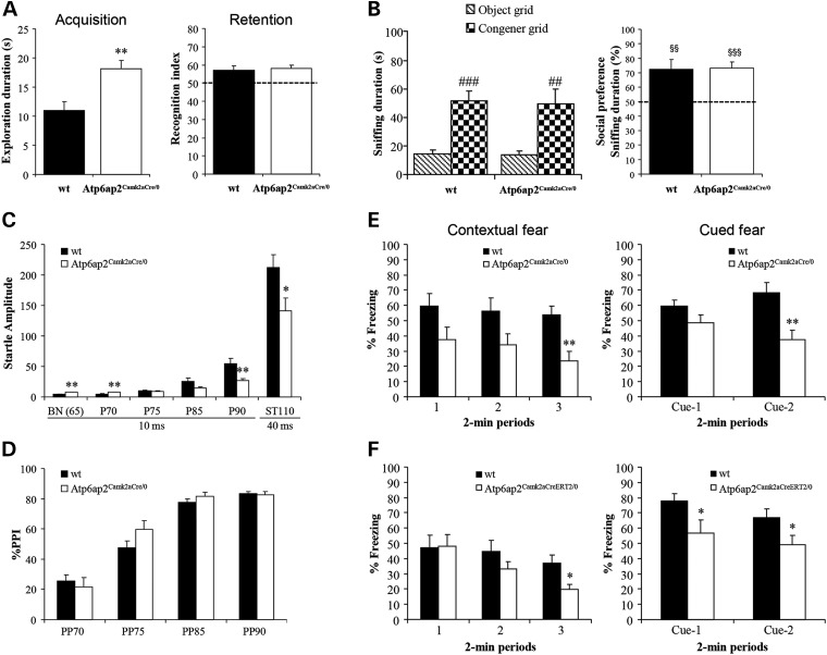
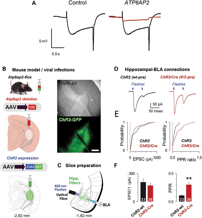
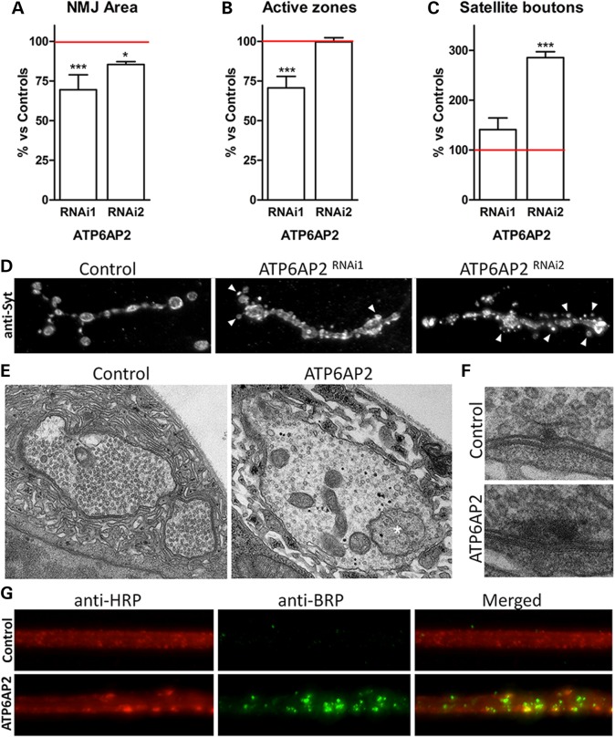
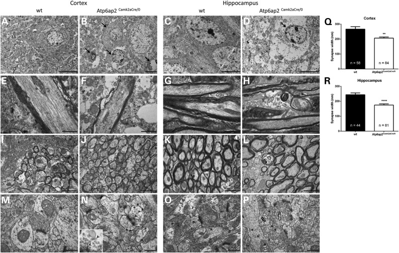
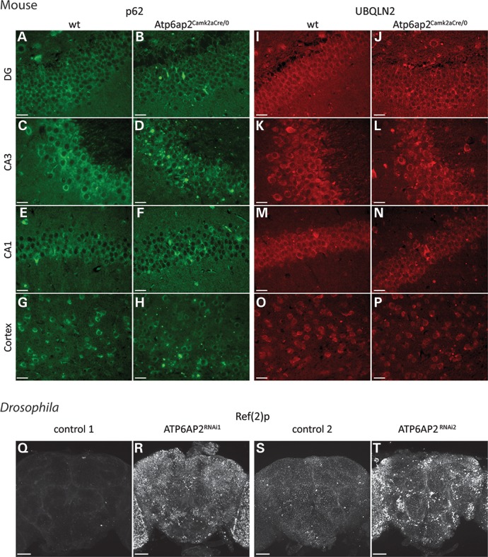
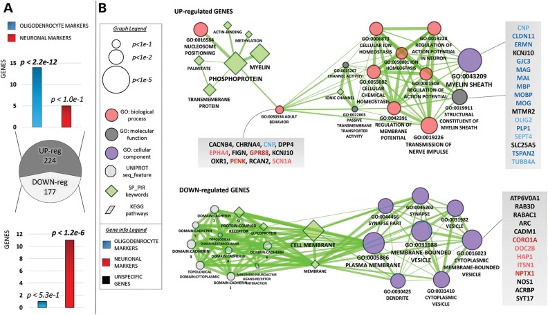
ATP6AP2, an essential accessory component of the vacuolar H+ ATPase (V-ATPase), has been associated with intellectual disability (ID) and Parkinsonism. ATP6AP2 has been implicated in several signalling pathways; however, little is known regarding its role in the nervous system. To decipher its function in behaviour and cognition, we generated and characterized conditional knockdowns of ATP6AP2 in the nervous system of Drosophila and mouse models. In Drosophila, ATP6AP2 knockdown induced defective phototaxis and vacuolated photoreceptor neurons and pigment cells when depleted in eyes and altered short- and long-term memory when depleted in the mushroom body. In mouse, conditional Atp6ap2 deletion in glutamatergic neurons (Atp6ap2(Camk2aCre/0) mice) caused increased spontaneous locomotor activity and altered fear memory. Both Drosophila ATP6AP2 knockdown and Atp6ap2(Camk2aCre/0) mice presented with presynaptic transmission defects, and with an abnormal number and morphology of synapses. In addition, Atp6ap2(Camk2aCre/0) mice showed autophagy defects that led to axonal and neuronal degeneration in the cortex and hippocampus. Surprisingly, axon myelination was affected in our mutant mice, and axonal transport alterations were observed in Drosophila. In accordance with the identified phenotypes across species, genome-wide transcriptome profiling of Atp6ap2(Camk2aCre/0) mouse hippocampi revealed dysregulation of genes involved in myelination, action potential, membrane-bound vesicles and motor behaviour. In summary, ATP6AP2 disruption in mouse and fly leads to cognitive impairment and neurodegeneration, mimicking aspects of the neuropathology associated with ATP6AP2 mutations in humans. Our results identify ATP6AP2 as an essential gene for the nervous system.


Figures

Figure 1.
Figure 1.
Knockdown of Drosophila ATP6AP2 disrupted phototaxis and short- and long-term memory. (A) Distribution of adult flies in the phototaxis assay measuring their visually induced activity and resulting phototaxis indexes (PI). Control flies (GMR-Gal4/+;UAS-Dicer-2/+) and flies with either eye-specific knockdown of ATP6AP2 (UAS-ATP6AP2RNAi1/GMR-Gal4;UAS-Dicer-2/+) or Vha100-1 (GMR-Gal4/+;UAS-Dicer-2/Vha100-1RNAi) are shown. ATP6AP2 and Vha100-1 knockdown animals showed dramatically decreased PIs, revealing functional photoreceptor neuron defects. (B and C) Histological and high-magnification electron microscopy images of the same genotypes presented in A. The pigment cells surrounding the photoreceptor neurons were strongly vacuolated (asterisk). Vesicular structures were present in the PR cytoplasm (PRC) (arrowheads). Rhabdomeres (R), the apical photosensitive membrane domains of photoreceptors, were intact in ATP6AP2 and Vha100-1 knockdown eyes. (A–C) Note the strikingly similar phenotypes caused by knockdown of ATP6AP2 and Vha100-1. (D) Short-term and (E) long-term memory in control (w+,UAS-Dicer-2/Y;247-Gal4/+;+/+) and ATP6AP2 mushroom body-specific knockdown flies (w+,UAS-Dicer-2/Y; 247-Gal4/UAS-ATP6AP2RNAi1; +/+) in the courtship conditioning paradigm. Left panels show boxplots of the courtship indexes (CI) of naïve (N) and trained (T) males. Significance of courtship suppression upon training was assessed with ANOVA and post hoc Tukey's multiple comparison test. Panels on the right side represent the learning indexes, which were calculated from CIs as specified in the Material and Methods section. The loss of ATP6AP2 caused a statistically significant reduction in courtship-based short-term memory (STM, A) and long-term memory (LTM, B) compared with controls. Statistical test: bootstrap analysis with 10 000 replicates. *P < 0.05, **P < 0.01, ***P < 0.001.
Figure 2.
Figure 2.
Atp6ap2 is expressed in both glutamatergic and GABAergic neurons in the adult mouse brain. (A) Immunohistochemical analysis of adult mouse brain sections showed Atp6ap2 expression in most tested brain areas. Strong expression of Atp6ap2 was detected in the cortex, hippocampus, thalamus and hypothalamus of wt mice (A), and net decreases in Atp6ap2 expression were observed in the cortex and hippocampus of Atp6ap2Camk2aCre/0(B). Scale bars: 500 µm. (CH) High-magnification images of the cortex and the CA1 area corresponding to the dashed boxes drawn in A and B. Atp6ap2 is expressed in glutamatergic neurons as revealed by its colocalization with Camk2a (a marker of glutamatergic neurons)-positive cells in the cortex and in the CA1 area of wt mice (C and F, respectively). In Atp6ap2Camk2aCre/0 mice, almost no Camk2a-positive cells from the cortex and CA1 expressed Atp6ap2 (D and G, respectively). Atp6ap2 is also expressed in GABAergic cells as revealed by Atp6ap2 colocalization with GAD67 (a marker of GABAergic neurons)-positive cells in the cortex and in the CA1 area (E and H, respectively). Scale bars: 100 µm.
Figure 3.
Figure 3.
Atp6ap2Camk2aCre/0 mouse line showed increased spontaneous activity in different behavioural experiments. (AC) In the circadian activity test, Atp6ap2Camk2aCre/0 mice showed significantly increased locomotor activity specifically during the dark phase (A and C) and increased local activity in the front part of the cages throughout the light/dark cycle (B). (D) The distance travelled and the rear number in the open field significantly increased in Atp6ap2Camk2aCre/0 mice compared with wt. (E) In the Y-maze, working memory was comparable between genotypes, as reflected by the percentage of spontaneous alternations (SPAs). The number of visited arms significantly increased in mutants. *P < 0.05, **P < 0.01, ***P < 0.001 versus wt.
Figure 4.
Figure 4.
The Atp6ap2Camk2aCre/0 mouse line showed altered contextual and cued fear memory and increased startle responses. (A) In the object recognition task, the duration of object exploration during acquisition significantly increased in Atp6ap2Camk2aCre/0 mice. Object recognition performance was not affected in mutants. (B) In the social recognition test, both genotypes spent more time exploring the congener than an object and showed comparable preference indexes for the congener. (C) Atp6ap2Camk2aCre/0 mutants showed significantly lower startle reactivity for higher pre-pulses and lower startle responses for high sudden acoustic stimulus. (D) The level of PPI was comparable between genotypes. (E) In the fear conditioning paradigm, when animals were re-exposed in the same context 24 h after conditioning, contextual freezing performance significantly decreased in Atp6ap2Camk2aCre/0 mutants. When the animals were placed in a new context, Atp6ap2Camk2aCre/0 mice also showed lower cued freezing performance than wt. (F) Atp6ap2Camk2aCreERT2/0 mice also showed decreased contextual and cued freezing performances (P < 0.05). *P < 0.05, **P < 0.01 versus wt; ##P < 0.01, ###P < 0.001 versus the object grid; §§P < 0.01, §§§P < 0.001 versus the chance level.
Figure 5.
Figure 5.
ATP6AP2 deletion induced presynaptic transmission defects in fly and mouse models. (A) Electroretinograms (ERGs) from control flies (GMR-Gal4/+; UAS-Dicer-2/+) in black and from flies with eye-specific knockdown of ATP6AP2 (ATP6AP2 RNAi1/GMR-Gal4; UAS-Dicer-2/+) in red. ATP6AP2 knockdown caused a complete absence of ‘on’ and ‘off’ transients and diminished amplitude photoreceptor de- and repolarization. (B) Specific presynaptic deletion of Atp6ap2 was achieved by the simultaneous viral infection of AAV-Cre and AAV-ChR2-GFP constructs within the caudal hippocampus. An in situ control of Atp6ap2 deletion in the GFP area was tested using immunohistochemistry. Scale bar: 500 µm. (C) Excitatory synaptic transmission at hippocampal to BLA projections was examined using coronal amygdala-containing acute slices. Synaptic transmission was evoked by short flashes of 460-nm light delivered through an optical fibre. (D) Postsynaptic responses to paired light stimulations separated by 50 ms were used to test for release probability at hippocampal terminals. Recordings were performed in Atp6ap2 flx mice infected solely with AAV-ChR2 (ChR2) or AAV-ChR2 and AAV-Cre (ChR2/Cre). (D and E) In the absence of presynaptic Atp6ap2, we observed a change in the EPSC2/EPSC1 ratio (PPR ratio). Statistics includes results from three experimental replicates. The number of recorded cells is indicated.
Figure 6.
Figure 6.
Presynaptic knockdown of Drosophila ATP6AP2 disrupted synapse morphology and ultrastructural organization and affected BRP transport. (AC) Quantitative assessment of morphological parameters at the NMJ upon presynaptic ATP6AP2 knockdown (ATP6AP2RNAi1: UAS-Dicer-2/+; elav-Gal4/UAS-ATP6AP2RNAi1, ATP6AP2RNAi2:UAS-Dicer-2/+; elav-Gal4/UAS-ATP6AP2RNAi2). The area of synaptic terminals, number of active zones and number of satellite boutons, as quantified based on NMJs immunostained with anti-DLG (postsynaptic) and anti-BRP (active zone) markers (see Supplementary Material, Fig. S6), were normalized against appropriate genetic background controls (UAS-Dicer-2/+; elav-Gal4/+). Bar graphs represent averages, and error bars represent SEM. Significance was determined using a one-way ANOVA and post hoc Tukey's multiple comparison test. *P < 0.5, **P < 0.01, ***P < 0.001. (D) Representative NMJs of control, ATP6AP2RNAi1 and ATP6AP2RNAi2 animals labelled with anti-synaptotagmin (anti-syt). This marker readily revealed evident abnormal bouton morphology and a highly increased number of satellite buttons (arrowheads) in both ATP6AP2 knockdown conditions. (E) Ultrastructural analysis of ATP6AP2RNAi1 versus control synaptic boutons showed the appearance of large multivesicular body-like structures (asterisk) and (F) abnormal T-bar morphology. (E and F) Images were acquired at 15 000× and 30 000× magnification, respectively. (G) Motor axons of control and ATP6AP2RNAi1 animals were immunolabelled with anti-HRP (red) and anti-BRP (nc82 anti-bruchpilot, green). ATP6AP2RNAi1 showed abnormal accumulation of the presynaptic marker BRP, which is indicative of axonal transport defects. All analyses were performed in third instar larvae muscle 4 type 1b NMJs.
Figure 7.
Figure 7.
Axonal and neuronal degeneration in the Atp6ap2Camk2aCre/0 mouse cortex and hippocampus. Electron microscopy analysis of adult cortical and hippocampal sections revealed several alterations in the Atp6ap2Camk2aCre/0 mice. (AD) We observed the presence of degenerating neurons (black arrows) in the cortex and hippocampus of Atp6ap2Camk2aCre/0 mice (B and D, respectively) compared with the morphology of neurons from the cortex and hippocampus of wt mice (A and C, respectively). Image B shows neurons at different stages of degeneration. Scale bar: 5 µm. (EL) Myelinated axons showed decreased myelin thickness owing to a reduction in the number of myelin sheaths in both the Atp6ap2Camk2aCre/0 cortex and hippocampus (F and H, respectively) compared with the number of myelin sheaths in the cortex and hippocampus of wt mice (E and G, respectively). Some axons also had disturbed microtubules (F). A considerable number of axons were degenerating (white arrows) or already disappeared in Atp6ap2Camk2aCre/0 mice (J and L, respectively). Scale bars: 1 µm (E–H), 2 µm (I–L). (MP) High-magnification images showed the normal morphology of synapses from the cortex and hippocampus of wt mice (M and O, respectively) and the altered morphology of synapses from the cortex and hippocampus of Atp6ap2Camk2aCre/0 mice with synapse width reduction and synapse splitting (N and P, respectively). Some synapses in mutant mice presented strange elongated vesicles (arrowhead) (N) that were absent in the synapses of wt mice (M). The number of vacuoles (asterisk) increased in the cortex and hippocampus of Atp6ap2Camk2aCre/0 mice (N and P, respectively) compared with the number of vacuoles in the cortex and hippocampus of wt mice (M and O, respectively). Scale bar: 500 nm. Analysis of high-magnification electron microscopy images of synapses from wt and Atp6ap2Camk2aCre/0 mice confirmed the reduction in the synapses width in the cortex (Q) and hippocampus (R) of mutant mice. Bar graphs represent averages, and error bars represent SEM. Significance was determined using unpaired non-parametric t-test. **P < 0.01, ***P < 0.001 versus wt.
Figure 8.
Figure 8.
Deletion of ATP6AP2 leads to defective autophagy in neurons. Immunohistochemical analysis of adult mouse brain sections using the autophagy markers p62 and UBQLN2 revealed perturbed autophagy in the Atp6ap2Camk2aCre/0 mouse cortex and hippocampus. (AH) Immunohistochemical analysis revealed increased p62 staining in Atp6ap2Camk2aCre/0 mice. The region most affected was the CA3 region, with massive increases in p62 staining and cell death (D) that were not observed in the CA3 region of wt mice (C). The DG was also highly affected, with a considerable increase in p62 staining (B). The CA1 (F) and the cortex (H) were less affected, with small increases in p62 staining compared with p62 staining in those regions in wt mice (A, E and G, respectively). (IP) Immunohistochemistry with the marker UBQLN2 also showed increases in staining, with a massive increase in the CA3 area (L), a strong increase in the DG (J) and a small increase in the CA1 area (N) and cortex (P) compared with UBQLN2 staining in those regions in wt mice (K, I, M and O, respectively). Scale bar (A–P): 100 µm. Immunohistochemical analysis of Drosophila adult brains using the p62 orthologue Ref(2)p as autophagy marker in pan-neuronally induced ATP6AP2 knockdown (ATP6AP2RNAi1: UAS-Dicer-2/+; elav-Gal4/UAS-ATP6AP2RNAi1, ATP6AP2RNAi2: UAS-Dicer-2/+; elav-Gal4/UAS-ATP6AP2RNAi2) and their appropriate genetic background controls. Representative images are shown for the indicated genotypes in (QT). In ATP6AP2 knockdown brains, a significant increase in the intensity of Ref(2)p signal is observed, revealing widespread autophagy defects. Scale bar (Q–T): 40 µm. A minimum of 14 images was collected per genotype. Quantitative assessments and statistical analysis of the images are shown in Supplementary Material, Figure S7.
Figure 9.
Figure 9.
Transcriptome analysis of the Atp6ap2Camk2aCre/0 mouse hippocampus showed the deregulation of genes related to myelination, behaviour and membrane-bound vesicles. (A) Atp6ap2Camk2aCre/0 hippocampus RNA-seq. The pie chart shows the number of dysregulated genes, with P < 0.05 (DEseq2). The bar charts display the enrichments of up-regulated and down-regulated genes with either neuronal- or oligodendrocyte-specific markers (i.e. genes expressed 16-fold higher in the latter cells compared with other cell types). (B) Visual representation of the GO enrichments for the Atp6ap2Camk2aCre/0 up-regulated and down-regulated genes. The network connectivity is shaped using an overlap score that highlights term similarities; thicker edges represent higher similarity between connected terms/functions. The single genes underlying the primary enrichments are listed in the grey shaded boxes; the gene names are coloured in blue (light blue) when expressed 16-fold (4-fold) more times in oligodendrocytes compared with other cell types. Neuronal markers are coloured in red (light red) using the same principle.

References

    1. Hedera P., Alvarado D., Beydoun A., Fink J.K. (2002) Novel mental retardation-epilepsy syndrome linked to Xp21.1-p11.4. Ann. Neurol., 51, 45–50. - PubMed
    1. Ramser J., Abidi F.E., Burckle C.A., Lenski C., Toriello H., Wen G., Lubs H.A., Engert S., Stevenson R.E., Meindl A. et al. (2005) A unique exonic splice enhancer mutation in a family with X-linked mental retardation and epilepsy points to a novel role of the renin receptor. Hum. Mol. Genet., 14, 1019–1027. - PubMed
    1. Piton A., Redin C., Mandel J.L. (2013) XLID-causing mutations and associated genes challenged in light of data from large-scale human exome sequencing. Am. J. Hum. Genet., 93, 368–383. - PMC - PubMed
    1. Poorkaj P., Raskind W.H., Leverenz J.B., Matsushita M., Zabetian C.P., Samii A., Kim S., Gazi N., Nutt J.G., Wolff J. et al. (2010) A novel X-linked four-repeat tauopathy with Parkinsonism and spasticity. Mov. Disord., 25, 1409–1417. - PMC - PubMed
    1. Korvatska O., Strand N.S., Berndt J.D., Strovas T., Chen D.H., Leverenz J.B., Kiianitsa K., Mata I.F., Karakoc E., Greenup J.L. et al. (2013) Altered splicing of ATP6AP2 causes X-linked parkinsonism with spasticity (XPDS). Hum. Mol. Genet., 22, 3259–3268. - PMC - PubMed

Publication types

MeSH terms