. 2015 Sep 17:14:169.
doi: 10.1186/s12943-015-0440-z.

The polarity protein Scrib mediates epidermal development and exerts a tumor suppressive function during skin carcinogenesis

Affiliations

The polarity protein Scrib mediates epidermal development and exerts a tumor suppressive function during skin carcinogenesis

Helen B Pearson et al. Mol Cancer. .

Abstract

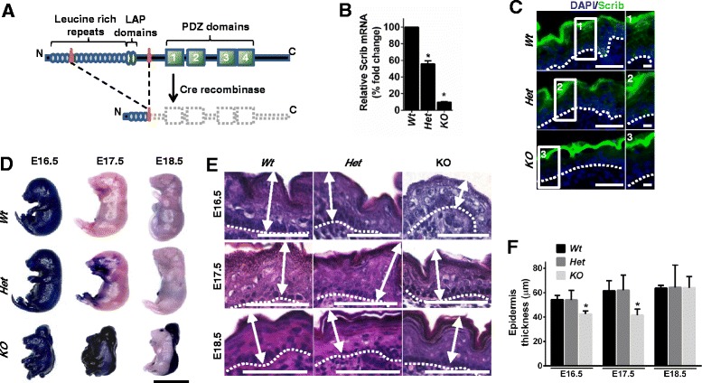
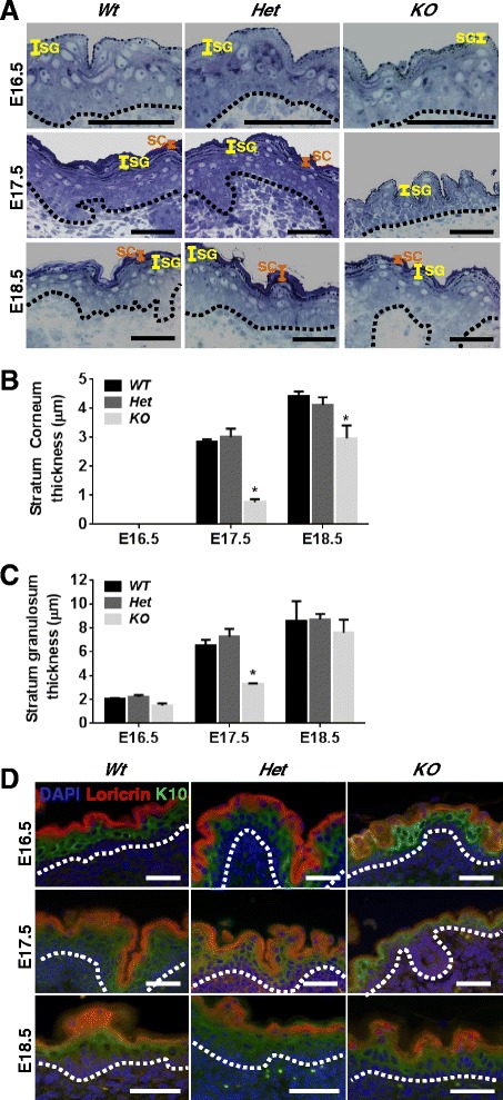
Background: The establishment and maintenance of polarity is vital for embryonic development and loss of polarity is a frequent characteristic of epithelial cancers, however the underlying molecular mechanisms remain unclear. Here, we identify a novel role for the polarity protein Scrib as a mediator of epidermal permeability barrier acquisition, skeletal morphogenesis, and as a potent tumor suppressor in cutaneous carcinogenesis.

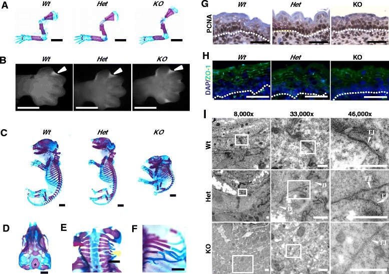
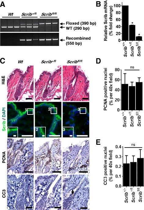
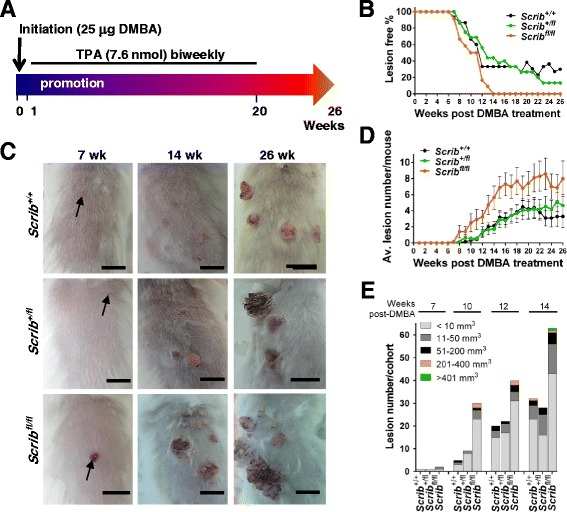
Methods: To explore the role of Scrib during epidermal development, we compared the permeability of toluidine blue dye in wild-type, Scrib heterozygous and Scrib KO embryonic epidermis at E16.5, E17.5 and E18.5. Mouse embryos were stained with alcian blue and alizarin red for skeletal analysis. To establish whether Scrib plays a tumor suppressive role during skin tumorigenesis and/or progression, we evaluated an autochthonous mouse model of skin carcinogenesis in the context of Scrib loss. We utilised Cre-LoxP technology to conditionally deplete Scrib in adult epidermis, since Scrib KO embryos are neonatal lethal.

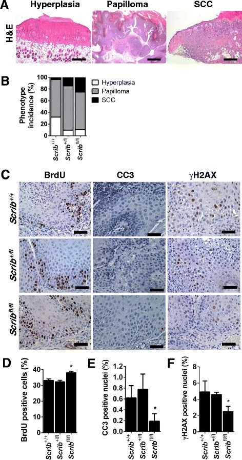
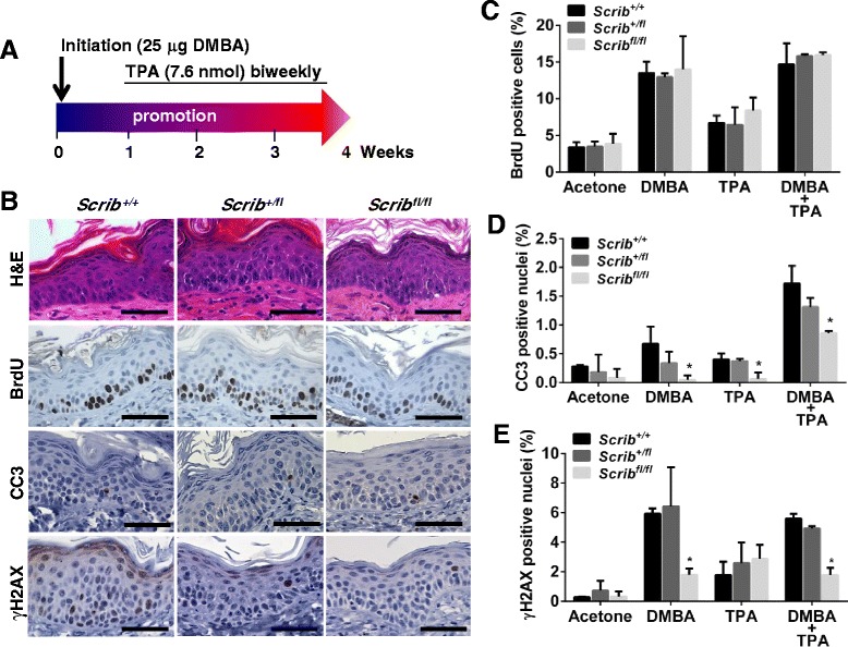
Results: We establish that Scrib perturbs keratinocyte maturation during embryonic development, causing impaired epidermal barrier formation, and that Scrib is required for skeletal morphogenesis in mice. Analysis of conditional transgenic mice deficient for Scrib specifically within the epidermis revealed no skin pathologies, indicating that Scrib is dispensable for normal adult epidermal homeostasis. Nevertheless, bi-allelic loss of Scrib significantly enhanced tumor multiplicity and progression in an autochthonous model of epidermal carcinogenesis in vivo, demonstrating Scrib is an epidermal tumor suppressor. Mechanistically, we show that apoptosis is the critical effector of Scrib tumor suppressor activity during skin carcinogenesis and provide new insight into the function of polarity proteins during DNA damage repair.

Conclusions: For the first time, we provide genetic evidence of a unique link between skin carcinogenesis and loss of the epithelial polarity regulator Scrib, emphasizing that Scrib exerts a wide-spread tumor suppressive function in epithelia.


Figures

Fig. 1
Fig. 1
Scrib KO embryos display a transient delay in epidermal permeability barrier acquisition. a Schematic representation of floxed targeting construct and mutated Scrib alleles. LoxP sites are represented by orange triangles. Cre-mediated recombination results in excision of exons 4–13, which introduces a frame-shift mutation that truncates the protein. b qRT-PCR to detect Scrib mRNA in embryonic skin at E16.5 (n = 3). Error bars: SD, *p < 0.0001 (unpaired t-test). c IF to detect Scrib (green) and DAPI (blue) in embryonic flank epidermis at E18.5 (n = 3 per genotype, scale bar = 50 μm. Inserts 1–3 scale bar = 10 μm). Scrib staining was detected along the membrane cortex of epidermal epithelial cells in Wt and Het embryonic epidermis and was absent in Scrib KO embryos. All genotypes displayed non-specific epidermal surface staining. d Representative images of toluidine blue skin permeability barrier assay (scale bar: 1 cm, n = 4–10 per genotype/time point). e H&E images of Wt, Het and KO epidermis at E16.5, E17.5 and E18.5, dashed line represents basement membrane, arrows indicate width of epidermis measured (n = 8–29 per genotype/time point, scale bar: 50 μm). f Quantitation of embryonic epidermal thickness. Error bars: SD, *p ≤ 0.0227 (2-way ANOVA with Tukey correction), n = 3 per genotype at each time point
Fig. 2
Fig. 2
Scrib KO embryos display normal developmental timing, proliferation and tight junction distribution. a Analysis of forelimb skeletal morphology following alcian blue and alizarin red staining at E17.5. No obvious developmental delay was observed in Scrib KO forelimbs when compared with Wt or Het littermates (scale bar = 2 mm, n = 3–6). b At E13.5, the characteristic pattern of interdigital apoptosis as revealed by Lysotracker incorporation (white arrowheads) indicates no obvious developmental delay in Scrib KO embryos when compared with Wt or Het littermates (scale bar = 1 mm, n = 3). c Whole E17.5 embryo skeletal analysis (scale bar: 5 mm, n = 3–6) reveals extensive dysmorphology in Scrib KO embryos, including (d) loss of dorsal cranial bones such that the basi-occipital bone (asterix) visible in dorsal view, (e) vertebral fusion (e, red arrowhead), hemivertebra (e, yellow arrowhead) and (f) distal rib fusion . Scale bar in d-f = 1 mm, n = 3. g IHC to detect PCNA in Scrib Wt, Het and KO embryonic epidermis at E16.5 (scale bar: 50 μm, n = 3, dashed line represents basement membrane). h IF images to detect ZO-1 (green) in Scrib Wt, Het and KO embryonic epidermis at E17.5 (scale bar = 50 μm, DAPI = blue, n = 3, dashed line represents basement membrane). i Tight junction ultrastructure analysis within the granular layer of Scrib Wt, Het and KO embryonic epidermis at E17.5 at 8,000x, 33,000x and 46,000x magnification (scale bar = 0.5 μm, TJ = tight junction and D = desmosome, n = 3–6). Scrib Wt, Het and KO embryos displayed a similar frequency of TJs with normal morphology within the SG
Fig. 3
Fig. 3
Scrib KO embryos display a transient delay in keratinocyte maturation. a Toluidine blue stained ultramicrotome sections of Scrib Wt, Het and KO embryonic flank epidermis at E16.5, E17.5 and E18.5 indicating stratum corneum (SC, orange) and stratum granulosum (SG, yellow) thickness (scale bar = 50 μm, dashed line represents basement membrane, n = 3). Quantitation of stratum corneum (b) and stratum granulosum (c) thickness in Scrib Wt, Het and KO embryonic flank epidermis at E16.5, E17.5 and E18.5 (*P ≤ 0.0066, two-way ANOVA with Tukey correction, error bars = SD, n = 3). d IF to detect K10 (green), loricrin (red) and DAPI (blue) in Scrib Wt, Het and KO embryonic flank epidermis at E16.5, E17.5 and E18.5 (scale bar = 100 μm, dashed line represents basement membrane, n = 3)
Fig. 4
Fig. 4
Scrib is dispensable for the maintenance of normal adult epidermal homeostasis. a PCR analysis of genomic DNA to detect Wt (290 bp), floxed (390 bp) and recombined (550 bp) Scrib alleles in Scrib +/+, Scrib +/fl and Scrib fl/fl adult dorsal epidermis (n = 3). b qRT-PCR for Scrib mRNA confirmed a significant reduction in Scrib +/fl and Scrib fl/fl adult dorsal epidermis compared to Scrib +/+ controls (*p ≤ 0.0001, unpaired t-test, n = 3). c Representative H&E (n = 10), Scrib IF (Scrib = green, DAPI = blue), PCNA IHC and CC3 IHC images of Scrib +/+, Scrib +/fl and Scrib fl/fl adult dorsal epidermis (n = 3). Scale bar = 50 μm (insert 1–3: scale bar = 10 μm). Arrow represents positive CC3 staining. Quantitation of nuclear PCNA (d) and CC3 (e) positive cells in adult dorsal epidermis shows no significant difference in Scrib-deficient epidermis compared to Scrib +/+ controls (p ≥ 0.6188, unpaired t-test, n = 3). Adult mice were 100 day old. Error bars = SD
Fig. 5
Fig. 5
Scrib deficiency facilitates DMBA/TPA-induced epidermal lesion growth. a Diagram illustrating long-term two-step DMBA/TPA carcinogenesis approach initiated at 8 weeks of age. b Lesion-free percentage, (c) representative photographs of dorsal skin at 7, 14 and 26 weeks post-DMBA (scale bar: 1 cm, arrows indicate lesion onset), (d) lesion multiplicity plot (error bars = SEM) and (e) histogram to illustrate volume distribution of DMBA/TPA-induced epidermal lesions in Scrib +/+, Scrib +/fl and Scrib fl/fl mice (n = 12–16)
Fig. 6
Fig. 6
Scrib depletion promotes DMBA/TPA-induced epidermal tumor progression. a Representative H&E images of mouse epidermis displaying cutaneous hyperplasia, benign papilloma formation and invasive SCC following long-term DMBA/TPA treatment (scale bar = 1 mm). b Percentage phenotype incidence at end-point (P = 0.0117, two-way ANOVA, n = 12–16) in Scrib +/+ , Scrib +/fl and Scrib fl/fl mice that have undergone long-term DMBA/TPA treatment. Scrib +/+ Scrib +/fl and Scrib fl/fl respective phenotype incidence; hyperplasia (32.1 %, 9.7 % and 10.7 %), benign papillomas (64.2 %, 75.8 %, 64.3 %) and invasive SCC (3.8 %, 14.4 % and 25 %) . c IHC images for BrdU, CC3 and γH2AX (scale bar = 50 μm, n = 3) and quantitation of the number of (d) BrdU, (e) CC3 and (f) γH2AX positive cells in Scrib +/+, Scrib +/fl and Scrib fl/fl mice that have undergone long-term DMBA/TPA treatment (% per 40x magnification field, *p ≤ 0.0486, unpaired t-test, error bars = SD, n = 3)
Fig. 7
Fig. 7
Scrib depletion sensitises the DMBA/TPA-induced pre-neoplastic epidermis to papillomagenesis by inhibiting apoptosis. a Diagram illustrating short-term DMBA/TPA administration initiated at 8 weeks of age. b Representative H&E images and IHC images for BrdU, CC3, and γH2AX following short-term application of DMBA/TPA in Scrib +/+, Scrib +/fl and Scrib fl/fl mice (scale bar = 50 μm, n = 3). Quantitation of (c) BrdU (d) CC3 and (e) γH2AX IHC in Scrib +/+, Scrib +/fl and Scrib fl/fl epidermis (per 40x magnification field) that have undergone short-term treatment with either acetone, DMBA, TPA or DMBA/TPA (*p ≤ 0.0402, two-way ANOVA with Tukey correction, error bars = SD, n = 3). Acetone topical applications served as a control

Similar articles

Cited by

References

    1. Humbert PO, Grzeschik NA, Brumby AM, Galea R, Elsum I, Richardson HE. Control of tumourigenesis by the Scribble/Dlg/Lgl polarity module. Oncogene. 2008;27(55):6888–907. doi: 10.1038/onc.2008.341. - DOI - PubMed
    1. Iden S, Collard JG. Crosstalk between small GTPases and polarity proteins in cell polarization. Nat Rev Mol Cell Biol. 2008;9(11):846–59. doi: 10.1038/nrm2521. - DOI - PubMed
    1. Muthuswamy SK, Xue B. Cell polarity as a regulator of cancer cell behavior plasticity. Annu Rev Cell Dev Biol. 2012;28:599–625. doi: 10.1146/annurev-cellbio-092910-154244. - DOI - PMC - PubMed
    1. Godde NJ, Pearson HB, Smith LK, Humbert PO. Dissecting the role of polarity regulators in cancer through the use of mouse models. Exp Cell Res. 2014;328(2):249–57. doi: 10.1016/j.yexcr.2014.08.036. - DOI - PubMed
    1. Bilder D, Perrimon N. Localization of apical epithelial determinants by the basolateral PDZ protein Scribble. Nature. 2000;403(6770):676–80. doi: 10.1038/35001108. - DOI - PubMed

Publication types

MeSH terms