

Atomoxetine Treatment Strengthens an Anti-Correlated Relationship between Functional Brain Networks in Medication-Naïve Adults with Attention-Deficit Hyperactivity Disorder: A Randomized Double-Blind Placebo-Controlled Clinical Trial
- PMID: 26377368
- PMCID: PMC4815465
- DOI: 10.1093/ijnp/pyv094
Atomoxetine Treatment Strengthens an Anti-Correlated Relationship between Functional Brain Networks in Medication-Naïve Adults with Attention-Deficit Hyperactivity Disorder: A Randomized Double-Blind Placebo-Controlled Clinical Trial
Abstract
Background: Although atomoxetine demonstrates efficacy in individuals with attention-deficit hyperactivity disorder, its treatment effects on brain resting-state functional connectivity remain unknown. Therefore, we aimed to investigate major brain functional networks in medication-naïve adults with attention-deficit hyperactivity disorder and the efficacy of atomoxetine treatment on resting-state functional connectivity.
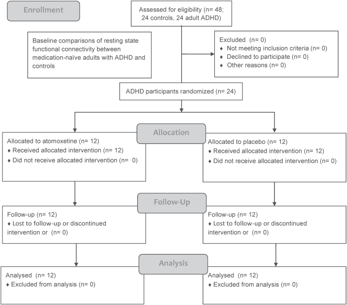
Methods: After collecting baseline resting-state functional MRI scans from 24 adults with attention-deficit hyperactivity disorder (aged 18-52 years) and 24 healthy controls (matched in demographic characteristics), the participants with attention-deficit hyperactivity disorder were randomly assigned to atomoxetine (n=12) and placebo (n=12) arms in an 8-week, double-blind, placebo-controlled trial. The primary outcome was functional connectivity assessed by a resting-state functional MRI. Seed-based functional connectivity was calculated and compared for the affective, attention, default, and cognitive control networks.
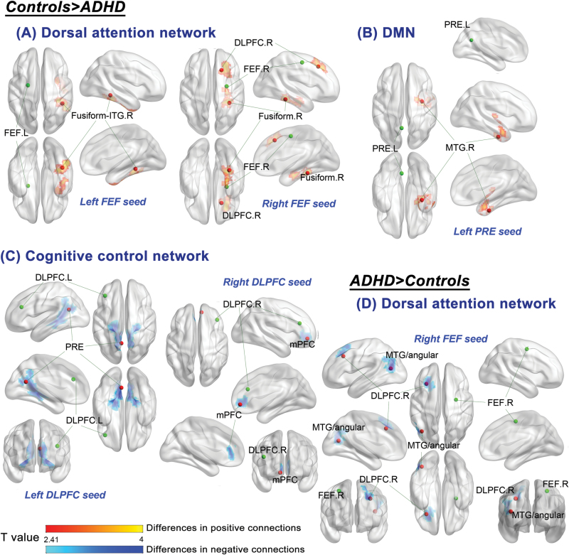
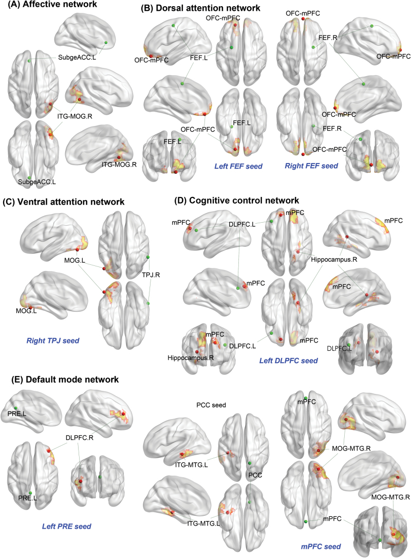
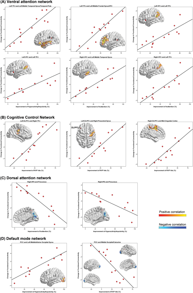
Results: At baseline, we found atypical cross talk between the default, cognitive control, and dorsal attention networks and hypoconnectivity within the dorsal attention and default networks in adults with attention-deficit hyperactivity disorder. Our first-ever placebo-controlled clinical trial incorporating resting-state functional MRI showed that treatment with atomoxetine strengthened an anticorrelated relationship between the default and task-positive networks and modulated all major brain networks. The strengthened anticorrelations were associated with improving clinical symptoms in the atomoxetine-treated adults.
Conclusions: Our results support the idea that atypical default mode network task-positive network interaction plays an important role in the pathophysiology of adult attention-deficit hyperactivity disorder. Strengthening this atypical relationship following atomoxetine treatment suggests an important pathway to treat attention-deficit hyperactivity disorder.
Trial registration: ClinicalTrials.gov NCT00917371.
Keywords: adult; atomoxetine; attention-deficit hyperactivity disorder; randomized double-blind placebo-controlled clinical trial; resting-state fMRI.
© The Author 2015. Published by Oxford University Press on behalf of CINP.
Figures
Similar articles
-
Neural correlates of atomoxetine improving inhibitory control and visual processing in Drug-naïve adults with attention-deficit/hyperactivity disorder.Hum Brain Mapp. 2017 Oct;38(10):4850-4864. doi: 10.1002/hbm.23683. Epub 2017 Jun 28. Hum Brain Mapp. 2017. PMID: 28657141 Free PMC article. Clinical Trial.
-
Atomoxetine Increased Effect over Time in Adults with Attention-Deficit/Hyperactivity Disorder Treated for up to 6 Months: Pooled Analysis of Two Double-Blind, Placebo-Controlled, Randomized Trials.CNS Neurosci Ther. 2016 Jul;22(7):546-57. doi: 10.1111/cns.12533. Epub 2016 Feb 28. CNS Neurosci Ther. 2016. PMID: 26922462 Free PMC article.
-
Differential effects of methylphenidate and atomoxetine on intrinsic brain activity in children with attention deficit hyperactivity disorder.Psychol Med. 2016 Nov;46(15):3173-3185. doi: 10.1017/S0033291716001938. Epub 2016 Aug 30. Psychol Med. 2016. PMID: 27574878
-
Atomoxetine for attention deficit hyperactivity disorder in children and adolescents with autism: A systematic review and meta-analysis.Autism Res. 2019 Apr;12(4):542-552. doi: 10.1002/aur.2059. Epub 2019 Jan 17. Autism Res. 2019. PMID: 30653855
-
Impairments of large-scale functional networks in attention-deficit/hyperactivity disorder: a meta-analysis of resting-state functional connectivity.Psychol Med. 2019 Nov;49(15):2475-2485. doi: 10.1017/S003329171900237X. Epub 2019 Sep 10. Psychol Med. 2019. PMID: 31500674 Review.
Cited by
-
Correlation of altered intrinsic functional connectivity with impaired self-regulation in children and adolescents with ADHD.Eur Arch Psychiatry Clin Neurosci. 2024 Jun 22. doi: 10.1007/s00406-024-01787-y. Online ahead of print. Eur Arch Psychiatry Clin Neurosci. 2024. PMID: 38906983
-
The Significance of the Default Mode Network (DMN) in Neurological and Neuropsychiatric Disorders: A Review.Yale J Biol Med. 2016 Mar 24;89(1):49-57. eCollection 2016 Mar. Yale J Biol Med. 2016. PMID: 27505016 Free PMC article. Review.
-
The Mechanism, Clinical Efficacy, Safety, and Dosage Regimen of Atomoxetine for ADHD Therapy in Children: A Narrative Review.Front Psychiatry. 2022 Feb 9;12:780921. doi: 10.3389/fpsyt.2021.780921. eCollection 2021. Front Psychiatry. 2022. PMID: 35222104 Free PMC article. Review.
-
Differences in Disrupted Dynamic Functional Network Connectivity Among Children, Adolescents, and Adults With Attention Deficit/Hyperactivity Disorder: A Resting-State fMRI Study.Front Hum Neurosci. 2021 Oct 5;15:697696. doi: 10.3389/fnhum.2021.697696. eCollection 2021. Front Hum Neurosci. 2021. PMID: 34675790 Free PMC article.
-
Quasi-periodic patterns contribute to functional connectivity in the brain.Neuroimage. 2019 May 1;191:193-204. doi: 10.1016/j.neuroimage.2019.01.076. Epub 2019 Feb 10. Neuroimage. 2019. PMID: 30753928 Free PMC article.
References
-
- Biederman J, Monuteaux MC, Mick E, Spencer T, Wilens TE, Silva JM, Snyder LE, Faraone SV. (2006) Young adult outcome of attention deficit hyperactivity disorder: a controlled 10-year follow-up study. Psychol Med 36:167–179. - PubMed
Publication types
MeSH terms
Substances
Associated data
LinkOut - more resources
Full Text Sources
Other Literature Sources
Medical
