. 2015 Sep 16;35(37):12869-89.
doi: 10.1523/JNEUROSCI.1164-15.2015.

Prox1 Regulates the Subtype-Specific Development of Caudal Ganglionic Eminence-Derived GABAergic Cortical Interneurons

Affiliations

Prox1 Regulates the Subtype-Specific Development of Caudal Ganglionic Eminence-Derived GABAergic Cortical Interneurons

Goichi Miyoshi et al. J Neurosci. .

Abstract

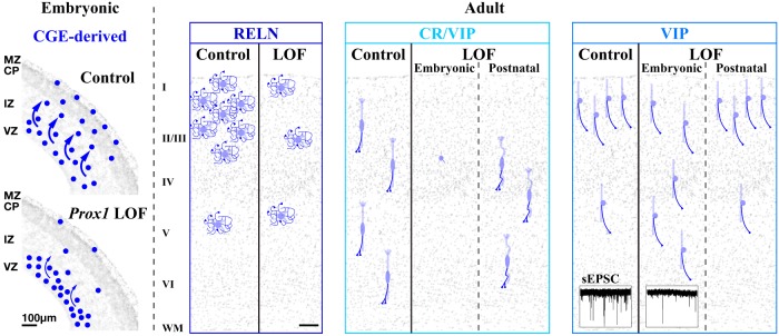
Neurogliaform (RELN+) and bipolar (VIP+) GABAergic interneurons of the mammalian cerebral cortex provide critical inhibition locally within the superficial layers. While these subtypes are known to originate from the embryonic caudal ganglionic eminence (CGE), the specific genetic programs that direct their positioning, maturation, and integration into the cortical network have not been elucidated. Here, we report that in mice expression of the transcription factor Prox1 is selectively maintained in postmitotic CGE-derived cortical interneuron precursors and that loss of Prox1 impairs the integration of these cells into superficial layers. Moreover, Prox1 differentially regulates the postnatal maturation of each specific subtype originating from the CGE (RELN, Calb2/VIP, and VIP). Interestingly, Prox1 promotes the maturation of CGE-derived interneuron subtypes through intrinsic differentiation programs that operate in tandem with extrinsically driven neuronal activity-dependent pathways. Thus Prox1 represents the first identified transcription factor specifically required for the embryonic and postnatal acquisition of CGE-derived cortical interneuron properties.

Significance statement: Despite the recognition that 30% of GABAergic cortical interneurons originate from the caudal ganglionic eminence (CGE), to date, a specific transcriptional program that selectively regulates the development of these populations has not yet been identified. Moreover, while CGE-derived interneurons display unique patterns of tangential and radial migration and preferentially populate the superficial layers of the cortex, identification of a molecular program that controls these events is lacking.Here, we demonstrate that the homeodomain transcription factor Prox1 is expressed in postmitotic CGE-derived cortical interneuron precursors and is maintained into adulthood. We found that Prox1 function is differentially required during both embryonic and postnatal stages of development to direct the migration, differentiation, circuit integration, and maintenance programs within distinct subtypes of CGE-derived interneurons.

Keywords: RELN; VIP; bipolar; mouse genetics; neurogliaform; transcription.


Figures

Figure 1.
Figure 1.
CGE-derived but not MGE-derived GABAergic cortical interneurons maintain expression of the transcription factor Prox1 during their development. A, B, Immunohistochemistry for Prox1 reveals expression within the SVZs of the ventral eminences (MGE, LGE, and CGE) as early as E10.5. Expression of Nkx2-1 largely demarcates the MGE domain. DAPI nuclear counterstain is shown in white. C, D, GABAergic neuronal precursors in the forebrain are labeled by EGFP through combinatorial use of a Dlx5/6-Flpe driver and an RCE:FRT reporter at E14.5. High levels of Prox1 expression are found in the SVZs of the MGE and CGE. Some non-GABAergic populations in the ventral forebrain also express Prox1 (open arrowheads). D′, A higher magnification picture of the neocortex in D is shown. Most Prox1-expressing cells are found to be GABAergic in the cortical plate (CP), intermediate zone (IZ), and subventricular/ventricular zones (SVZ/VZ). Non-GABAergic Prox1-expressing cells (red without green) within the marginal zone (MZ) are Cajal–Retzius cells and Prox1 expression within this population shuts off by E18.5 (confirmed by Wnt3a-Cre driver fate mapping; data not shown). E, E′, At E16.5, Prox1 expression is largely confined to GABAergic neuronal precursors within the cortex (E) and overlaps with Sp8 (E′). F, G, MGE-derived GABAergic populations are labeled with EGFP in Lhx6BAC-Cre; Dlx5/6-Flpe; RCE:dual animals. MGE-derived cortical interneuron precursors at E16.5 do not express Prox1 (F, F′). At P21, Prox1 remains absent from MGE-derived interneurons (G–G″). H–J, Analysis of Prox1 expression in specific CGE-derived cortical interneuron subtypes in Dlx6a-Cre; Prox1-C:EGFP/+ P21 brains. H, RELN-expressing interneurons that are negative for SST originate from the embryonic CGE and Prox1 expression (EGFP) are observed in 84.3% of this population albeit at variable levels (arrowheads; open arrowhead indicates Prox1-negative RELN-positive cell). SST-positive cells do not express Prox1 (asterisk). I, J, The majority of CR(Calb2)/VIP (double arrowheads) and VIP-single (single arrowhead) populations express Prox1. Most CR-positive cells lacking VIP are MGE derived (K) and do not express Prox1 (asterisk). K, A schematic correlating the molecular expression and the embryonic origin of GABAergic interneurons within the P21 mouse somatosensory barrel cortex (S1BF; adapted from Miyoshi et al., 2010). Prox1 is expressed within CGE-derived but not MGE-derived cortical interneuron subtypes. GP, globus paliidus; HC, hippocampus; Str, striatum; Th, thalamus. Scale bars: A–D, F, 200 μm; E–J, 50 μm.
Figure 2.
Figure 2.
Prox1 promotes the superficial layer positioning of CGE-derived interneurons by regulating embryonic tangential migration. Localization of EGFP(Prox1)-labeled control (Dlx6a-Cre; Prox1-C:EGFP/+) and Prox1 loss-of-function (Dlx6a-Cre; Prox1-C:EGFP/F) GABAergic cells were analyzed within the cortex (shown in black). Nuclear counter staining by DAPI is shown in blue. A, B, While we found no delay in the initiation of tangential migration from the CGE into the cortex of Prox1 loss-of-function at E14.5 (Fig. 3A,B), by E16.5, we observed a decrease in Prox1-null EGFP-labeled cells reaching the marginal zone (MZ) and cortical plate (CP). Comparable numbers of EGFP-labeled cells are found in the intermediate and subventricular/ventricular zones (IZ and SVZ/VZ). C, Bar graphs comparing the cortical area normalized distribution of EGFP-labeled (GABAergic Prox1) cells at E16.5 in the control (filled) and Prox1 loss-of-function (LOF, shaded) experiments. Two-tailed t test: p = 0.0255*(MZ), p = 0.0172*(CP), p = 0.425(IZ/SVZ/VZ). D–F, By E18.5, in addition to the loss in marginal zone and cortical plate, more EGFP-labeled cells were observed within the intermediate zone and SVZ/VZ of the conditional Prox1 loss-of-function cortices. For the sake of clarity, hippocampal areas are cropped from the figures (bottom). Two-tailed t test: p = 0.0156*(MZ), p = 0.00893**(CP), p = 0.132(IZ/SVZ/VZ). G–I, At P7, mutant cells were decreased in superficial (I–III) layers, but increased in deep (V, VI) layers compared with the control cortex. Two-tailed t test: p = 0.00693**(I), p = 0.00960**(II/III), = 0.837(IV), p = 0.0842(V/VI). In addition, the population of cells with vertically oriented processes that is normally found in the superficial layers (G) was less obvious in the Prox1 loss-of-function cortex (H). All error bars indicate SEM. Scale bar, 50 μm.
Figure 3.
Figure 3.
Embryonic (Dlx6a-Cre) Prox1 loss-of-function does not affect the interneuron precursors for their tangential migration from the CGE into the developing cortex as well as the expression of Sp8 and CoupTFII transcription factors. We performed embryonic (Dlx6a-Cre) control (A, C, E) and Prox1 loss-of-function (Prox1-C:EGFP/+ and /F) experiments (B, D, F). A, B, At E14.5, about the time the earliest cohort of CGE-derived interneuron precursors is first observed tangentially migrating through the intermediate zone (IZ) of the cortex (Miyoshi et al., 2010), we found no obvious differences between control and Prox1-null cells labeled with EGFP (shown in black) with regard to their migration pattern or distance from the CGE. Nuclear counter staining by DAPI is shown in blue. Double arrowheads indicate the CGE-derived interneuron precursors that have reached the border area between the hippocampus (HC) and cortex (Ctx). While control EGFP(Prox1)-labeled cells are positioned laterally to the developing globus pallidus (GP) and do not express CoupTFII (Nr2f2), in the Prox1 loss-of-function ventral telencephalon, very few EGFP(Prox1)-labeled cells are observed in the comparable domain. C, D, In addition to its expression within cortical progenitors, at E16.5, the Sp8 expression pattern within EGFP(Prox1)-labeled cells is comparable between the control and Prox1 loss-of-function cortex. E, F, Higher magnification pictures of the cortical subventricular/ventricular zones (SVZ/VZ) in Figure 2D and E. While there are significantly more EGFP-labeled cells found in the SVZ/VZ of the Prox1 loss-of-function cortex at E18.5 (see also Fig. 2F), the proportion of CoupTFII expression within EGFP(Prox1)-labeled cells is not changed. M, MGE; L, LGE; Th, thalamus; MZ, marginal zone; CP, cortical plate. Scale bars: A–D, 200 μm; E, F, 50 μm.
Figure 4.
Figure 4.
Subtype-specific requirement for Prox1 in CGE-derived GABAergic cortical interneuron development. A, B, Immunohistochemistry for RELN/SST on EGFP(Prox1)-labeled cells in control and Prox1 loss-of-function (Dlx6a-Cre; Prox1-C:EGFP/+ and /F) cortices at P21. While RELN-expressing cells (negative for SST) expressed EGFP at variable levels (arrowheads), they were generally reduced in the superficial layers of conditional Prox1 loss-of-function cortices (B). Open arrowhead indicates the EGFP(Prox1)-negative RELN cells and double arrowheads indicate MGE-derived SST/RELN-expressing interneurons. C, D, Characterization of CR, VIP, and EGFP-positive cell profiles at P21. Double and single arrowheads indicate CR/VIP or VIP-single interneurons, respectively. While CR/VIP cells were severely reduced in mutants, the total numbers of VIP-single cells was unchanged. Furthermore, the remaining CR/VIP cells in mutants (D, double arrowheads) did not exhibit the characteristic bipolar morphology observed in controls (C). E–H, To better visualize the morphologies in Prox1-null CR/VIP interneurons, stacked views from confocal microscopy images were generated. CR/VIP-expressing cells (double arrowhead and inset) are found with bipolar morphology (E). In contrast, none of the CR/VIP-expressing cells remaining in the Prox1 loss-of-function cortex exhibit typical bipolar morphology (F–H). The best example of a mutant cell that we found to retain bipolar morphology to some extent is shown in H, but in most cases, obvious processes protruding from the soma could not be found (F, G). I, Layer distribution of EGFP(Prox1)-labeled control (filled bars) and Prox1 loss-of-function (LOF) cells (shaded bars) at P21. At P21, EGFP-labeled Prox1-null cells were displaced within deeper layers of the cortex, similar to the phenotype observed at P7 (Fig. 2I). Two-tailed t test: p = 0.0133*(I), p = 0.000380***(II/III), p = 0.00376**(IV), p = 0.463(V), p = 0.0148*(VI). J, Molecular expression profiles of interneurons in the P21 somatosensory barrel cortex for control and Prox1 loss-of-function experiments. The total numbers of PV(Pvalb)-expressing (red) and SST-expressing (orange) interneurons were not altered in the mutant. RELN (SST-negative) cell numbers were reduced to ∼40%, CR/VIP-interneurons were severely reduced, and the VIP-single population was unaltered. Overall, EGFP(Prox1)-expressing cells were reduced to 75% in the Prox1 loss-of-function mutant at P21. Two-tailed t test: p = 0.880(PV), p = 0.0905(SST), p = 0.000334***(RELN), p = 0.0236*(CR/VIP), p = 0.403(VIP), p = 0.00169***(EGFP). K, The laminar distribution of each RELN, CR/VIP, and VIP-single interneuron subtype is shown. In addition to control (filled bars) and Prox1 loss-of-function (shaded bars), gray bars indicate the EGFP-negative population of each subtype. While the RELN-positive population was primarily reduced in the superficial (I–IV) layers, CR/VIP-expressing cells were reduced in all layers. The VIP-single population was reduced in superficial (I–III) and increased in deep (IV–VI) layers and thus was ectopically displaced into deeper layers. Statistics are shown for the marker-positive profiles including both EGFP-positive and EGF-negative cells. Two-tailed t test: p = 0.00437**, p = 0.00127**, p = 0.1841, p = 0.6411, p = 0.551 (I–VI, RELN), p = 0.188, p = 0.0369*, p = 0.0136*, p = 0.0371*, p = 0.316 (I–VI, CR/VIP), p = 0.216, p = 0.148, p = 0.414, p = 0.0507, p = 0.0164* (I–VI, VIP). All error bars are SEM. Scale bars, 50 μm.
Figure 5.
Figure 5.
Late embryonic (Htr3ABAC-Cre) Prox1 loss-of-function (LOF) in CGE-derived interneurons shows similar but milder phenotypes compared with pan-GABAergic early embryonic (Dlx6a-Cre) removal. We used the Htr3ABAC-Cre driver to restrict recombination to CGE-derived lineages. This driver recombines the Prox1-C:EGFP allele at somewhat later stages of embryonic cell migration than Dlx6a-Cre (Figs. 2–4) in a subpopulation of CGE-derived cells. A–C, Recombination efficiency of the Htr3ABAC-Cre driver was addressed by analyzing crosses with Prox1-C:EGFP/+ and Ai9 reporter (R26R-CAG-loxPstop-tdTomato-WPRE polyA) lines in the same animal at E14.5 (A) and E18.5 (B, C). At E14.5, the Htr3ABAC-Cre driver (A) resulted in fewer EGFP-expressing cells within the cortex compared with the Dlx6a-Cre driver (Fig. 3A). At E18.5, substantial numbers of EGFP-labeled cells were evident in the cortex, although to a lesser extent when compared with the Dlx6a-Cre driver (Fig. 2D). C, C′, A higher magnification view of B also shown for EGFP signals only (C′). The Htr3ABAC-Cre driver also targets non-GABAergic populations, most likely the Cajal–Retzius and subplate cells (red without green), in addition to CGE-derived interneuron precursors. D–G, Comparison of control and Prox1 loss-of-function (Htr3ABAC-Cre; Prox1-C:EGFP/+ and /F) cells at P21. E, G, Higher magnification of the areas in D and F are presented and EGFP signals are shown in black (E′, G′). Double arrowheads indicate CR/VIP-expressing cells and single arrowheads indicate VIP-single (CR-negative) cells. In contrast to what we observed in the early embryonic (Dlx6a-Cre) Prox1 loss-of-function study, in the Htr3ABAC-Cre mutant, not all of the remaining CR/VIP cells lost their characteristic bipolar morphologies (G′). While the CR/VIP Prox1-null cell in the middle has a round morphology with no obvious processes, the cell at the bottom exhibits a bipolar morphology (G′). H, A graph indicating the layering of late embryonic (Htr3ABAC-Cre) Prox1 loss-of-function cells in P21 cortex. Note that the decrease (Layers I–III) and increase (Layers V/VI) found in the EGFP-expressing cells of Prox1 loss-of-function cortex are consistent but milder compared with the results observed from the early embryonic (Dlx6a-Cre) loss-of-function (Fig. 4I). Two-tailed t test: p = 0.0636(I), p = 0.212(II/III), p = 0.359(IV), p = 0.0412*(V), p = 0.0205*(VI). I, Cell numbers of CR/VIP-expressing and VIP-single populations in the P21 cortex of control and late embryonic Prox1 loss-of-function animals (shaded bars). EGFP(Prox1)-negative populations are shown in gray bars. Consistent with the early embryonic (Dlx6a-Cre) loss-of-function, we also observed a reduction in CR/VIP-expressing cells, with no obvious change in the VIP-single population following late embryonic (Htr3ABAC-Cre) loss-of-function. Note that the recombination mediated by the Htr3ABAC-Cre driver does not take place in all CGE-derived population at P21 in either control or Prox1 loss-of-function animals (gray bars) compared with that observed when the Dlx6a-Cre driver was used (Figs. 1I–K, 4K). Two-tailed t test: p = 0.0852(CR/VIP), p = 0.0786(VIP). Error bars indicate SEM. Scale bars: A–C, 200 μm; D–G, 50 μm.
Figure 6.
Figure 6.
Reduction of excitatory events in VIP-single cortical interneurons following embryonic (Dlx6a-Cre) Prox1 removal. Control and embryonic (Dlx6a-Cre) Prox1 loss-of-function (LOF) cells labeled with EGFP were recorded in brain slices of the somatosensory cortex (P16–P20). Subsequent to electrophysiological analysis and biocytin filling, post hoc immunohistochemistry for RELN/VIP or CR/VIP was performed. A, A control cell that was identified by post hoc analysis to be positive for RELN but not VIP showed characteristic LS firing at near threshold (top, black trace; red trace is at subthreshold) and received sEPSCs at a frequency of 3 Hz (bottom trace). B, A Prox1-null RELN-positive VIP-negative cell showed a similar LS action potential discharge and received sEPSCs at a comparable frequency and amplitude to that of control cells (A). C, Analysis of the frequency (left) and amplitude (right) of sEPSCs in the control (n = 6) and Prox1 loss-of-function LS cells (n = 4, shaded bars) shows that these interneurons do not require Prox1 for the establishment of proper excitatory inputs. Mann–Whitney: p = 0.187(frequency), p = 0.442(amplitude). D, A representative example of a VIP-single (CR-negative) control cell with dNFS3 firing properties, with the sEPSCs it receives shown at the bottom. E, A trace of a dNFS3 cell (VIP-positive CR-negative) lacking Prox1. F, In contrast to RELN-positive LS cells (C), Prox1-null VIP-single interneurons (n = 4) show a dramatic reduction in the frequency of sEPSCs (left) compared with control cells (n = 4). Nevertheless, the amplitude of sEPSCs shows no change (right). Mann–Whitney: p = 0.0397*(frequency), p = 0.191(amplitude). Scale bar, 50 μm.
Figure 7.
Figure 7.
Postnatally (Vip-Cre), Prox1 is required for the differentiation and survival of CR/VIP interneurons. Postnatal conditional loss-of-function (LOF) of Prox1 was performed specifically within VIP-expressing interneurons with the Vip-Cre driver. Vip-positive cells were labeled independently of Prox1 expression by combining the tdTomato reporter (Ai9) with the control or Prox1 loss-of-function (Prox1-C:EGFP/+ or /F) alleles. This Vip-Cre driver initiates recombination in the Prox1-C:EGFP allele only after E18.5 (data not shown), thus allowing us to perform postnatal loss-of-function studies. A, B, By P7, we observed efficient recombination in both the tdTomato reporter and conditional Prox1-C:EGFP alleles in control and mutant cortices. In controls, VIP-positive interneurons exhibited a mature profile of CR (Calb2) expression by P7 (A, inset, double arrowheads), whereas in mutants, fewer cells expressed CR (B, inset). Note that layer Va pyramidal neurons have not downregulated CR at P7. C–G, Still, by P21, mutants exhibited a reduction in CR expression compared with controls, and those that remained did not show the similar characteristic bipolar morphologies observed in controls (C, D), but had kinked processes (E–G, double arrowheads). To better visualize cell morphology, EGFP is shown in black to the right of C–G (C′–G′). H, The laminar distribution of control and postnatal (Vip-Cre) Prox1 loss-of-function cells in the cortex at P7 and P21. While there was no obvious difference between the control and postnatal Prox1 conditional mutant at P7, by P21, cells were specifically reduced in layers II/III of the mutant. EGFP(Prox1) labeling and tdTomato expression (Ai9 reporter) are shown in green and red, respectively, and the overlap is indicated by yellow. Two-tailed t test: p = 0.127, p = 0.620, p = 0.577, p = 0.756(I–V/VI, P7), p = 0.953, p = 0.0301*, p = 0.735, p = 0.467, p = 0.648(I–VI, P21), p = 0.700, p = 0.121(total numbers for P7 and P21). Error bars indicate SEM for total number of recombined cells in each layer. I, CR and VIP expressions were analyzed in postnatal (Vip-Cre) Prox1 conditional mutants (labeled green and/or red) at P7 and P21. Values are shown as the proportion of labeled cells normalized to the total number of control cells at each age. While there was no change in the reporter-positive cell numbers at P7 (H), CR expression was decreased in the mutant compared with control at both ages. VIP expression was delayed in onset (P7) but recovered by P21 (the loss of VIP at P21 matches the cell loss in H, bottom). Reporter-negative CR-expressing cells (orange), most of which are derived from the MGE and coexpress SST, showed no change at P21. Two-tailed t test: p = 0.0143*(CR P7), p = 0.0120*(CR P21), p = 0.383(VIP P7), p = 0.198(VIP P21), p = 0.562(CR, no reporter). Error bars represent SEM. Scale bars: 50 μm.
Figure 8.
Figure 8.
Cell migration and bipolar differentiation of CR/VIP-expressing interneurons is independently and cell-autonomously controlled by Prox1. A, A schematic of heterochronic transplantation experiments. E14.5 CGE tissue from Dlx6a-Cre control and Prox1 conditional loss-of-function (LOF) embryos (Prox1-C:EGFP/+ and /F) was dissected, dissociated, and subsequently transplanted into WT host P3 cortices, allowing CGE-derived interneuron precursors to bypass the tangential migration phase and to directly differentiate inside the cortical plate. B–D, Representative examples of transplanted control EGFP(Prox1)-labeled cells at P21 that are positive for VIP-single (B) or CR/VIP (C, D) with EGFP labeling shown in black. E–H, Prox1 loss-of-function EGFP-labeled transplanted cells positive for VIP-single (E, F) or CR/VIP (G, H) at P21. Transplantation did not rescue the embryonic (Dlx6a-Cre) Prox1 loss-of-function phenotype, as only a few EGFP(Prox1)-labeled mutant cells expressed CR/VIP and these did not possess the characteristic bipolar morphologies of this subtype (G, H). I, Bar graphs representing the proportion of CR/VIP or VIP-single-labeled populations in cells labeled by EGFP(Prox1) expression. The values obtained from the heterochronic transplantation experiments (transplant+) resembled the results without transplantation (transplant−; see also Fig. 4J) for both control and Prox1 loss-of-function. Two-tailed t test for transplant-negative versus positive: p = 0.191(VIP control), p = 0.264(CR/VIP control), p = 0.129(VIP mutant), p = 0.628(CR/VIP mutant). Error bars represent SEM. Scale bar, 50 μm.
Figure 9.
Figure 9.
Prox1 regulates multiple developmental steps of CGE-derived interneurons in a subtype-specific manner. Left, Embryonic, Prox1 is selectively expressed within CGE-derived cortical interneuron precursors. During embryonic stages, Prox1 facilitates the transition of tangentially migrating cells from the intermediate zone (IZ) into the cortical plate (CP) and marginal zone (MZ). This step is critical for the positioning of CGE-derived interneuron precursors into the superficial layers of the neocortex. Adult, Embryonic (Dlx6a-Cre) and postnatal (Vip-Cre) loss-of-function studies reveal that Prox1 function is differentially required in a stage- and subtype-specific manner. (1) RELN-positive cells were reduced by ∼40%. (2) CR/VIP double-positive cells were mostly eliminated from the cortex and the remaining cells failed to acquire their characteristic bipolar morphology. Sustained expression of Prox1 during postnatal stages is required for the maintenance and survival of this cell class. (3) The VIP-single population was displaced into deep cortical layers. Prox1 is further required for this interneuron class to receive proper excitatory inputs and to integrate into superficial neocortical circuits. LOF, loss-of-function; VZ, ventricular zone.

Similar articles

Cited by

References

    1. Anderson SA, Eisenstat DD, Shi L, Rubenstein JL. Interneuron migration from basal forebrain to neocortex: dependence on Dlx genes. Science. 1997;278:474–476. doi: 10.1126/science.278.5337.474. - DOI - PubMed
    1. Anderson SA, Marín O, Horn C, Jennings K, Rubenstein JL. Distinct cortical migrations from the medial and lateral ganglionic eminences. Development. 2001;128:353–363. - PubMed
    1. Azim E, Jabaudon D, Fame RM, Macklis JD. SOX6 controls dorsal progenitor identity and interneuron diversity during neocortical development. Nat Neurosci. 2009;12:1238–1247. doi: 10.1038/nn.2387. - DOI - PMC - PubMed
    1. Balamotis MA, Tamberg N, Woo YJ, Li J, Davy B, Kohwi-Shigematsu T, Kohwi Y. Satb1 ablation alters temporal expression of immediate early genes and reduces dendritic spine density during postnatal brain development. Mol Cell Biol. 2012;32:333–347. doi: 10.1128/MCB.05917-11. - DOI - PMC - PubMed
    1. Bartolini G, Ciceri G, Marín O. Integration of GABAergic interneurons into cortical cell assemblies: lessons from embryos and adults. Neuron. 2013;79:849–864. doi: 10.1016/j.neuron.2013.08.014. - DOI - PubMed

Publication types

MeSH terms

LinkOut - more resources