. 2015 Dec 1;309(11):R1347-57.
doi: 10.1152/ajpregu.00164.2015. Epub 2015 Sep 16.

Cardiac responses to hypoxia and reoxygenation in Drosophila

Affiliations

Cardiac responses to hypoxia and reoxygenation in Drosophila

Rachel Zarndt et al. Am J Physiol Regul Integr Comp Physiol. .

Abstract

An adequate supply of oxygen is important for the survival of all tissues, but it is especially critical for tissues with high-energy demands, such as the heart. Insufficient tissue oxygenation occurs under a variety of conditions, including high altitude, embryonic and fetal development, inflammation, and thrombotic diseases, often affecting multiple organ systems. Responses and adaptations of the heart to hypoxia are of particular relevance in human cardiovascular and pulmonary diseases, in which the effects of hypoxic exposure can range in severity from transient to long-lasting. This study uses the genetic model system Drosophila to investigate cardiac responses to acute (30 min), sustained (18 h), and chronic (3 wk) hypoxia with reoxygenation. Whereas hearts from wild-type flies recovered quickly after acute hypoxia, exposure to sustained or chronic hypoxia significantly compromised heart function upon reoxygenation. Hearts from flies with mutations in sima, the Drosophila homolog of the hypoxia-inducible factor alpha subunit (HIF-α), exhibited exaggerated reductions in cardiac output in response to hypoxia. Heart function in hypoxia-selected flies, selected over many generations for survival in a low-oxygen environment, revealed reduced cardiac output in terms of decreased heart rate and fractional shortening compared with their normoxia controls. Hypoxia-selected flies also had smaller hearts, myofibrillar disorganization, and increased extracellular collagen deposition, consistent with the observed reductions in contractility. This study indicates that longer-duration hypoxic insults exert deleterious effects on heart function that are mediated, in part, by sima and advances Drosophila models for the genetic analysis of cardiac-specific responses to hypoxia and reoxygenation.

Keywords: Drosophila; genetic selection; heart; hypoxia; hypoxia-inducible factor; reoxygenation; sima.


Figures

Fig. 1.
Fig. 1.
Response of wild-type (w1118) controls to acute hypoxia exposure. A: schematic showing the acute hypoxia/reoxygenation protocol (acute H/R). B: M-mode records (15 s) from movies of beating hearts taken before (T = 0), at 30 min during (T = 30), and at two time points (T = 60, 90) following hypoxia treatments. Left: representative M-mode showing changes in heart beat in response to 4% O2. Right: representative M-modes showing changes in heart beat in response to 1% O2. C: incidence of nonbeating or “asystolic” hearts in response to hypoxia exposure, expressed as a percentage of total hearts examined. Quantification of changes in heart period (D) and fractional shortening (E) under 4% or 1% hypoxia (T = 30) relative to prehypoxia exposure baseline. All values are mean percent change ± SE (w1118: n = 19 at 21% O2, n = 10 at 4% O2, n = 9 at 1% O2). Data were analyzed by one-way ANOVA and Tukey's multiple-comparisons post hoc test; *P < 0.05, **P < 0.01, ***P < 0.001. See Supporting Information for raw data.
Fig. 2.
Fig. 2.
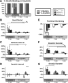
Acute cardiac response to 30 min 1% O2 acute H/R in wild-type and sima mutant Drosophila. A: incidence of nonbeating or “asystolic” hearts (>25 s without contractions) in response to hypoxia exposure, expressed as a percentage of total hearts examined. Nearly half of wild-type and sima mutant hearts entered prolonged periods of asystole at 30 min in 1% O2, and all resumed beating on reoxygenation. B–G: data are presented as the percent change from the prehypoxia baseline during and after hypoxia treatment for wild-type, sima+/−, and sima−/−. B: in hearts that beat, heart period (HP) is increased 100% or greater under 1% O2 hypoxia exposure (T = 30), and this reached significance in hearts from wild-type flies. HP returned to baseline levels exhibited by normoxia controls in all genotypes after hypoxia exposure (T = 90). C: diastolic intervals (DI) increased in wild-type and sima−/− under 1% O2 (T = 30) and then returned to baseline levels upon reoxygenation (T = 60, 90). D: systolic intervals did not change significantly across treatments and genotypes. E: for all hearts, fractional shortening was significantly reduced under 1% O2, but reductions were greatest in wild-type and greater in sima+/− than in sima−/− (T = 30). F: diastolic diameters increased significantly in both sima+/− and sima−/− under 1% O2 and remained larger at 30 min after hypoxia exposure compared with controls. G: systolic diameters were significantly increased during acute hypoxia in both wild-type and sima+/−. All values are mean percent change ± SE (n = 10 for w1118; n = 12 for sima+/−; n = 16 for sima−/−). For heart period, systolic and diastolic interval, asystolic hearts were excluded, while all hearts are included in fractional shortening and diastolic and systolic diameter. Data were analyzed by Kruskal-Wallis test (B, C) and Dunn multiple-comparison post hoc test (D–F), and two-way ANOVA and Tukey's multiple-comparisons post hoc test. n.s., no statistical significance. *P < 0.05, **P < 0.01, ***P < 0.001; n.s., not significant. Changes at T = 30 between genotypes were analyzed by Sidak-Bonferroni t-test. tP < 0.05; ttP < 0.01. See Supporting Information for raw numbers.
Fig. 3.
Fig. 3.
Cardiac response to sustained H/R in wild-type and sima mutants. A: protocol for sustained hypoxia treatment (sustained H/R). B: percent of hearts beating during normoxia and post-18 h 1% hypoxia. C: heart period significantly shortened (rate increased) upon exposure to sustained H/R in both wild-type and sima+/− hearts. D: exposure to sustained H/R leads to significant reductions in diastolic interval in both wild-type and sima+/− hearts. E: sustained H/R caused a significant increase in systolic intervals in hearts from wild-type controls, whereas sima+/− hearts exhibited significant decreases in systolic interval compared with normoxic conditions. F: fractional shortening decreased in wild type in response to sustained H/R; in sima+/− hearts, this decrease was significantly exacerbated compared with controls. G: these changes in fractional shortening were due, in part, to significant decreases in diastolic diameters in both lines of flies. H: there is no change in systolic diameter in sima+/−, whereas the systolic diameter is significantly reduced in wild-type controls in response to sustained H/R. All values are mean percent change ± SE for hearts that beat on reperfusion after a sustained hypoxia exposure (w1118: n = 59 normoxia, n = 24 hypoxia; sima+/−: n = 17 normoxia, n = 20 hypoxia). Data were analyzed by Kruskal-Wallis test and Dunn multiple-comparisons post hoc test (C, D) and two-way ANOVA with Tukey's multiple-comparison post hoc test (E–H). *P < 0.05, **P < 0.01, ***P < 0.001. See Supporting Information for raw data.
Fig. 4.
Fig. 4.
Response to chronic H/R in wild-type and sima mutants. A: protocol for chronic hypoxia treatment (chronic H/R). B: chronic H/R exposure significantly increased heart period in hearts from w1118 but not in hearts from sima+/− flies compared with normoxia controls. C: diastolic intervals were also significantly increased by chronic H/R in w1118 but not sima+/− compared with normoxia controls. D: systolic intervals were unchanged following chronic H/R in w1118 but were significantly decreased in sima+/− compered to normoxia controls. E: fractional shortening was also unchanged following chronic H/R in w1118 but decreased significantly in sima+/− compared with normoxia controls. F: diastolic diameters were unchanged in following chronic H/R in w1118 but were significantly reduced in sima+/− compared with normoxia controls. G: systolic diameters were unaffected by chronic H/R in both genotypes compared with their normoxia controls. All values are mean percent change ± SE (w1118: n = 36 under normoxia, n = 41 under hypoxia; sima+/−: n = 17 under normoxia, n = 20 under hypoxia). Data were analyzed by Kruskal-Wallis test and Dunn multiple-comparisons post hoc test (B and C) and two-way ANOVA with Tukey's multiple-comparison post hoc test (D–G). *P < 0.05, **P < 0.01, ***P < 0.001. See Supporting Information file for raw data.
Fig. 5.
Fig. 5.
Cardiac parameters of hypoxia-selected and normoxia control populations. A: representative 15-s M-modes showing heart wall movements in three normoxia controls and three hypoxia-selected fly hearts at their relative normoxia. Left: 21% O2 for normoxia controls. Right: 4% O2 for hypoxia-selected. B: under their relative normoxic conditions, heart period is significantly longer in hypoxia-selected flies compared with normoxia controls. C: diastolic intervals are significantly longer in hypoxia-selected flies compared with normoxia controls. D: systolic intervals are significantly decreased in hypoxia-selected flies compared with normoxia controls. E: fractional shortening is significantly reduced in hypoxia-selected flies, due to reductions in both diastolic diameter (F) and systolic diameter (G) compared with control populations. All values are expressed as means ± SE for hearts. ***P < 0.001, Mann-Whitney unpaired t-test; normoxia controls n = 87, hypoxia-selected n = 72.
Fig. 6.
Fig. 6.
Hypoxia-induced alterations in cardiac structure and extracellular matrix. Exposed hearts in semi-intact preparations were stained for F-actin with phalloidin (A–C, H, I) and for collagen IV with anti-pericardin antibodies (D–F, J, K). In all images, the ventral view of the heart is shown, anterior to the left. A: Heart from a 2-wk old w1118 fly exhibits the tightly packed, circumferential arrangement of F-actin-stained myofibrils within the myocardial cells. This structure is not affected by sustained H/R (B) or chronic H/R (C). D: pericardin (collagen IV homolog) staining reveals the “fish net” pattern of fibers that associate with sarcomeric structures. E: collagen network appears denser and is moderately disorganized following sustained H/R and is more disorganized following chronic H/R (F). G: pericardin staining that was quantified from Z stacks of immunostained hearts shows an increase in intensity following both sustained and chronic H/R. Data are expressed as the percent of total pixels with an intensity above threshold (see methods; number of hearts (l to r) = 13, 11, 7). Data were analyzed by one-way ANOVA and Tukey's multiple-comparisons post hoc test. *P < 0.05, **P < 0.01. H: phalloidin staining of a normoxia control heart showing tightly spaced circumferential myofibrils of the heart tube with overlying longitudinal fibers (nonmyocardial). I: phalloidin staining of a heart from a hypoxia-selected fly showing smaller diameter and disorganized myofibrils. J: immunostaining of pericardin (collagen IV) in lower-magnification images of the terminal region of a normoxia control heart reveals a compact distribution around the region of the outflow tract. K: pericardin immunostaining of the same terminal region from a hypoxia-selected fly showing an expanded and disorganized deposition of collagen IV. Scale bars: 50 μm.
Fig. 7.
Fig. 7.
A summary of relative changes in cardiac function observed between genetic backgrounds relative to their normoxia baseline or control population for varying durations of hypoxia or reoxygenation is shown.

Comment in

Similar articles

Cited by

References

    1. Akasaka T, Klinedinst S, Ocorr K, Bustamante EL, Kim SK, Bodmer R. The ATP-sensitive potassium (KATP) channel-encoded dSUR gene is required for Drosophila heart function and is regulated by tinman. Proc Natl Acad Sci USA 103: 11,999–12,004, 2006. - PMC - PubMed
    1. Azad P, Haddad GG. Survival in acute and severe low o environment: use of a genetic model system. Ann NY Acad Sci 1177: 39–47, 2009. - PubMed
    1. Azad P, Zhou D, Zarndt R, Haddad GG. Identification of genes underlying hypoxia tolerance in Drosophila by a P-element Screen. G3 2: 1169–1178, 2012. - PMC - PubMed
    1. Bacon NC, Wappner P, O'Rourke JF, Bartlett SM, Shilo B, Pugh CW, Ratcliffe PJ. Regulation of the Drosophila bHLH-PAS protein Sima by hypoxia: functional evidence for homology with mammalian HIF-1 alpha. Biochem Biophys Res Commun 249: 811–816, 1998. - PubMed
    1. Brusselmans K, Compernolle V, Tjwa M, Wiesener MS, Maxwell PH, Collen D, Carmeliet P. Heterozygous deficiency of hypoxia-inducible factor-2α protects mice against pulmonary hypertension and right ventricular dysfunction during prolonged hypoxia. J Clin Invest 111: 1519–1527, 2003. - PMC - PubMed

MeSH terms

LinkOut - more resources