. 2015 Nov;138(Pt 11):3206-20.
doi: 10.1093/brain/awv250. Epub 2015 Sep 15.

Brain endothelial dysfunction in cerebral adrenoleukodystrophy

Affiliations

Brain endothelial dysfunction in cerebral adrenoleukodystrophy

Patricia L Musolino et al. Brain. 2015 Nov.

Abstract

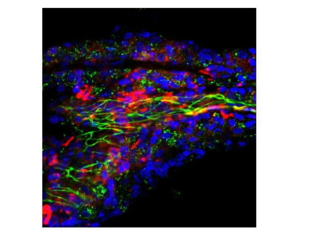
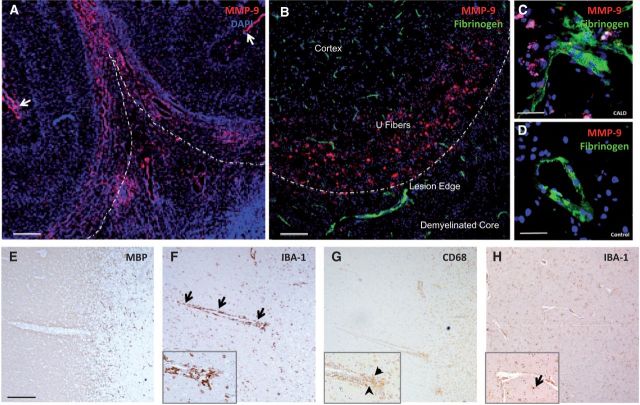
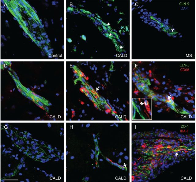
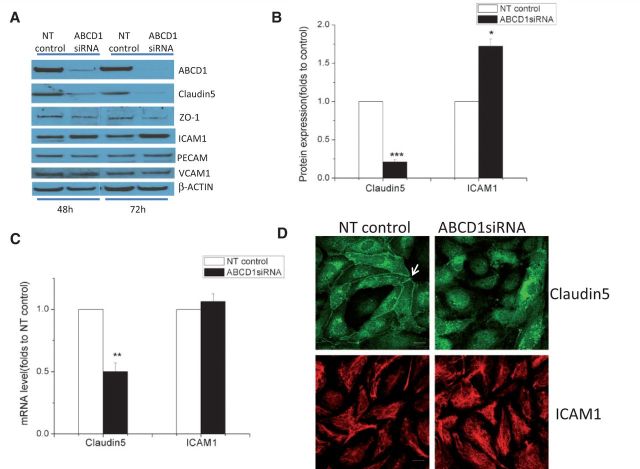
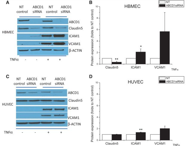
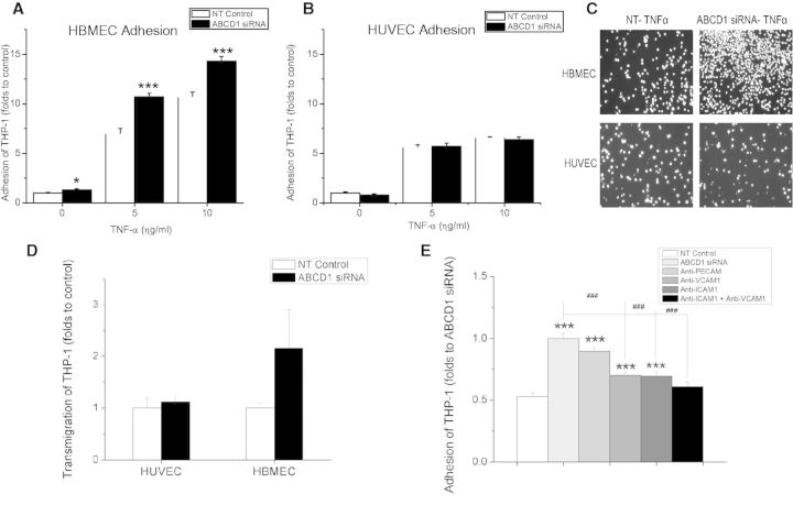
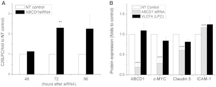
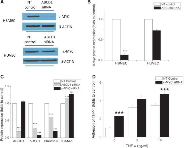
See Aubourg (doi:10.1093/awv271) for a scientific commentary on this article.X-linked adrenoleukodystrophy is caused by mutations in the ABCD1 gene leading to accumulation of very long chain fatty acids. Its most severe neurological manifestation is cerebral adrenoleukodystrophy. Here we demonstrate that progressive inflammatory demyelination in cerebral adrenoleukodystrophy coincides with blood-brain barrier dysfunction, increased MMP9 expression, and changes in endothelial tight junction proteins as well as adhesion molecules. ABCD1, but not its closest homologue ABCD2, is highly expressed in human brain microvascular endothelial cells, far exceeding its expression in the systemic vasculature. Silencing of ABCD1 in human brain microvascular endothelial cells causes accumulation of very long chain fatty acids, but much later than the immediate upregulation of adhesion molecules and decrease in tight junction proteins. This results in greater adhesion and transmigration of monocytes across the endothelium. PCR-array screening of human brain microvascular endothelial cells after ABCD1 silencing revealed downregulation of both mRNA and protein levels of the transcription factor c-MYC (encoded by MYC). Interestingly, MYC silencing mimicked the effects of ABCD1 silencing on CLDN5 and ICAM1 without decreasing the levels of ABCD1 protein itself. Together, these data demonstrate that ABCD1 deficiency induces significant alterations in brain endothelium via c-MYC and may thereby contribute to the increased trafficking of leucocytes across the blood-brain barrier as seen in cerebral adrenouleukodystrophy.

Keywords: blood–brain barrier; demyelination; genetics; leukodystrophy; neurodegeneration; neuroinflammation.


Figures

None
See Aubourg (doi:10.1093/awv271) for a scientific commentary on this article. Cerebral adrenoleukodystrophy is a neuroinflammatory demyelinating disease caused by mutations in ABCD1. Musolino et al. report that the progressive demyelination coincides with blood-brain barrier disruption and endothelial changes. Silencing endothelial ABCD1 in vitro upregulates expression of adhesion molecules and downregulates expression of tight-junction proteins and c-MYC, promoting monocyte transmigration.
Figure 1
Figure 1
Blood–brain barrier disruption in cerebral adrenoleukodystrophy. (A) Microphotographs of brain specimen from patients with CALD shows disruption of the blood–brain barrier in perilesional white matter as evidenced by increased MMP9 expression extending beyond the inflammatory active zone at low magnification as well as (B and C) leakage of fibrinogen (an exclusively intravascular protein) into the perivascular space. Subarachnoid vessels also expressed MMP9 (arrows). (D) Control brain specimen. Anatomical regions in the upper panel are marked as cortex, U fibres, lesion edge (dashed line) and demyelinated core. Lower panel shows vessels moving from normal white matter into the demyelinated lesion apparent by the lack of myelin staining with MBP (E). Representative images show microglia (IBA1+) surrounding the entire course of the vessels (arrows) at low and high magnification (inset) in F as well as macrophages-monocytes (CD68+) clustered on the lesion end of the vessel (arrowheads) (G). This pattern of perivascular microglia activation was not observed around vessels of normal-appearing white matter of control (H) or multiple sclerosis specimens (not shown). Scale bars = 2000 µm (A), 300 µm (B) and 50 µm (C and D), 500 µm (E and H).
Figure 2
Figure 2
Abnormal localization of CLDN5 and ZO1 expression in CALD and multiple sclerosis. Microphotographs of representative confocal imaging of CLDN5 immunostaining show intact structures of vessels and CLDN5 in the endothelial membrane in the cortex (A). Perilesional white matter vessels of CALD (B) and demyelinated plaque of multiple sclerosis brain (C) showed disrupted vascular structure and displaced CLDN5 (arrowhead) expression from tight junction to cytoplasm and perivascular space. Co-staining of CLDN5 and CD68 indicates some co-localization of CLN5 with monocytes (CD68+) (arrows and inset) in perilesional (E) and core (F) white matter vessels but not in the cortex (D) in CALD. Co-staining of ZO1 and IBA1 indicates some colocalization of ZO1 with microglia (IBA1+) (arrows) in CALD brain (arrows) in the core of the lesion (I) but not in perilesional white matter (H) or cortical vessels (G). Scale bar = 50 µm.
Figure 3
Figure 3
Effect of ABCD1 silencing on the expression of tight-junction proteins and adhesion molecules. (A) Changes in tight-junction proteins (CLDN5 and ZO1) and adhesion molecules (ICAM1, VCAM1 and PECAM) detected by western blot in HBMECs silenced with either non-targeting (NT) control or ABCD1 siRNA for 48 h. Differences in CLDN5 and ICAM1 protein expression (B) and mRNA (C) quantification using ImageJ are shown as mean ± SEM. (D) CLDN5 and ICAM1 immunofluorescence staining in HBMECs silenced with non-targeting control or ABCD1 siRNA. White arrow indicates membrane CLDN5 staining which is diminished and displaced from the membrane in ABCD1 siRNA treated HBMECs. Scale bar = 20 µm. Images are representative of three different experiments where *P < 0.05, **P < 0.01 and ***P < 0.001.
Figure 4
Figure 4
Comparison of tight-junction protein and adhesion molecule expression after ABCD1 silencing between HUVECs and HBMECs at both basal and TNFα-stimulated condition. Western blot depicting ABCD1, CLDN5, ICAM1, VCAM1 in HBMECs (A) or HUVEC (C) after silencing with non-targeting (NT) control or ABCD1 siRNA for 48 h followed by vehicle or 10 ng/ml TNFα treatment for 24 h and their quantification by ImageJ (B and D). Data are mean ± SEM of three different experiments normalized to non-targeting control and *P < 0.05, **P < 0.01 and ***P < 0.001.
Figure 5
Figure 5
Effects of ABCD1 silencing on THP-1 adhesion and transmigration in HBMECs and HUVECs. Adhesion of calcein AM labelled THP-1 cells to HBMECs (A) and HUVECs (B) after silencing with either non-targeting (NT) control or ABCD1 siRNA for 48 h and treatment with 10 ng/ml TNFα for 4 h. (C) Microphotographs of adherent fluorescent THP-1 cells to endothelial monolayers. (D) Transmigration of THP-1 cell through activated endothelium with 10 ng/ml TNFα stimulation for 4 h and 2 h of 100 ng/ml MCP1 as a chemoattractant. Transmigrated THP-1 cells were measured using flow cytometry and data is shown as mean ± SEM of two different experiments normalized to non-targeting control. (E) Effect of blocking monoclonal antibodies targeting different adhesion molecules upon adhesion of THP-1 cells to ABCD1 siRNA silenced HBMECs compared to NT-control. Data are the mean ± SEM of three different experiments normalized to non-targeting control. *P < 0.05, ***P < 0.001 as compared to non-targeting control and ###P < 0.001 as compared to ABCD1 siRNA group.
Figure 6
Figure 6
VLCFA level in ABCD1 silenced HBMECs and its effect on protein marker expression. (A) HBMECs were silenced with non-targeting (NT) control or ABCD1 siRNA for 48 h, 72 h and 96 h, respectively, and then cells were collected for C26:0 lysophosphatidylcholine (LPC) measurement using LC-MS. Data are mean ± SEM of three or four different samples. **P < 0.01. (B) ABCD1, c-MYC, CLDN5 and ICAM1 protein detection by western blot in HBMECs treated with either non-targeting control, ABCD1 siRNA or VLCFA (C26:0 lysophosphatidylcholine, 30 μg/ml) for 48 h. Protein was quantified using ImageJ software. Data are the mean ± SEM of three different experiments at 48 h normalized to non-targeting control and ***P < 0.001.
Figure 7
Figure 7
ABCD1 silencing changes protein expression and function of HBMECs through c-MYC. c-MYC expression by western blot (A) and its quantification using ImageJ (B) in HBMECs and HUVECs treated with either non-targeting (NT) control or ABCD1 siRNA for 48 h. (C) ABCD1, c-MYC, CLDN5 and ICAM1 protein quantification of western blots in HBMECs following 48 h of either non-targeting control, ABCD1 siRNA or MYC siRNA. (D) Adhesion of calcein AM labelled THP-1 cells to TNFα (10 ng/ml) activated HBMECs after silencing with MYC siRNA normalized to non-targeting control. Data are the mean ± SEM of three different experiments normalized to non-targeting control and ***P < 0.001.

Comment in

References

    1. Ajami B, Bennett JL, Krieger C, McNagny KM, Rossi FM. Infiltrating monocytes trigger EAE progression, but do not contribute to the resident microglia pool. Nat Neurosci 2011; 14: 1142–9. - PubMed
    1. Barr TL, Latour LL, Lee KY, Schaewe TJ, Luby M, Chang GS, et al. Blood-brain barrier disruption in humans is independently associated with increased matrix metalloproteinase-9. Stroke 2010; 41: e123–8. - PMC - PubMed
    1. Bastaki M, Nelli EE, Dell'Era P, Rusnati M, Molinari-Tosatti MP, Parolini S, et al. Basic fibroblast growth factor-induced angiogenic phenotype in mouse endothelium. A study of aortic and microvascular endothelial cell lines. Arterioscler Thromb Vasc Biol 1997; 17: 454–64. - PubMed
    1. Baudino TA, McKay C, Pendeville-Samain H, Nilsson JA, Maclean KH, White EL, et al. c-Myc is essential for vasculogenesis and angiogenesis during development and tumor progression. Genes Dev 2002; 16: 2530–43. - PMC - PubMed
    1. Berger J, Forss-Petter S, Eichler FS. Pathophysiology of X-linked adrenoleukodystrophy. Biochimie 2014; 98: 135–42. - PMC - PubMed

Publication types

MeSH terms

Substances