. 2015 Sep 16:15:18.
doi: 10.1186/s12900-015-0043-3.

An intact helical domain is required for Gα14 to stimulate phospholipase Cβ

Affiliations

An intact helical domain is required for Gα14 to stimulate phospholipase Cβ

Dawna H T Kwan et al. BMC Struct Biol. .

Abstract

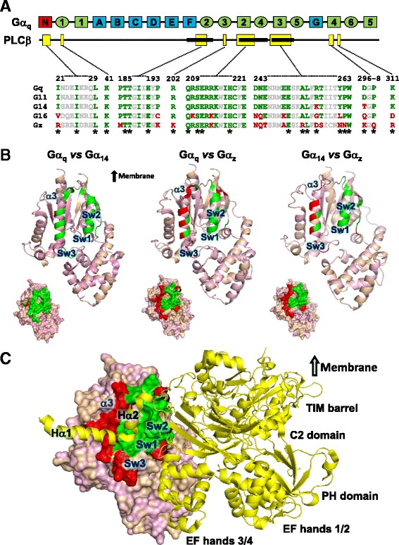
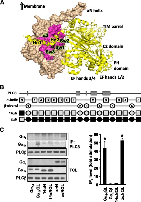
Background: Stimulation of phospholipase Cβ (PLCβ) by the activated α-subunit of Gq (Gαq) constitutes a major signaling pathway for cellular regulation, and structural studies have recently revealed the molecular interactions between PLCβ and Gαq. Yet, most of the PLCβ-interacting residues identified on Gαq are not unique to members of the Gαq family. Molecular modeling predicts that the core PLCβ-interacting residues located on the switch regions of Gαq are similarly positioned in Gαz which does not stimulate PLCβ. Using wild-type and constitutively active chimeras constructed between Gαz and Gα14, a member of the Gαq family, we examined if the PLCβ-interacting residues identified in Gαq are indeed essential.

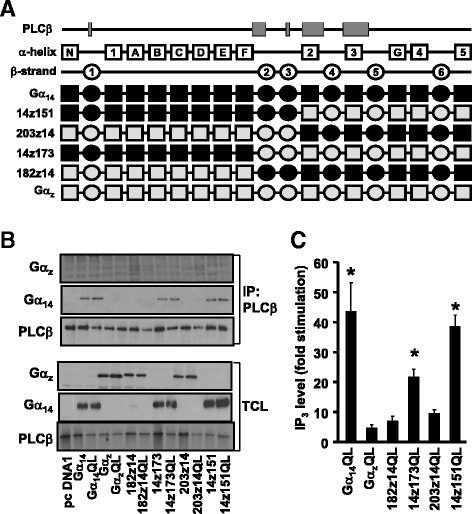
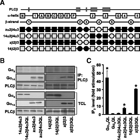
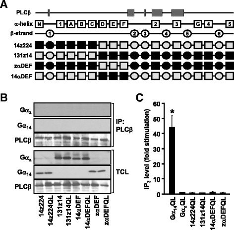
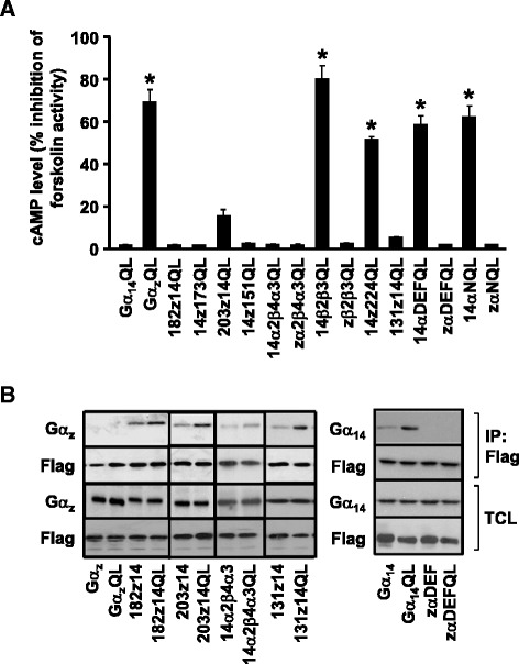
Results: Four chimeras with the core PLCβ-interacting residues composed of Gαz sequences were capable of binding PLCβ2 and stimulating the formation of inositol trisphosphate. Surprisingly, all chimeras with a Gαz N-terminal half failed to functionally associate with PLCβ2, despite the fact that many of them contained the core PLCβ-interacting residues from Gα14. Further analyses revealed that the non-PLCβ2 interacting chimeras were capable of interacting with other effector molecules such as adenylyl cyclase and tetratricopeptide repeat 1, indicating that they could adopt a GTP-bound active conformation.

Conclusion: Collectively, our study suggests that the previously identified PLCβ-interacting residues are insufficient to ensure productive interaction of Gα14 with PLCβ, while an intact N-terminal half of Gα14 is apparently required for PLCβ interaction.


Figures

Fig. 1
Fig. 1
Alignment of PLCβ-interacting residues in Gαq family and Gαz. a Schematic view of Gαq divided into helical (light blue) and GTPase (light green) domains with α-helices and β-strands represented by rectangles and ovals, respectively. Interacting domains of Gαq with PLCβ are indicated by yellow boxes below the Gαq sequence; the three bold segments indicate the relative positions of the three switch regions (Sw1 to Sw3 from left to right). Sequence alignment of PLCβ-interacting domains in the Gαq family as compared to that of Gαz; conserved (green) or divergent (red) PLCβ-interacting residues are interspersed by conserved residues which are not implicated in interaction with PLCβ (grey). Residues forming direct interactions with PLCβ3 as identified by Waldo et al. [13] are indicated by an asterisk. b Structural representation of Gαq, Gα14, and Gαz alignments with switch regions (Sw1-3) and the α3 region. PLCβ3-interacting residues revealed in the sequence alignment are colored as indicated in A. Space filling models are showing interacting surfaces. Structural models of Gα14 and Gαz are generated based on Gαq-PLCβ3 (PDB code: 3OHM) using SWISS-MODEL [65, 66]. Structure alignments are carried out with PyMOL (The PyMOL Molecular Graphics System, Version 1.3 Schrödinger, LLC). c Complex of Gαq/Gαz-PLCβ3. The Gαq/Gαz aligned model is represented as indicated in (B). PLCβ3 (yellow) is depicted as a cartoon ribbon, containing the helix-turn-helix segment (Hα1/Hα2), the N-terminal PH domain, four EF hands, the catalytic TIM barrel, and a C2 domain. The switch regions of Gαq interact with PLCβ3 through an extended loop region between EF hands 3/4 and the region between the catalytic TIM barrel and C2 domain. The helix-turn-helix segment (Hα1/Hα2) at the C-terminus of PLCβ3 resides on the surface region formed by switch 2 and α3 of Gαq. The αN helix of Gα proteins and carboxy-terminal (CT) domain of PLCβ3 are not included in the structural models
Fig. 2
Fig. 2
The putative PLCβ domain of Gα14 is not required for PLCβ interaction and activation. a Schematic representation of the 14z151, 203z14, 14z173, and 182z14 chimeras. Predicted secondary structures are illustrated as boxes (α helices) or circles (β strands) above the chimeras. Black areas represent human Gα14 sequence while those in grey signify the corresponding sequence of human Gαz. b HEK293 cells were co-transfected with PLCβ2 and the indicated Gα protein or chimeras. Cell lysates from the transfectants were immunoprecipitated by anti-PLCβ2 antiserum. The immunoprecipitates were immunoblotted with anti-Gα14, anti-Gαz or anti-PLCβ2 antiserum. Aliquots of cell lysates were used to detect the expression levels of Gα14, Gαz and PLCβ by Western blot analysis (TCL). Data shown represent one of three sets of immunoblots; two other sets yielded similar results. c HEK293 cells were transiently transfected with wild-type or constitutively active mutants (QL) of Gα protein or chimeras. Cells were then labelled and assayed for IP3 formation. Fold stimulations were calculated as the ratios of QL-induced to wild-type IP3 accumulations. Data represent the mean ± S.E.M. of three independent experiments, n = 3. *, IP3 production was significantly enhanced as compared to corresponding wild-type transfected cells; Dunnett t test, p < 0.05
Fig. 3
Fig. 3
Role of β2-β3 and α2-β4-α3 regions of Gα14 in interaction and activation of PLCβ. a Schematic representation of zα2β4α3, 14α2β4α3, zβ2β3 and 14β2β3 chimeras. b, Cells were co-transfected with PLCβ2 and Gα protein or the indicated chimeras. Co-immunoprecipitation assays were performed and analyzed as in Fig. 2. Data shown represent one of three sets of immunoblots; two other sets yielded similar results. c HEK293 cells were transiently transfected with the wild-type or constitutively active mutants (QL) of Gα proteins or chimeras and then subjected to IP3 accumulation assay and analyzed as in Fig. 2. *, IP3 production was significantly enhanced as compared to corresponding wild-type transfected cells; Dunnett t test, p < 0.05
Fig. 4
Fig. 4
An intact N-terminal and helical domain are required for Gα14 mediated PLCβ interaction and activation. a Schematic representation of the 14z224, 131z14, 14αDEF and zαDEF chimeras. b, Cells were co-transfected with PLCβ2 and the indicated chimeras. Co-immunoprecipitation assays were performed and analyzed as in Fig. 2. Data shown represent one of three sets of immunoblots; two other sets yielded similar results. c HEK293 cells were transiently transfected with the wild-type or constitutively active mutants (QL) of Gα protein or the indicated chimeras and then subjected to IP3 accumulation assay and analyzed as in Fig. 2. *, IP3 production was significantly enhanced as compared to corresponding wild-type transfected cells; Dunnett t test, p < 0.05
Fig. 5
Fig. 5
Role of the N-terminal helix (αN) in the Gαq-PLCβ3 complex. a The model of Gαq (light orange) is shown as a space filling structure and contains the αN-helix and other regions as indicated. PLCβ3 (yellow) is depicted as a cartoon ribbon, containing the helix-turn-helix segment (Hα1/Hα2), the N-terminal PH domain, four EF hands, the catalytic TIM barrel, and a C2 domain. PLCβ3-interacting residues of Gαq are colored in magenta. The carboxy-terminal (CT) domain of PLCβ3 is not included in the structural model. The structure of the αN-helix is generated by replacing the amino acid sequence of Gαi (Gαiβ1γ2, PDB code: 1GP2) with the Gαq sequence. The final model is generated by alignment of Gαq-PLCβ3 (PDB code: 3OHM) and the modified heterotrimer Gαqβ1γ2 using PyMOL (The PyMOL Molecular Graphics System, Version 1.3 Schrödinger, LLC). The orientation of the αN-helix represents the conformation in the heterotrimer and is not optimized for the Gαq-PLCβ3 complex. In this case, the αN-helix points towards the cell membrane and clashes with PLCβ3, but in fact may exist in a conformation which interacts with PLCβ3. b Schematic representation of 14αΝ and zαN chimeras. c Cells were co-transfected with PLCβ2 and Gα protein or the indicated chimeras. Co-immunoprecipitation assays were performed and analyzed as in Fig. 2. Data shown represent one of three sets of immunoblots; two other sets yielded similar results. For the IP3 accumulation assay, HEK293 cells were transiently transfected with the wild-type or constitutively active mutants (QL) of Gα proteins or chimeras and analyzed as in Fig. 2. *, IP3 production was significantly enhanced as compared to corresponding wild-type transfected cells; Dunnett t test, p < 0.05
Fig. 6
Fig. 6
Ability of different chimeras to interact with AC and TPR1. a HEK293 cells were transiently transfected with the wild-type or constitutively active mutants of Gα protein and chimeras indicated in the figure. The transfectants were labelled with [3H]adenine (1 μCi/ml) in 1 % FBS/MEM. The labelled cells were treated with 50 μM of FSK for 30 min before subjected to cAMP accumulation assay. cAMP fold inhibition was calculated as the ratios of QL-induced to wild-type cAMP inhibition. Data represent the mean ± S.E.M. of three independent experiments, n = 3. *, cAMP accumulation was significantly inhibited as compared to corresponding wild-type transfected cells; Dunnett t test, p < 0.05. b HEK293 cells were transiently co-transfected with FLAG-TPR1 in combination with Gα proteins and the indicated chimeras. Cell lysates were immunoprecipitated by anti-FLAG affinity agarose gel. The immunoprecipitates were immunoblotted with anti-Gα14, anti-Gαz or anti-FLAG antiserum. Crude lysates were used to examine the expression levels of Gα14, Gαz,14/Gαz chimeras or FLAG-TPR1 by Western blot analysis. The immunoblots shown represent one of three sets of immunoblots; two other sets yielded similar results

Similar articles

Cited by

References

    1. Oldham WM, Hamm HE. Heterotrimeric G protein activation by G-protein-coupled receptors. Nat Rev Mol Cell Biol. 2008;9(1):60–71. doi: 10.1038/nrm2299. - DOI - PubMed
    1. Tesmer JJ. The quest to understand heterotrimeric G protein signaling. Nat Struct Mol Biol. 2010;17(6):650–652. doi: 10.1038/nsmb0610-650. - DOI - PMC - PubMed
    1. Sandal M, Paltrinieri D, Carloni P, Musiani F, Giorgetti A. Structure/function relationships of phospholipases C Beta. Curr Protein Pept Sci. 2013;14(8):650–657. doi: 10.2174/13892037113146660085. - DOI - PubMed
    1. Jhon DY, Lee HH, Park D, Lee CW, Lee KH, Yoo OJ, et al. Cloning, sequencing, purification, and Gq-dependent activation of phospholipase C-beta 3. J Biol Chem. 1993;268(9):6654–6661. - PubMed
    1. Lyon AM, Tesmer JJ. Structural insights into phospholipase C-β function. Mol Pharmacol. 2013;84(4):488–500. doi: 10.1124/mol.113.087403. - DOI - PMC - PubMed

Publication types

MeSH terms

Substances

LinkOut - more resources