. 2015 Sep 17:12:172.
doi: 10.1186/s12974-015-0391-8.

Inhibition of NOX2 reduces locomotor impairment, inflammation, and oxidative stress after spinal cord injury

Affiliations

Inhibition of NOX2 reduces locomotor impairment, inflammation, and oxidative stress after spinal cord injury

Guzal Khayrullina et al. J Neuroinflammation. .

Abstract

Background: Spinal cord injury (SCI) results in the activation of the NADPH oxidase (NOX) enzyme, inducing production of reactive oxygen species (ROS). We hypothesized that the NOX2 isoform plays an integral role in post-SCI inflammation and functional deficits.

Methods: Moderate spinal cord contusion injury was performed in adult male mice, and flow cytometry, western blot, and immunohistochemistry were used to assess NOX2 activity and expression, inflammation, and M1/M2 microglia/macrophage polarization from 1 to 28 days after injury. The NOX2-specific inhibitor, gp91ds-tat, was injected into the intrathecal space immediately after impact. The Basso Mouse Scale (BMS) was used to assess locomotor function at 24 h post-injury and weekly thereafter.

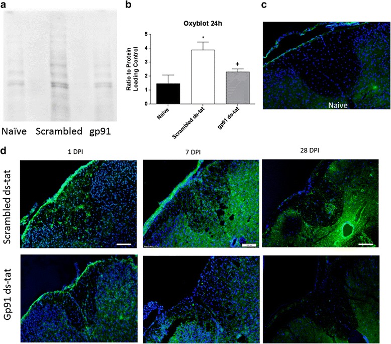
Results: Our findings show that gp91ds-tat treatment significantly improved functional recovery through 28 days post-injury and reduced inflammatory cell concentrations in the injured spinal cord at 24 h and 7 days post-injury. In addition, a number of oxidative stress markers were reduced in expression at 24 h after gp91ds-tat treatment, which was accompanied by a reduction in M1 polarization marker expression.

Conclusion: Based on our findings, we now conclude that inhibition of NOX2 significantly improves outcome after SCI, most likely via acute reductions in oxidative stress and inflammation. NOX2 inhibition may therefore have true potential as a therapy after SCI.


Figures

Fig. 1
Fig. 1
Inhibition of NOX2 improves motor function. Gross hindlimb motor function was scored using the BMS (a), with further analysis of hindpaw function with the BMS subscore (b). Mice were tested at days 1, 7, 14, 21, and 28 post-injury. Those treated with gp91ds-tat showed significant increases in BMS score and BMS subscore at 7 days; this improvement was sustained through 28 days in the BMS score. Points represent mean ± SEM. N = 9/group. *p < 0.05, repeated measures ANOVA
Fig. 2
Fig. 2
Acute inhibition of NOX2 reduces markers of oxidative stress. OxyBlot Protein Oxidation assay was used to detect protein carbonylation in naïve (n = 8), scrambled ds-tat- (n = 4), and gp91ds-tat-treated mice (n = 4) at 24 h post-injury (a). Densitometry demonstrated a significant increase in protein carbonylation in scrambled ds-tat-treated mice, which was significantly reduced to naïve levels with gp91ds-tat treatment (b). At these same time points, spinal cord tissue was immunolabeled for 3-nitrotyrosine (green), a marker of nitrosylated protein (scrambled ds-tat: n = 4/time point; gp91ds-tat: n = 4/24 h, 3/7 days; 4/28 days). DAPI nuclear stain is shown in blue. Naïve (n = 4) tissue is shown in c. Qualitative analysis shows that scrambled ds-tat-treated tissue had elevated 3NT immunolabeling in comparison to naïve tissue, primarily in gray matter (d). This elevation grew through 7 days and was lessened at 28 days. Treatment with gp91ds-tat appeared to reduce this immunolabeling at all time points. Bar = 100 μm. *p < 0.05 vs naïve; +p < 0.05 vs scrambled; one-way ANOVA with Tukey’s post-test. Bars represent mean ± SEM
Fig. 3
Fig. 3
Acute inhibition of NOX2 using gp91ds-tat reduces p47PHOX expression and/or phosphorylation. Phosphorylated p47PHOX (p-p47) and GAPDH were evaluated at 24 h, 7 days, and 28 days post-injury in naïve (N; n = 8), scrambled ds-tat- (S; n = 4/24 h; 6/7 days; 3/28 days), and gp91ds-tat-treated samples (G; n = 4/24 h; 5/7 days; 3/28 days). Bands were observed at 47 and 38 kDa, respectively, in a representative western blot (a). Pixel densitometry for p-p47PHOX (b) showed significant induction of phosphorylation of p47PHOX at 24 h. NOX2 inhibition reduced p-p47PHOX levels at 7 days to a point that was not significantly greater than sham. **p < 0.01 vs naïve. +p < 0.01 vs scrambled; one-way ANOVA with Tukey’s post-test. Bars represent mean ± SEM
Fig. 4
Fig. 4
Acute inhibition of NOX2 reduces neutrophil populations by 24 h post-injury. Neutrophils were characterized as alive, CD45+/CD11b+/GR-1+ population (a). b Quantitation shows that gp91ds-tat treatment significantly reduced this population. *p < 0.05 vs scrambled ds-tat, Student’s t test. Bars represent mean ± SEM
Fig. 5
Fig. 5
Acute NOX2 inhibition reduces inflammatory response by 7 days post-injury. All listed cell types were under a live CD45+ gate. Microglia/macrophage were further isolated as CD11b+Gr-1 (a), neutrophils as CD11b+Gr-1+ (c), and T cells as CD3+ (e). The first column in each row indicates an isotype control. Quantitation shows that gp91ds-tat treatment significantly reduced the macrophage/microglia (b) and neutrophil (d) populations but had no significant effect on the T cell population (f). *p < 0.05 vs scrambled ds-tat, Student’s t test. Bars represent mean ± SEM
Fig. 6
Fig. 6
Acute NOX2 inhibition specifically targets microglia. Flow cytometry was used to analyze macrophage and microglia populations in the injured region. Live cells were first gated on and further differentiated based on CD11b+ and CD45+. CD45 high, macrophages, and CD45 low, microglia, were arbitrarily chosen by a researcher blinded to study (a). Quantitation demonstrated no significant difference in macrophages at 7 days post-injury between scrambled ds-tat- and gp91ds-tat-treated tissue. Microglia, on the other hand, showed a significant decrease in tissue treated with gp91ds-tat compared to scrambled ds-tat (b). *p < 0.05 vs scrambled ds-tat, Student’s t test. Bars represent mean ± SEM
Fig. 7
Fig. 7
Acute NOX2 inhibition reduces microglia/macrophage presence in injured spinal cord tissue. Gp91ds-tat- and scrambled ds-tat-treated tissue at 24 h (a, b; n = 4/group) and 7 days (c, d; n = 3 gp91ds-tat, 4 scrambled ds-tat) post-injury was stained for the pan microglia/macrophage marker Iba1 (red). DAPI nuclear stain is shown in blue. At 24 h post-injury, the scrambled ds-tat-treated tissue (a) have a greater number of activated microglia compared to gp91ds-tat-treated tissue as seen in ×10 magnification (b). The activated morphology includes a more amoeboid body with retracted dendrites (which can be better observed in the higher magnification inset image (×20)). At the same time point, the microglia present in gp91ds-tat tissue have long processes and smaller bodies. This difference is greater at the 7-day time point when microglia are at their peak (c, d). The gp91ds-tat-treated microglia at 7 days (×20) are activated, but the number are still downregulated compared to those treated with scrambled-tat. Lower magnification and high-magnification images shown; bar (×20) = 50 μm; bar (×10) = 100 μm
Fig. 8
Fig. 8
Acute NOX2 inhibition increases the M2 marker CD206 by 7 days post-injury. Cells were gated on live CD45+CD11b+Gr-1CD206+ population (a). Quantitation shows that gp91ds-tat treatment significantly increased the CD206 population (b). *p < 0.05 vs scrambled ds-tat, Student’s t test. Bars represent mean ± SEM
Fig. 9
Fig. 9
Acute inhibition of NOX2 using gp91ds-tat alters microglia/macrophage polarization marker expression. Protein samples (25 μg) were probed for CD206, iNOS, CD86, and GAPDH at 24 h, 7 days, and 28 days post-injury in naïve (n = 8), scrambled ds-tat- (n = 4/24 h; 6/7 days; 3/28 days), and gp91ds-tat-treated samples (n = 4/24 h; 5/7 days; 3/28 days). Pixel densitometry for CD206 (a), CD86 (b), and iNOS (c) showed significant alteration with gp91ds-tat treatment in comparison to scrambled ds-tat treatment. The M2 marker CD206 was elevated in both groups at 24 h in comparison to naïve, but remained elevated only in the gp91ds-tat-treated group at 7 days post-injury, before returning to baseline levels by 28 days. In contrast, at 24 h, the inhibition of NOX2 significantly reduced the expression of the M1 marker CD86, although by 7 and 28 days, no significant difference was observed between groups. Finally, a second M1 marker, iNOS, was elevated in both groups at 24 h post-injury, and gp91ds-tat treatment only prevented this induction by 7 days post-injury. *p < 0.05 vs naïve; +p < 0.05 vs scrambled; one-way ANOVA with Tukey’s post-test. Bars represent mean ± SEM

Similar articles

Cited by

References

    1. Fleming JC, Norenberg MD, Ramsay DA, Dekaban GA, Marcillo AE, Saenz AD, et al. The cellular inflammatory response in human spinal cords after injury. Brain. 2006;129:3249–69. doi: 10.1093/brain/awl296. - DOI - PubMed
    1. Kroner A, Greenhalgh AD, Zarruk JG, Passos Dos Santos R, Gaestel M, David S. TNF and increased intracellular iron alter macrophage polarization to a detrimental M1 phenotype in the injured spinal cord. Neuron. 2014;83:1098–116. doi: 10.1016/j.neuron.2014.07.027. - DOI - PubMed
    1. Yao A, Liu F, Chen K, Tang L, Liu L, Zhang K, et al. Programmed death 1 deficiency induces the polarization of macrophages/microglia to the M1 phenotype after spinal cord injury in mice. Neurotherapeutics. 2014;11:636–50. doi: 10.1007/s13311-013-0254-x. - DOI - PMC - PubMed
    1. Kigerl KA, Gensel JC, Ankeny DP, Alexander JK, Donnelly DJ, Popovich PG. Identification of two distinct macrophage subsets with divergent effects causing either neurotoxicity or regeneration in the injured mouse spinal cord. J Neurosci. 2009;29:13435–44. doi: 10.1523/JNEUROSCI.3257-09.2009. - DOI - PMC - PubMed
    1. Choi SH, Aid S, Kim HW, Jackson SH, Bosetti F. Inhibition of NADPH oxidase promotes alternative and anti-inflammatory microglial activation during neuroinflammation. J Neurochem. 2012;120:292–301. doi: 10.1111/j.1471-4159.2011.07572.x. - DOI - PMC - PubMed

Publication types

MeSH terms