. 2015 Sep 15;43(3):515-26.
doi: 10.1016/j.immuni.2015.08.016.

Nociceptive Sensory Fibers Drive Interleukin-23 Production from CD301b+ Dermal Dendritic Cells and Drive Protective Cutaneous Immunity

Affiliations

Nociceptive Sensory Fibers Drive Interleukin-23 Production from CD301b+ Dermal Dendritic Cells and Drive Protective Cutaneous Immunity

Sakeen W Kashem et al. Immunity. .

Erratum in

  • Immunity. 2015 Oct 20;43(4):830

Abstract

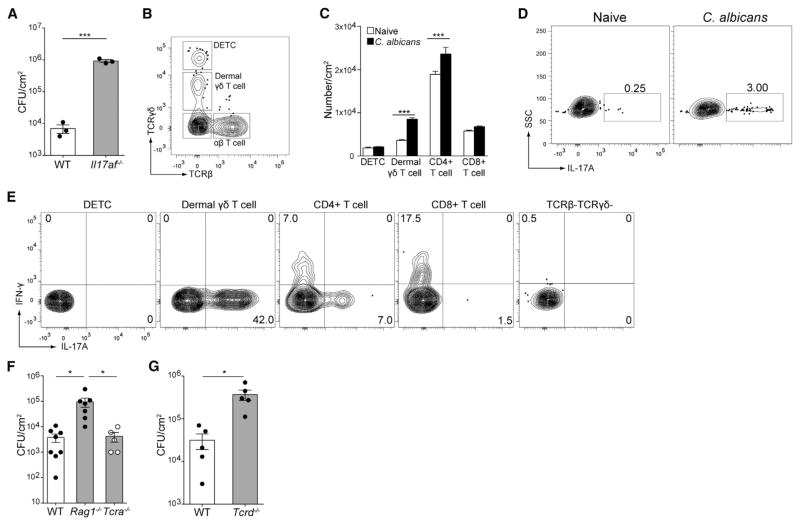
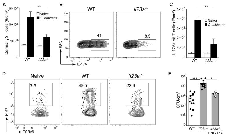
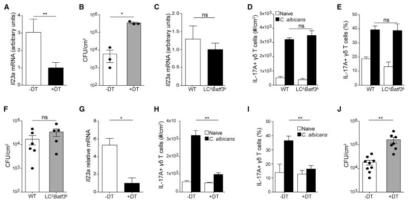
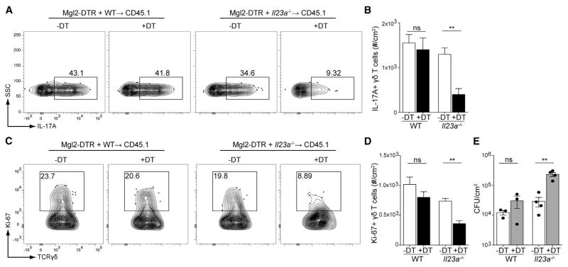
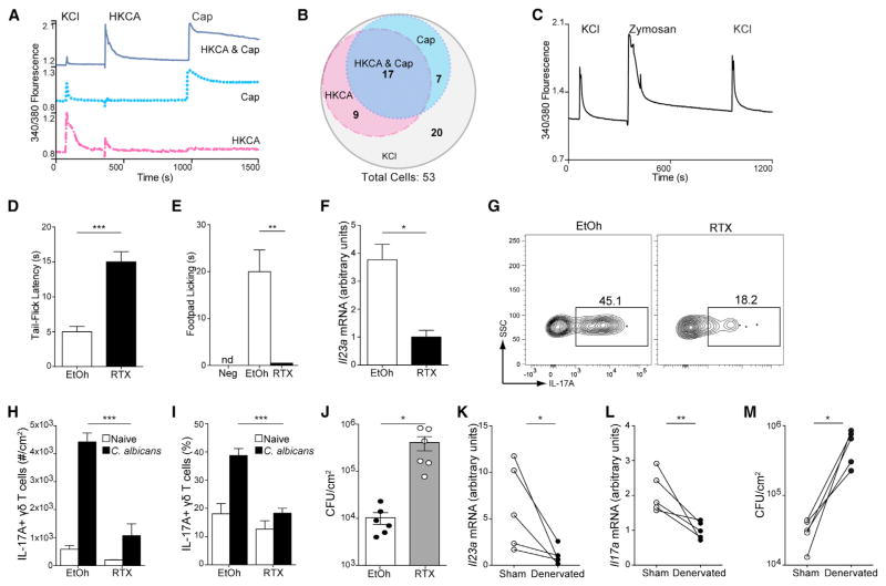
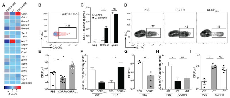
Innate resistance to Candida albicans in mucosal tissues requires the production of interleukin-17A (IL-17A) by tissue-resident cells early during infection, but the mechanism of cytokine production has not been precisely defined. In the skin, we found that dermal γδ T cells were the dominant source of IL-17A during C. albicans infection and were required for pathogen resistance. Induction of IL-17A from dermal γδ T cells and resistance to C. albicans required IL-23 production from CD301b(+) dermal dendritic cells (dDCs). In addition, we found that sensory neurons were directly activated by C. albicans. Ablation of sensory neurons increased susceptibility to C. albicans infection, which could be rescued by exogenous addition of the neuropeptide CGRP. These data define a model in which nociceptive pathways in the skin drive production of IL-23 by CD301b(+) dDCs resulting in IL-17A production from γδ T cells and resistance to cutaneous candidiasis.


Figures

Figure 1
Figure 1. Dermal γδ T Cells, but Not αβ T Cells, Are Required for IL-17-Mediated Protection against C. albicans
Cohorts of mice were infected on their shaved dorsum with 2 × 10⁸ CFU C. albicans. (A) Day 3 whole skin homogenates from wild-type or Il17af−/− infected mice were plated onto YPAD agar plates and incubated at 30°C for 48 hr. The number of colony-forming units (CFU) per cm2 of skin is shown. (B) Single-cell suspensions from whole skin was obtained by enzymatic digestion and analyzed by flow cytometry gated as singlets, live/dead excluded, CD90.2+, and Lineage (B220CD11cCD11bF4/80). Expression of TCRβ and TCRγδ is used to identify lymphoid subsets in the skin. (C) The numbers in each lymphocyte subset as in (B) in naive (white) and C. albicans-infected (black) mice are shown. (D) Cells isolated from skin were re-stimulated with PMA and ionomycin in the presence of monensin and gated as in (B). Expression of IL-17A by CD90.2+ lymphocytes is shown. (E) Representative expression of IL-17A and interferon-γ (IFN-γ) by individual subsets from (D) are shown. (F) C. albicans CFUs isolated from the skin of WT, Rag1−/−, and Tcra−/− mice 3 days after infection is shown. (G) Lethally irradiated B6.SJL-Ly5.2 were reconstituted with bone marrow from WT C57BL/6 or Tcrd−/− mice and rested for 12 weeks. C. albicans CFUs 3 days after infection is shown. Error bars represent mean ± SEM. Student’s unpaired t test representative of *p < 0.05, **p < 0.01, ***p < 0.001. Data points in (A), (F), and (G) represent individual animals. Data are representative of at least three mice per group and at least three independent experiments.
Figure 2
Figure 2. IL-23 Is Required for IL-17 Secretion and Proliferation of Dermal γδ T Cells
Mice were infected on their dorsum with 2 × 10⁸ CFUs C. albicans. Three days later, single-cell suspensions from whole skin was obtained by enzymatic digestion and subjected to flow cytometry. (A) Total number of dermal γδ T cells in WT or Il23a−/− mice is shown in naive (white) or infected (black) mice. (B) As in (A), cells were re-stimulated with PMA and ionomycin in the presence of monensin and analyzed for IL-17 expression. (C) The number of IL-17A-expressing dermal γδ T cells in (B) is shown in naive (white) or infected (black) mice. (D) Dermal γδ T cells in WT or Il23a−/− mice were analyzed for proliferation by nuclear Ki-67 staining compared to naive mice. (E) C. albicans CFU from day 3 skin homogenates from WT, Il23a−/−, or Il23a−/− mice administered 1 μg intradermal recombinant IL-17A (rIL-17A) 1 day prior to and 1 day after infection. Error bars represent mean ± SEM. Student’s unpaired t test representative of *p < 0.05, **p < 0.01, ***p < 0.001. Data points in (E) represent individual animals. Data are representative of at least three independent experiments with cohorts of at least six mice.
Figure 3
Figure 3. CD301b+ Dermal DCs Are Required for IL-23-Mediated Protection against C. albicans
CD11c-DTR mice were treated with 1 μg of diphtheria toxin (DT) or vehicle 1 day before infection with 2 × 10⁸ CFU C. albicans. (A) Il23a expression in whole skin tissue 16 hr after infection as analyzed by qRT-PCR is shown. (B) C. albicans CFUs in whole skin from control and DC-depleted mice 3 days after infection is shown. (C–F) Mice lacking LCs and CD103+ dDCs (LCΔBatf3Δ) were infected with C. albicans. (C) Il23a expression in skin was analyzed 16 hr after infection by qRT-PCR. (D and E) Number (D) and percent (E) of IL-17A-expressing dermal γδ T cells in mice kept naive (white) or infected with C. albicans (black) is shown. (F) Skin CFUs 3 days after C. albicans infection in LCΔBatf3Δ depleted mice is shown. (G–J) Mgl2-DTR mice were treated with 1 μg of DT or vehicle 1 day before infection with C. albicans. (G) Il23a expression in skin was assessed by qRT-PCR 16 hr after infection. (H and I) Number (H) and percent (I) of IL-17A-expressing dermal γδ T cells in mice kept naive (white) or infected with C. albicans (black) is shown. (J) CFUs in skin 3 days after infection is shown. Error bars represent mean ± SEM. Student’s unpaired t test representative of *p < 0.05, **p < 0.01, ***p < 0.001. Data points in (B), (F), and (J) represent individual animals. Data are representative of at least three independent experiments with cohorts of at least three animals.
Figure 4
Figure 4. IL-23 from CD301b+ Dermal DCs Is Necessary for IL-17 Secretion and Proliferation of Dermal γδ T Cells
Lethally irradiated B6.SJL-Ly5.2 (CD45.1) mice were re-constituted with 1:1 ratio of bone marrow from Mgl2-DTR mice and either WT or Il23a−/− mice. After 12 weeks, mice were treated with 1 μg of diphtheria toxin (DT) or vehicle 1 day before infection with 2 × 10⁸ CFU C. albicans. (A) IL-17A expression of dermal γδ T cells in mice 3 days after C. albicans infection is shown. (B) The number of IL-17-expressing dermal γδ T cells in the skin from (A) is shown. (C and D) As in (A), Ki-67 staining of dermal γδ T cells (C) and quantification of Ki-67+ dermal γδ T cells (D) 3 days after infection is shown. (E) C. albicans skin CFUs from mixed bone marrow chimeric mice 3 days after infection. Student’s unpaired t test representative of *p < 0.05, **p < 0.01, ***p < 0.001. Error bars represent mean ± SEM. Data points in (E) represent individual animals. Data are representative of at least three independent experiments with cohorts of at least three mice.
Figure 5
Figure 5. Nociception and Sensory Nerves Are Crucial Mediators of Anti-fungal Immunity
(A) Sensory neurons were dissociated from dorsal root ganglion of WT mice and cultured for 18–24 hr before being loaded with the ratiometric intracellular calcium indicator Fura2-AM. Cultured neurons were stimulated sequentially with 30 mM KCl (10 s), heat-killed 1 × 10⁷ CFU C. albicans (HKCA; 15 s), and 500 mM capsaicin (Cap; 10 s). Traces showing calcium flux as indicated by the 340/380 ratio visualized by microscopy from three individual cells are shown. Dark blue line represents cell that responded to HKCA and Cap, dotted blue line represents cell that responded to Cap but not HKCA, and red dashed line represents cell that responded to HKCA but not Cap. Data are representative of 53 cells tested. (B) Scaled venn diagram representation of the 53 DRG neuron responsiveness to different stimuli. (C) Cultured neurons were stimulated sequentially with 30 mM KCl (10 s), 1 × 10⁷ CFU zymosan (15 s), and 30 mM KCl (10 s). Traces showing calcium flux as indicated by the 340/380 ratio visualized by microscopy from a single neuronal cell representative of ten cells. (D–J) Cohorts of WT mice were injected s.c. with resineferatoxin (RTX) or vehicle (EtOh). (D) Mice were checked for their latency to tail-flick after submersion in a 52°C waterbath. (E and F) Nocifensive behavior (E) was assessed for 10 min after injection of vehicle or 5 μg of capsaicin into the hindpaws of EtOh- or RTX-treated mice. Neg is defined as EtOH mice treated with vehicle. Mice were infected with C. albicans and (F) Il23a expression was quantified by qRT-PCR in skin 16 hr later. (G) IL-17A expression by dermal γδ T cells 3 days after infection is shown. (H and I) The number (H) and percentage (I) of IL-17-expressing dermal γδ T cells is shown in naive (white) and infected (black) mice. (J) C. albicans CFU in vehicle- or RTX-treated mice 3 days after infection is shown. (K–M) Mice were surgically denervated on one lateral side of their dorsum and infected 7 days later with equal CFUs of C. albicans. (K and L) Il23a (K) and Il17a (L) expression in skin was quantified by qRT-PCR 16 hr and 60 hr later, respectively. (M) CFU from mechanically denervated side of infected mice and mock denervated side are shown. Data from individual mice are connected by a line. Error bars represent mean ± SEM. Student’s unpaired (D–F; H–J) or paired (K–M) t test representative of *p < 0.05, **p < 0.01, ***p < 0.001. Data are representative of at least three independent experiments with cohorts of at least five mice.
Figure 6
Figure 6. Neuropeptide CGRPα Drives IL-23 Response and Cutaneous Fungal Resistance
(A) Gene expression patterns of CGRP, substance P, neuropeptide Y, somatostatin, and vaso-active intestinal peptide receptors on the indicated skin dendritic cell subsets. Raw data were obtained from the Immunological Genome Consortium. (B) Expression of CGRP receptor CALCRL (red) with isotype control (blue) on skin CD11b+ DC subsets. (C) Cultured DRG neurons were incubated for 10 min in HEPES buffer to measure basal release (Neg) and then incubated for 10 min in HEPES buffer (release, open bars) or HKCA (release, black bars). CGRP levels in supernatants and cell lysates (lysate) were determined by ELISA. (D–I) Cohorts of WT mice were treated intradermally with either PBS or 0.5 μg of peptide CGRPα or inhibitor CGRP32-37 on days −1, 0, 1, and 2 of C. albicans infection. (D) IL-17A expression of PMA and ionomycin-stimulated dermal γδ T cells in mice 3 days after infection is shown. (E) Skin CFUs 3 days after infection from mice treated with PBS, CGRPα, or inhibitor CGRP32-37 is shown. (F and G) 4-week-old WT mice were injected with RTX or vehicle (EtOH) subcutaneously in flank. 4 to 6 weeks later, mice were treated with either PBS, CGRPα, or inhibitor CGRP32-37. (F) Expression of Il23a in skin 16 hr after infection was determined by qRT-PCR. (G) Skin CFUs 3 days after infection is shown. (H and I) Mgl2-DTR mice were treated with 1 μg of diphtheria toxin (DT) or vehicle 1 day before infection with C. albicans. Mice were given intradermal PBS or CGRPα on days −1, 0, 1, and 2 of C. albicans infection. (H) Expression of Il23a in skin 16 hr after infection was assessed by qRT-PCR. (I) Skin CFU 3 days after infection is shown. Student’s unpaired t test representative of *p < 0.05, **p < 0.01, ***p < 0.001. Data are representative of at least three independent experiments with cohorts of at least three mice.

Comment in

  • IL-17A Has Some Nerve!
    Sanford JA, Gallo RL. Sanford JA, et al. Immunity. 2015 Sep 15;43(3):414-5. doi: 10.1016/j.immuni.2015.08.025. Immunity. 2015. PMID: 26377893

References

    1. Assas BM, Miyan JA, Pennock JL. Cross-talk between neural and immune receptors provides a potential mechanism of homeostatic regulation in the gut mucosa. Mucosal Immunol. 2014;7:1283–1289. - PubMed
    1. Augustyniak D, Nowak J, Lundy FT. Direct and indirect antimicrobial activities of neuropeptides and their therapeutic potential. Curr Protein Pept Sci. 2012;13:723–738. - PMC - PubMed
    1. Awasthi A, Riol-Blanco L, Jäger A, Korn T, Pot C, Galileos G, Bettelli E, Kuchroo VK, Oukka M. Cutting edge: IL-23 receptor gfp reporter mice reveal distinct populations of IL-17-producing cells. J Immunol. 2009;182:5904–5908. - PMC - PubMed
    1. Bannon AW, Malmberg AB. Models of nociception: hot-plate, tail-flick, and formalin tests in rodents. Curr Protoc Neurosci. 2007;8:9. - PubMed
    1. Bär E, Whitney PG, Moor K, Reis e Sousa C, LeibundGut-Landmann S. IL-17 regulates systemic fungal immunity by controlling the functional competence of NK cells. Immunity. 2014;40:117–127. - PubMed

Publication types

MeSH terms