. 2015 Oct 27;6(33):34992-5003.
doi: 10.18632/oncotarget.5252.

Intracellular activation of EGFR by fatty acid synthase dependent palmitoylation

Affiliations

Intracellular activation of EGFR by fatty acid synthase dependent palmitoylation

Lakshmi Reddy Bollu et al. Oncotarget. .

Abstract

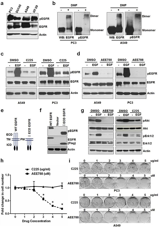
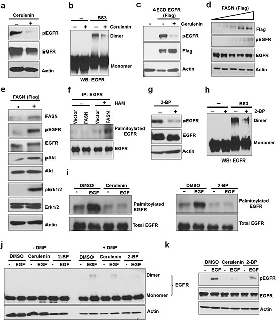
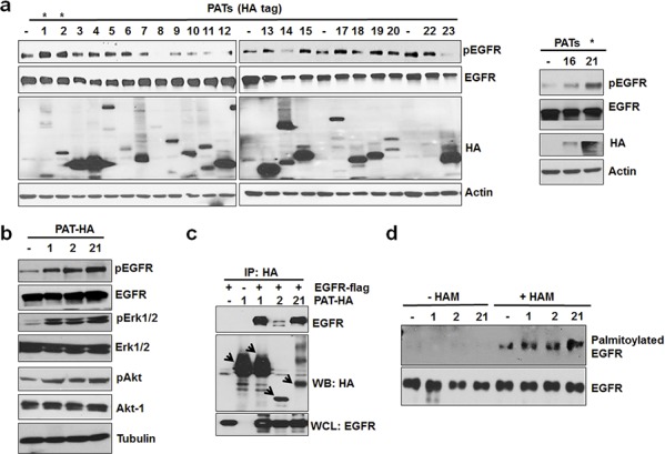
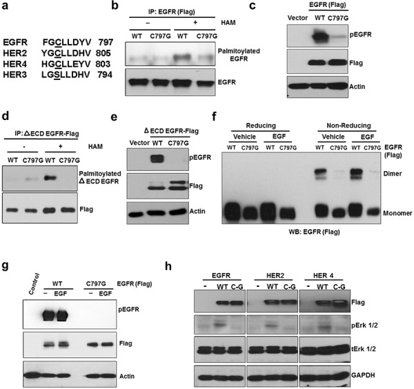
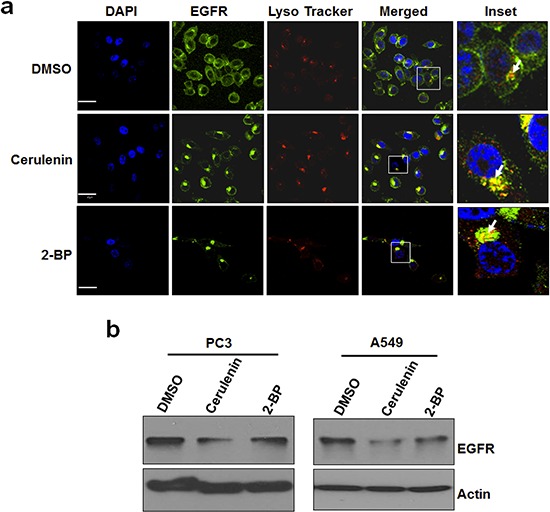
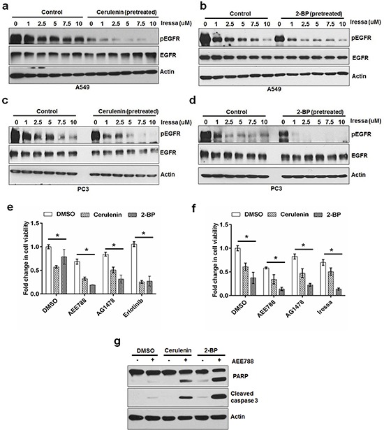
Epidermal growth factor receptor (EGFR) is an oncogenic receptor tyrosine kinase. Canonically, the tyrosine kinase activity of EGFR is regulated by its extracellular ligands. However, ligand-independent activation of EGFR exists in certain cancer cells, and the underlying mechanism remains to be defined. In this study, using PC3 and A549 cells as a model, we have found that, in the absence of extracellular ligands, a subpopulation of EGFR is constitutively active, which is needed for maintaining cell proliferation. Furthermore, we have found that fatty acid synthase (FASN)-dependent palmitoylation of EGFR is required for EGFR dimerization and kinase activation. Inhibition of FASN or palmitoyl acyltransferases reduced the activity and down-regulated the levels of EGFR, and sensitized cancer cells to EGFR tyrosine kinase inhibitors. It is concluded that EGFR can be activated intracellularly by FASN-dependent palmitoylation. This mechanism may serve as a new target for improving EGFR-based cancer therapy.

Keywords: EGFR; cancer; fatty acid synthase; palmitoyl transferases; palmitoylation.


Conflict of interest statement

CONFLICTS OF INTEREST

All authors declare no actual, potential, or perceived conflict of interest that would prejudice the impartiality of this article.

Figures

Figure 1
Figure 1. Constitutive activation of EGFR sustains cell proliferation in the absence of ligands
a. Serum starved PC3, DU145, A549 and HT-29 cells were tested for EGFR phosphorylation. b. PC3 and A549 cells grown in the absence of serum/ligands were tested for EGFR dimerization (crosslinked by DMP) and phosphorylation. Serum starved A549 and PC3 were treated with EGF +/− C225 at 2.5 ug/ml c. or AEE788 at 2.5 uM d. for 15 minutes and measured for EGFR phosphorylation, EGFR and Actin using Western blot. e. Schematic diagram of WT and extra cellular domain deleted EGFR (ΔECD-EGFR). f. Western blot analysis of protein samples for pEGFR and EGFR (Flag) isolated from HEK 293 cells transfected with vector alone, WT EGFR or ΔECD EGFR for 24 hours. g. Western blot analysis of protein samples for pAkt, Akt, pErk1/2, Erk1/2 and EGFR isolated from PC3 cells treated with EGF +/− AEE788 or EGF +/− C225 for 15 minutes similarly as cells used in c and d. h. A549 cells were treated with AEE788 or C225 for 5 days at the indicated concentrations and measured cell number. i. Colony formation assay on PC3 and A549 cells were treated with increasing concentrations of AEE788 or C225 as indicated in 6-well plate and colony formation was counted when the cells reached 80–90% confluence. Data are means of +/− SD of triplicates (Suppl Figure 1a and 1b). Asterisk indicates the statistical significance between treated group and DMSO (P-value ≤ 0.0001).
Figure 2
Figure 2. Activation of EGFR by de novo fatty acid synthesis dependent palmitoylation
a. Western blot analysis of protein samples for pEGFR and EGFR isolated from PC3 cells treated with FASN inhibitor, cerulenin, at 5ug/ml for 8 hours. b. PC3 cells were treated with cerulenin for 8 hours and isolated protein samples were cross linked with BS3 as described in the materials and methods section. The cross linked samples were analyzed using Western blot for EGFR dimers. For control group, protein samples were left untreated. c. HEK 293 cells were transfected with ΔECD EGFR for 24 hours followed by treatment with cerulenin in serum free medium for overnight before protein isolation and Western blot analysis. d. Western blot analysis of protein samples for pEGFR, EGFR and Actin isolated from MCF10A cells transfected with increasing FASN (Flag tagged) concentration for 24 hours followed by serum starvation for 12 hours. e. Western blot analysis of protein samples (for indicated proteins) isolated from PC3 cells transfected with vector alone or FASN flag for 24 hours followed by serum starvation for 12 hours. f. Acyl-Biotin Exchange assay (ABE) for EGFR palmitoylation. PC3 cells were transfected with vector alone or FASN (panel 2e) and isolated samples were subjected to ABE assay for EGFR palmitoylation. HAM stands for hydroxyl amine. ABE assay was performed as described in the methods. g. Western blotting analysis of protein samples for pEGFR, EGFR and actin isolated from PC3 cells treated with a palmitoylation inhibitor, 2-bromopalmitate (2-BP), at 8uM for 6–8 hours. h. PC3 cells were treated with or without 2-BP for 8 hours and EGFR dimers were detected using cross linking agent, BS3, followed western blot analysis i. Western blotting of ABE assay samples for EGFR palmitoylation prepared from PC3 cells treated with DMSO or EGF (10 ng/ ml) for 20 minutes in the presence or absence of cerulenin (pretreated at 5 ug/ml for 8 hours) (left panel) or 2-BP (pretreated at 6uM for 8 hours) (right panel). j. EGF induced EGFR dimerization was tested in the presence of cerulenin or 2-BP (pretreated for overnight). Protein samples were prepared in the presence or absence of cross linking agent (BS3 at 3 mM) for Western blot analysis of EGFR. k. Western blot analysis of protein samples for pEGFR, EGFR and Actin isolated from A549 cells treated with 5ng of EGF +/− Cerulenin (5 ug/ml) or 2-BP (8 uM) for 15 minutes.
Figure 3
Figure 3. PAT 1, 2 and 21 increases EGFR activation and palmitoylation
a. Screening of PAT enzymes for EGFR activation. A549 cells were transfected with individual PAT (HA-tagged) constructs for 24 hours followed by 12 hours of serum starvation. Isolated protein samples were tested for EGFR activation using pEGFR antibody. b. Western blot analysis of protein samples for pEGFR, EGFR, pAkt, Akt, pErk 1/2, Erk 1/2 and Actin isolated from A549 cells transfected with PAT plasmids 1, 2, 21 or vector alone for 24 hours followed by serum starvation for 12 hours. c. Western blot analysis of immunoprecipitated samples for HA and EGFR antibodies. HEK 293 cells were transfected with EGFR-flag alone or in combination with indicated PATs as shown in the Figure 3c and PAT enzymes were immunoprecipitated with HA antibody. d. Western blotting analysis of palmitoylated EGFR from PC3 cells transfected with vector alone or PATs.
Figure 4
Figure 4. C797 site is conserved in kinase active ErbB members and is important for constitutive activation and palmitoylation of EGFR
a. Comparison of palmitoylation site (C797) of EGFR with HER2, HER3 and HER4. b. Western blotting analysis for palmitoylated EGFR prepared from HEK293 cells transfected with EGFR WT or C797G mutant (Flag-tagged). c. C797G mutation blocks EGFR constitutive phosphorylation when expressed in HEK 293 cells. d. C797G mutation blocks palmitoylation of ΔECD EGFR in HEK 293 cells. e. C797G mutation blocks ΔECD EGFR constitutive phosphorylation in HEK 293 cells. f. HEK 293 cells were transfected with WT or C797G EGFR for 24 hours and treated with DMSO or EGF at 10 ng/ml for 20 minutes and protein samples were analyzed for EGFR dimers using Western blotting under reducing or non-reducing conditions. g. The same samples from panel f were subjected to Western blot analysis. h. Western blot analysis for Flag, pErk 1/2, Erk 1/2 and GAPDH. Protein samples were prepared from HEK 293 cells transfected with WT or cysteine mutants of ErbB members.
Figure 5
Figure 5. Inhibition of de novo fatty acid synthesis or palmitoylation alters EGFR cellular distribution and reduces total EGFR levels
a. Representative immuno fluorescent images of PC3 cells. PC3 cells were treated with DMSO or cerulenin at 5 ug/ml for 24 hours and stained for EGFR (green), lysosomes (red) and nucleus (DAPI, blue). Images were taken using Olympus confocal microscope using 60x objective. Scale bar is 30um. b. Western blot analysis of protein samples for EGFR and Actin isolated from PC3 cells and A549 cells treated with FASN inhibitor, cerulenin at 5ug/ml or 2-BP (6 uM) for 24 hours.
Figure 6
Figure 6. FASN or PAT inhibitor sensitizes cancer cells to EGFR TKIs
Western blot analysis of protein samples for pEGFR, EGFR and Actin isolated from A549 cells treated (30 minutes) with increasing concentrations of EGFR tyrosine kinase inhibitor in the presence or absence of cerulenin at 5 ug/ml a. or 2-BP at 8 uM b. The cells were pretreated with either cerulenin or 2-BP for 5 hours before the treatment with Iressa. Western blot analysis of protein samples for pEGFR, EGFR and Actin isolated from PC3 cells treated (30 minutes) with increasing concentrations of EGFR tyrosine kinase inhibitor in the presence or absence of cerulenin at 5 ug/ml c. or 2-BP at 8 uM d, e. MTS assay in PC3 cells treated with EGFR TKI alone or in combination with cerulenin or 2-BP for 48 hours. f. MTS assay in A549 cells treated with EGFR TKI alone or in combination with cerulenin or 2-BP for 48 hours. Data are means of +/− SD of triplicates. Asterisk indicates the statistical significance between treated group and DMSO (P-value ≤ 0.001). g. Western blotting analysis of protein samples for PARP cleavage, cleaved caspase 3 and actin.

References

    1. Hynes NE, Lane HA. ERBB receptors and cancer: the complexity of targeted inhibitors. Nature reviews. Cancer. 2005;5:341–354. doi: 10.1038/nrc1609. - DOI - PubMed
    1. Seshacharyulu P, et al. Targeting the EGFR signaling pathway in cancer therapy. Expert opinion on therapeutic targets. 2012;16:15–31. doi: 10.1517/14728222.2011.648617. - DOI - PMC - PubMed
    1. Chong CR, Janne PA. The quest to overcome resistance to EGFR-targeted therapies in cancer. Nature medicine. 2013;19:1389–1400. doi: 10.1038/nm.3388. - DOI - PMC - PubMed
    1. Dancey JE, Freidlin B. Targeting epidermal growth factor receptor—are we missing the mark? Lancet. 2003;362:62–64. doi: 10.1016/S0140-6736(03)13810-X. - DOI - PubMed
    1. Weiss J. First line erlotinib for NSCLC patients not selected by EGFR mutation: keep carrying the TORCH or time to let the flame die? Transl Lung Cancer Res. 2012;1:219–223. doi: 10.3978/j.issn.2218-6751.2012.08.03. - DOI - PMC - PubMed

Publication types