. 2015 Oct 15;195(8):3685-93.
doi: 10.4049/jimmunol.1500713. Epub 2015 Sep 16.

Deleted in Breast Cancer 1 Suppresses B Cell Activation through RelB and Is Regulated by IKKα Phosphorylation

Affiliations

Deleted in Breast Cancer 1 Suppresses B Cell Activation through RelB and Is Regulated by IKKα Phosphorylation

Sinyi Kong et al. J Immunol. .

Abstract

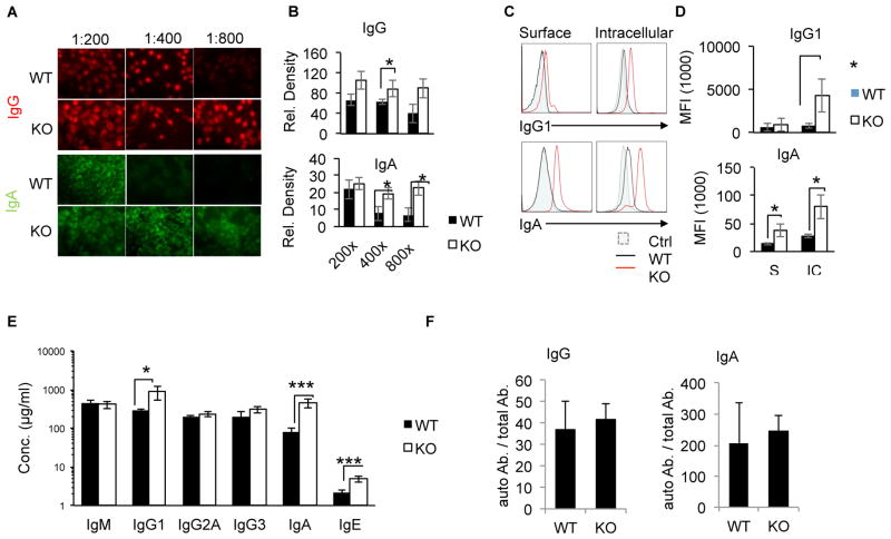
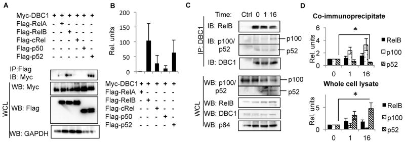
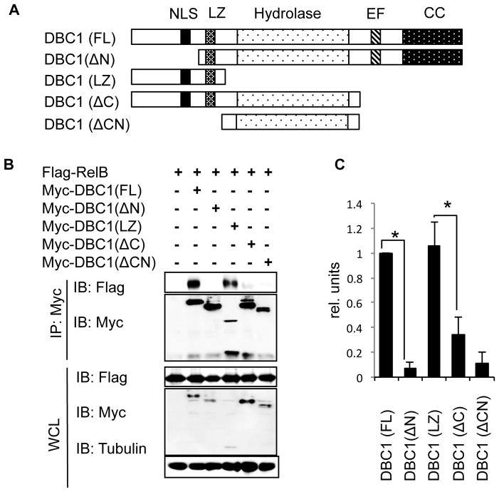
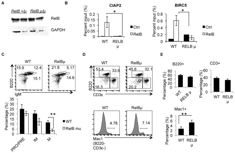
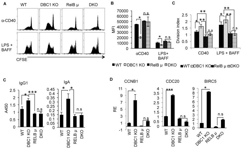
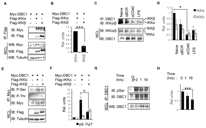
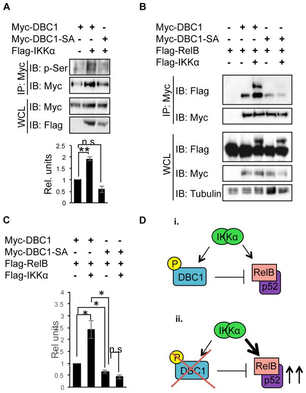
Alternative NF-κB signaling is crucial for B cell activation and Ig production, and it is mainly regulated by the inhibitor of κ B kinase (IKK) regulatory complex. Dysregulation of alternative NF-κB signaling in B cells could therefore lead to hyperactive B cells and Ig overproduction. In our previous, study we found that deleted in breast cancer 1 (DBC1) is a suppressor of the alternative NF-κB pathway to attenuate B cell activation. In this study, we report that loss of DBC1 results in spontaneous overproduction of Ig in mice after 10 mo of age. Using a double mutant genetic model, we confirm that DBC1 suppresses B cell activation through RelB inhibition. At the molecular level, we show that DBC1 interacts with alternative NF-κB members RelB and p52 through its leucine zipper domain. In addition, phosphorylation of DBC1 at its C terminus by IKKα facilitates its interaction with RelB and IKKα, indicating that DBC1-mediated suppression of alternative NF-κB is regulated by IKKα. Our results define the molecular mechanism of DBC1 inhibition of alternative NF-κB activation in suppressing B cell activation.


Conflict of interest statement

Disclosure of Conflicts of Interest. The authors declare no competing financial interests.

Figures

Figure 1
Figure 1. DBC1 KO mice spontaneously increase production of serum Ig at 10 months
(A) Autoreactive IgG (red) and IgA (green) from the sera of 10 month-old WT and DBC1 KO mice was detected by indirect Immunofluorescent (IF) staining using NIH 3T3 cell line. (B) Mean fluorescence density from (A). (C) Serum from WT and DBC1 KO mice were tested for IgG1 and IgA reactivity towards surface (top) and intracellular (bottom) autoantigens in EL4 murine T lymphoma cell line, and detected by flow cytometry. (D) Mean fluorescence intensity (MFI) of EL4- bound immunoglobulin as detected in (C). (E) Blood serum was isolated from 10 month-old WT and DBC1 KO mice, and serum immunoglobulin levels for the indicated isotypes were measured by ELISA. (F) Autoreactive IgG and IgA as measured in (A) normalized to total IgG and IgA levels (fluorescence density (A.U)/μg). N=5, error bars represent SEM. * p<0.05, ** p<0.01, ***P<0.001.
Figure 2
Figure 2. DBC1 interacts with RelB and p52
(A) Flag-tagged RelA, RelB, c-Rel, p50 and p52 were each co-transfected with myc-tagged DBC1 into HEK293T cells, cell lysates were then immunoprecipitated using anti-Flag antibody. Interaction with DBC1 was detected by immunoblotting with anti-Myc antibody. (B) Densitometry analysis of (A) based on 5 independent trials. (C) Splenic B cells from WT mice were activated with anti-CD40 for 0, 1 and 16 hours, then cell lysates were immunoprecipitated with anti-DBC1 antibody. Interaction between endogenous DBC1 and RelB and p52 were detected by immunoblotting. Normal rabbit IgG serves as negative control. (D) Densitometry analysis of (C) based on 6 independent trials. * p<0.05.
Figure 3
Figure 3. The LZ of DBC1 is necessary and sufficient for interaction with RelB
(A) Schematic of structure of full length DBC1 and generated truncated mutants. NLS: Nuclear Localization Signal, LZ: Leucine Zipper, Hydrolase: Putative hydrolase domain, EF: Inactive EF hand, CC: Coiled-coil. (B) Myc-tagged DBC1 and its truncated mutants were co-transfected with Flag-tagged RelB into HEK293T cells, and cell lysates were immunoprecipitated with anti-Myc antibody. Interaction with RelB was detected by Western Blot using anti-Flag antibody (top panel). Whole cell lysate (WCL) (bottom panel) serves as loading control. (C) Densitometry analysis of (B). RelB co-immunoprecipitated with full length DBC1 (lane 2 of (B)) was used to normalize RelB co-immunoprecipitate levels with truncated mutants. N=3, error bars represent SEM. *p<0.05.
Figure 4
Figure 4. RelB shep/shep mutation leads to loss-of-function RelB protein
(A) Western Blot analysis of RelB protein levels from 2 pairs of RelB +/shep and RelB shep/shep mice. GAPDH levels serves as loading control. (B) B cells from RelB +/shep (WT) and RelB shep/shep (RelB mu) mice were activated with anti-CD40 overnight and subjected to ChIP analysis. RelB-bound target gene promoters were quantified by qPCR. (C) Bone marrow cells were isolated from WT and RelBshep/shep mice and B220LO IgM, B220LO IgM+, and B220HI IgM+ cells analyzed by flow cytometry (top panel). Mean percentages of B cell subpopulations of WT and RelBshep/shep mice are shown (bar graph at bottom panel) (D) Splenocytes were isolated WT and RelBshep/shep mice and analyzed for B220+, CD3ε+ and B220CD3ε Mac1+ cells. (E) Mean percentages of B cells, T cells and macrophages/monocytes as analyzed in (D). For (A) and (B) N=5 from 2 independent experiments. For (C) – (E), N= 5. Error bars indicate S.E.M. *P<0.05.
Figure 5
Figure 5. Increased proliferation and Ig production in DBC1 KO B cells is abrogated by deletion of RelB
(A) Splenic B cells were isolated from WT, DBC1 KO, RelBshep/shep (RelB μ) and Dbc1−/− RelBshep/shep (DKO) mice. CFSE-stained B cells were stimulated with anti-CD40 or LPS+ BAFF for 5 days then subjected to flow cytometry for detection of proliferation. (B) Mean Fluorescence Intensity (MFI) of CFSE from (A). Reduction of MFI indicates increased proliferation. (C) Division index calculated from CFSE experiments as performed in (A). (D) ELISA measuring IgG1 and IgA levels in culture supernatants of WT, DBC1 KO, RelB μ, and DKO B cells stimulated as in (A). (E) Real Time PCR (qPCR) detection of NFκB target genes from WT, DBC1 KO, RelB μ and DKO B cells activated overnight with anti-CD40. N=5, error bars represent SEM. *p<0.05, ** p<0.01, *** p<0.001, n.s= not significant.
Figure 6
Figure 6. DBC1 interacts with IKKα and IKKβ and is Serine phosphorylated by IKKα
(A) Flag-tagged IKKα and IKKβ were each co-transfected with Myc-tagged DBC1, then cell lysates were immunoprecipitated with anti-Flag antibody. Interaction with DBC1 was detected by anti-Myc antibody. (B) Densitometry analysis of IKKα and IKKβ precipitates relative to IKKα in lane 2. (C) Primary B cells from WT mice were activated with the indicated stimuli for 1 hour. Cell lysates were then immunoprecipitated with anti-DBC1 antibody, and interaction with IKKα and β were detected by immunoblotting with antibody recognizing both IKKα and IKKβ. (D) Densitometry analysis of IKKα and IKKβ protein in the precipitates relative to IKKα levels at naïve state. (E) Myc-tagged DBC1 was co-transfected with Flag-tagged IKKα and β into HEK293T, cell lysates were then immunoprecipitated with anti-Myc, and phosphorylation of DBC1 detected using anti-phosphoSerine (α-pS) and anti-phosphoThreonine (α-pT) antibodies. (F) Densitometry analysis of pS and pT levels from (E) relative to lane 1 (overexpression of Myc-DBC1 alone). (G) Primary B cells were stimulated with α-CD40 for the indicated times, DBC1 was immunoprecipitated from cell lysates and phosphorylation of DBC1 was detected using α-pS antibody. Normal rabbit IgG serves as negative control, and whole cell lysate (WCL) as loading control. (H) Densitometry analysis of p-DBC1 as detected in (G) are normalized to lane 2 (naïve state). N=5, error bars represent SEM. *P<0.05, ** P<0.01, ***P<0.0001.
Figure 7
Figure 7. DBC1 Serine phosphorylation by IKKα is required for interaction with RelB and IKKα
(A) Myc-tagged DBC1 and Myc-tagged DBC1-SA mutant were each co-transfected with Flag-tagged IKKα. Both myc-DBC1 and myc-DBC1-SA were then immunoprecipitated using anti-Myc, and phosphorylation levels detected using α-pS antibody. Densitometry analysis of pS levels in the precipitate (bottom bar graph) is shown; pS-levels are relative to lane 1 (overexpression of WT DBC1 alone). (B) Myc-tagged DBC1 or DBC1-SA mutant was co-transfected with Flag-tagged RelB with or without flag-tagged IKKα. Cell lysates were then immunoprecipitated with anti-Myc antibody. Interaction between DBC1 and RelB or IKKα were detected by immunoblotting for Flag. (C) Densitometry analysis of precipitated RelB from (B) relative lane 2 (overexpression of WT DBC1 and RelB). (D) Schematic of proposed molecular mechanism of DBC1 suppression of B cells. i) DBC1 interacts with both IKKα and RelB in B cells and maintains the balance of active and inactive NFκB. ii) Loss of DBC1 suppression of RelB leads to imbalanced increase of alternative NFκB activity, leading to increased B cell proliferation and Ig production. N=5. Error bars represent SEM. *=P<0.05, **=P<0.01, ***=P<0.001.

Similar articles

Cited by

References

    1. Bonizzi G, Karin M. The two NF-[kappa]B activation pathways and their role in innate and adaptive immunity. Trends in Immunology. 2004;25:280–288. - PubMed
    1. Grossmann M, Metcalf D, Merryfull J, Beg A, Baltimore D, Gerondakis S. The combined absence of the transcription factors Rel and RelA leads to multiple hemopoietic cell defects. Proceedings of the National Academy of Sciences. 1999;96:11848–11853. - PMC - PubMed
    1. Hoffmann A, Leung TH, Baltimore D. Genetic analysis of NF-κB/Rel transcription factors defines functional specificities. The EMBO Journal. 2003;22:5530–5539. - PMC - PubMed
    1. Yao Z, Li Y, Yin X, Dong Y, Xing L, Boyce BF. NF-kappaB RelB negatively regulates osteoblast differentiation and bone formation. Journal of bone and mineral research: the official journal of the American Society for Bone and Mineral Research. 2014;29:866–877. - PMC - PubMed
    1. Franzoso G, Carlson L, Poljak L, Shores EW, Epstein S, Leonardi A, Grinberg A, Tran T, Scharton-Kersten T, Anver M, Love P, Brown K, Siebenlist U. Mice Deficient in Nuclear Factor (NF)-κB/p52 Present with Defects in Humoral Responses, Germinal Center Reactions, and Splenic Microarchitecture. The Journal of Experimental Medicine. 1998;187:147–159. - PMC - PubMed

Publication types