. 2015 Sep 29;112(39):12211-6.
doi: 10.1073/pnas.1512375112. Epub 2015 Sep 16.

Receptor-like cytoplasmic kinase MARIS functions downstream of CrRLK1L-dependent signaling during tip growth

Affiliations

Receptor-like cytoplasmic kinase MARIS functions downstream of CrRLK1L-dependent signaling during tip growth

Aurélien Boisson-Dernier et al. Proc Natl Acad Sci U S A. .

Abstract

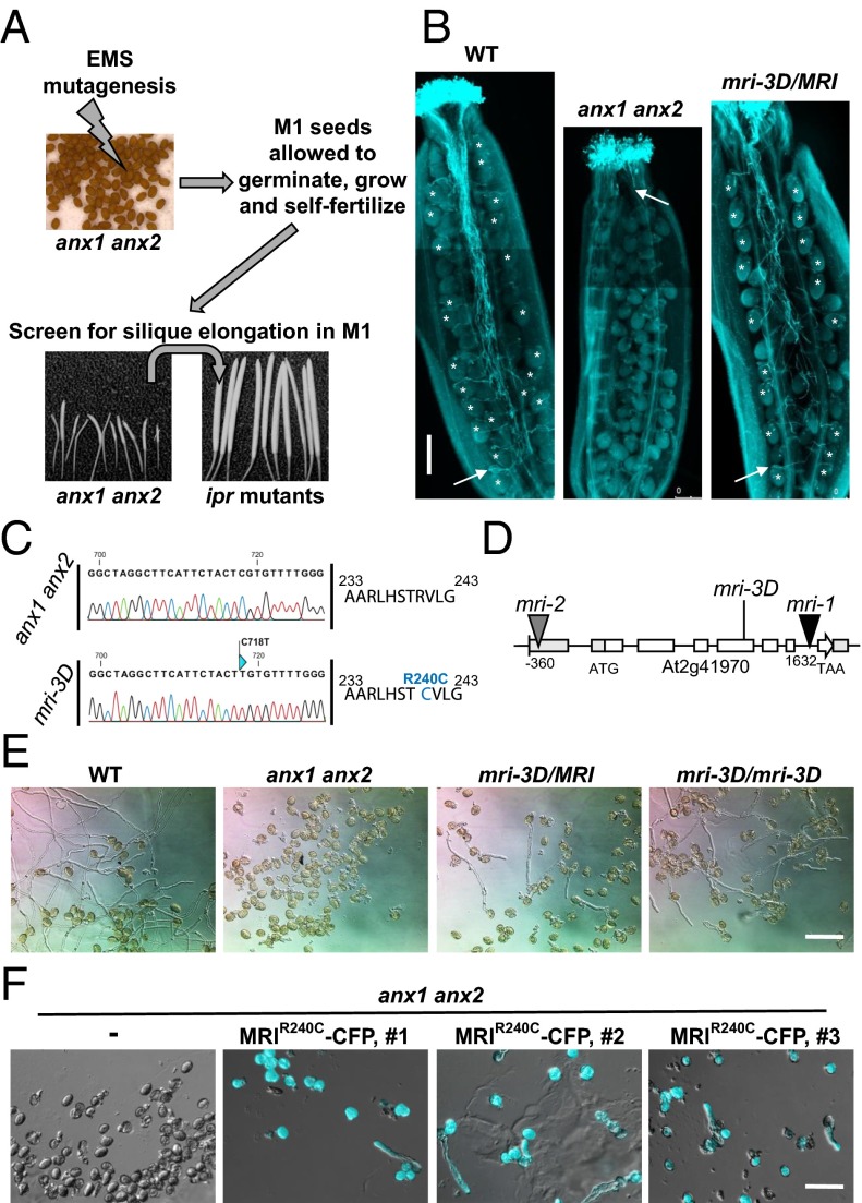
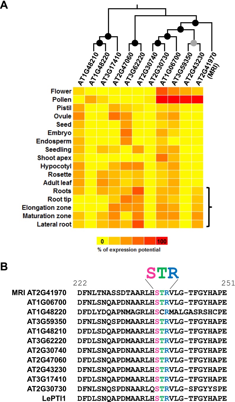
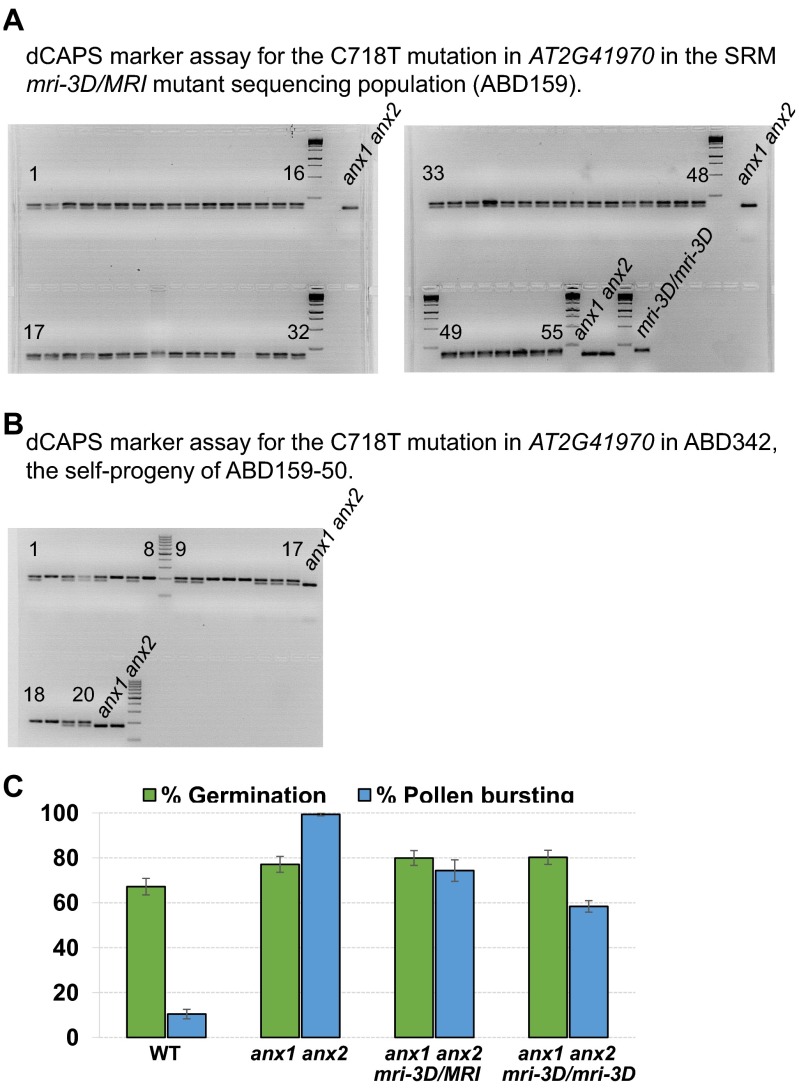
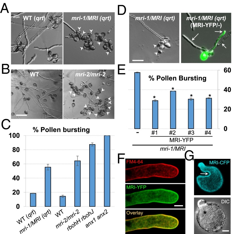
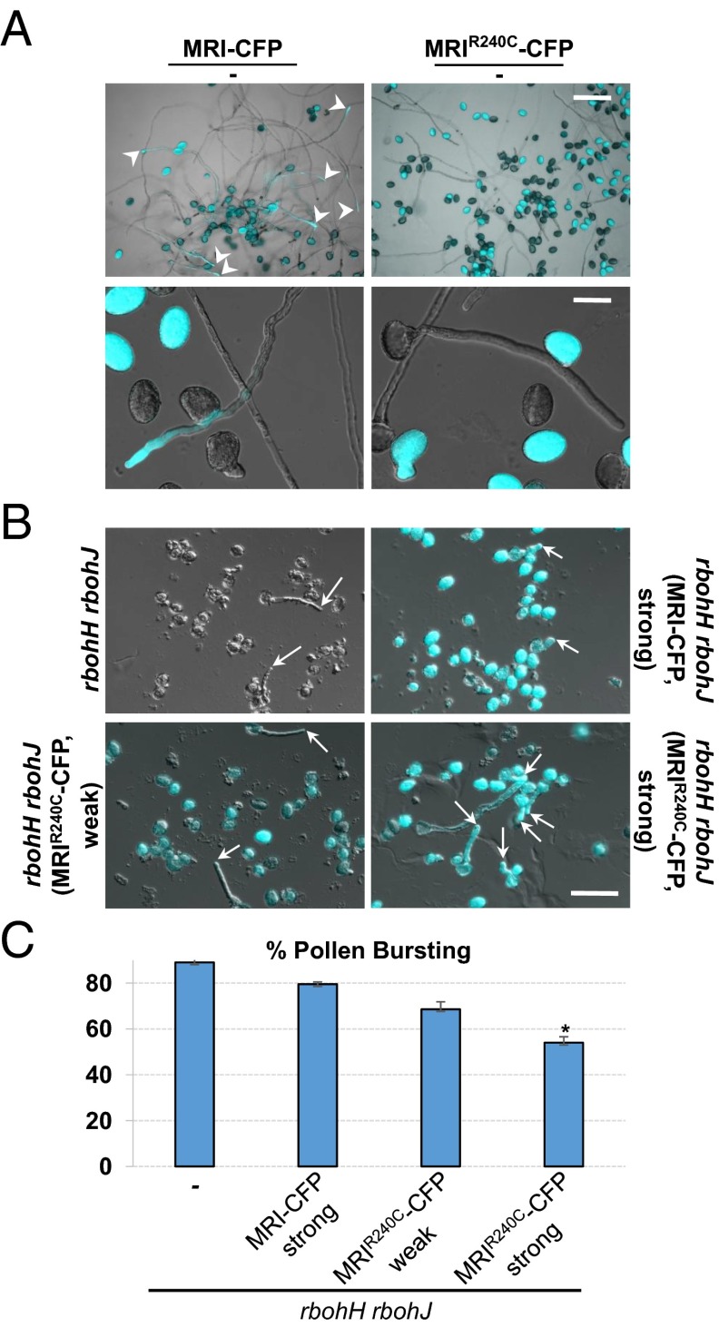
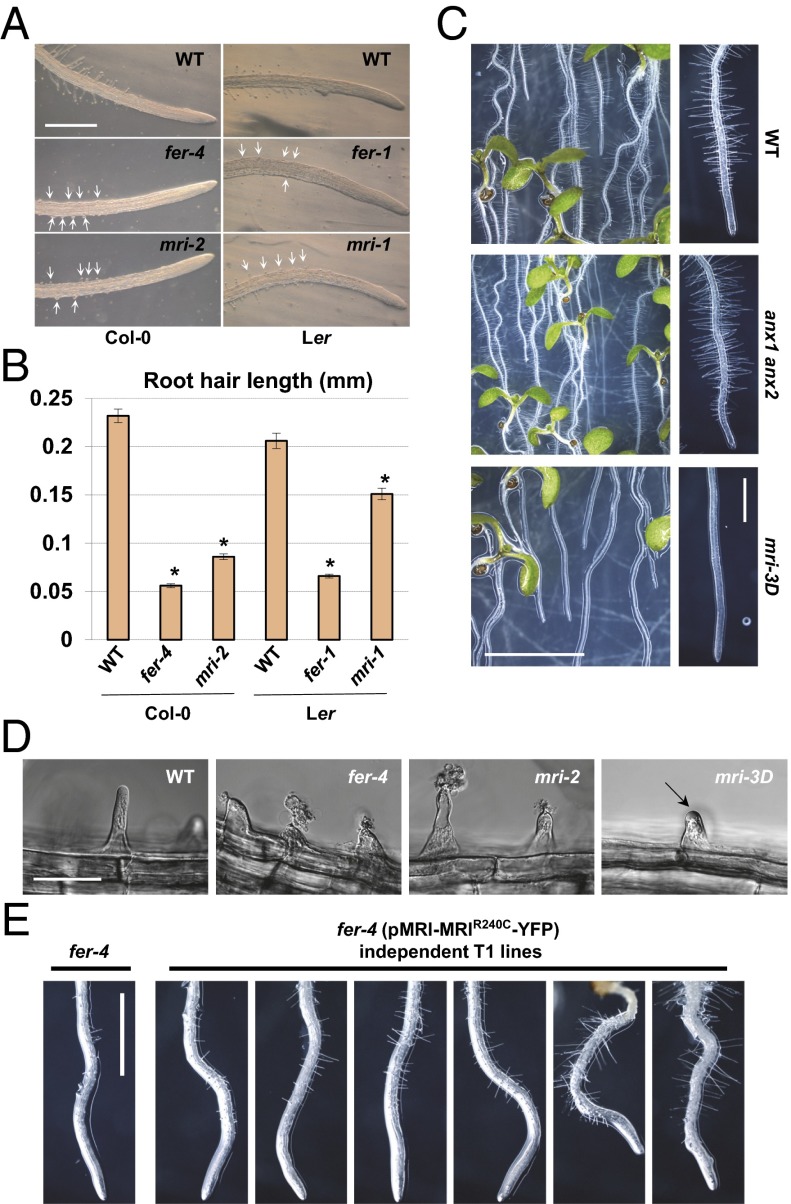
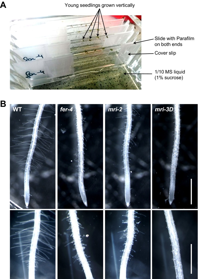
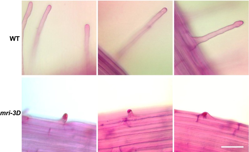
Growing plant cells need to rigorously coordinate external signals with internal processes. For instance, the maintenance of cell wall (CW) integrity requires the coordination of CW sensing with CW remodeling and biosynthesis to avoid growth arrest or integrity loss. Despite the involvement of receptor-like kinases (RLKs) of the Catharanthus roseus RLK1-like (CrRLK1L) subfamily and the reactive oxygen species-producing NADPH oxidases, it remains largely unknown how this coordination is achieved. ANXUR1 (ANX1) and ANX2, two redundant members of the CrRLK1L subfamily, are required for tip growth of the pollen tube (PT), and their closest homolog, FERONIA, controls root-hair tip growth. Previously, we showed that ANX1 overexpression mildly inhibits PT growth by oversecretion of CW material, whereas pollen tubes of anx1 anx2 double mutants burst spontaneously after germination. Here, we report the identification of suppressor mutants with improved fertility caused by the rescue of anx1 anx2 pollen tube bursting. Mapping of one these mutants revealed an R240C nonsynonymous substitution in the activation loop of a receptor-like cytoplasmic kinase (RLCK), which we named MARIS (MRI). We show that MRI is a plasma membrane-localized member of the RLCK-VIII subfamily and is preferentially expressed in both PTs and root hairs. Interestingly, mri-knockout mutants display spontaneous PT and root-hair bursting. Moreover, expression of the MRI(R240C) mutant, but not its wild-type form, partially rescues the bursting phenotypes of anx1 anx2 PTs and fer root hairs but strongly inhibits wild-type tip growth. Thus, our findings identify a novel positive component of the CrRLK1L-dependent signaling cascade that coordinates CW integrity and tip growth.

Keywords: Arabidopsis; cell wall integrity; pollen tube; receptor-like kinase signaling; root hair.


Conflict of interest statement

The authors declare no conflict of interest.

Figures

Fig. 1.
Fig. 1.
Identification of the ipr mutants, suppressors of anx1 anx2 male sterility, and characterization of maris-3D (mri-3D). (A) Scheme for the anx1 anx2 male sterility-suppressor screen. Reproduced from ref. . (B) Aniline blue staining reveals that mri-3D/MRI PTs are able to elongate in vivo and fertilize female gametophytes, unlike anx1 anx2 PTs, which burst and stop growth early in the style. White arrows indicate the tip of the longest PT. Asterisks indicate female gametophytes normally targeted by PTs. (Scale bar: 200 μm.) (C) The mri-3D mutant carries a C718T SNP in AT2G41970 that corresponds to an R240C mutation at the protein level. Shown are spectra from the Sanger sequencing reactions of the AT2G41970 cDNA amplified from anx1 anx2 (Upper) or mri-3D anx1 anx2 (Lower) flowers. (D) AT2G41970 locus with introns, exons, and positions of the mutant alleles. (E) In vitro PT growth assays of WT, anx1 anx2, heterozygous mri-3D/MRI anx1 anx2, and homozygous mri-3D/mri-3D anx1 anx2. Note how the mri-3D mutation partially rescues the anx1 anx2 pollen-bursting phenotype and allows PT growth. See Fig. S2C for corresponding quantification. (Scale bar: 100 μm.) (F) In vitro PT growth assays of untransformed anx1 anx2 and three independent anx1 anx2 transgenic lines expressing MRIR240C-CFP. DIC and CFP channels were overlaid for the transgenic lines. (Scale bar: 80 μm.)
Fig. S1.
Fig. S1.
Sequence alignment and phylogenic tree of the Arabidopsis RLCK-VIII homologs of the tomato Pto-interacting protein 1 to which MRI belongs. (A) Multiple alignments of Arabidopsis RLCK-VIII proteins were performed with ClustalW 2.0, and the phylogenetic tree was reconstructed with MEGA6 using the protein sequence parsimony method (bootstrap test, 1,000 replicates). Black and gray circles at nodes indicate bootstrap values of more than 900 and between 800 and 900, respectively. Then the tree was combined with the relative gene expression of Arabidopsis RLCK-VIII family members in various plant tissues according to the Genevestigator microarray database using the Meta-Profile Analysis tool, Anatomy Profile (45). Note the strong and preferential expression of MRI in pollen and roots (bracket). See Table S1, for more detailed gene-expression data in pollen and root hairs. (B) Multiple alignments of Arabidopsis RLCK-VIII protein sequences around the conserved STR motifs were performed with ClustalW 2.0.
Fig. S2.
Fig. S2.
(A) dCAPS marker assay for the C718T mutation in AT2G41970 in the mri-3D/MRI mutant sequencing population (ABD159). (B) dCAPS marker assay for the C718T mutation in AT2G41970 in population ABD342, the self-progeny of ABD159-50. Note that the gDNA of plant ABD159-13 was used as a control and labeled mri-3D/mri-3D in A. (C) Quantification of in vitro germination and bursting rates for WT, anx1 anx2, mri-3D/MRI anx1 anx2, and mri-3D/mri-3D anx1 anx2 plants. Data are mean ± SEM of three independent experiments with more than 150 pollen grains per genotype and experiment.
Fig. 2.
Fig. 2.
MARIS is a positive component of the CW-integrity signaling pathway. (A) In vitro PT growth assays of WT and mri-1/MRI in the qrt background. Arrowheads point to rupturing PTs discharging cytoplasm into the medium. (Scale bar: 25 μm.) (B) In vitro PT growth assays of WT and mri-2/mri-2; arrowheads point to cytoplasmic discharge. (Scale bar: 100 μm.) (C) Quantification of PT rupture in vitro for WT (qrt), mri-1/MRI (qrt), WT, mri-2, rbohH rbohJ, and anx1 anx2. (D) Pollen tetrad of untransformed mri-1/MRI (qrt) and mri-1/MRI (qrt) hemizygous for pLAT52-MRI-YFP. Arrowheads and arrows point to cytoplasmic discharge and PT, respectively. DIC and YFP channels were overlaid for the transgenic line. (Scale bar: 50 μm.) (E) Four independent T1 lines of mri-1/MRI (qrt) hemizygous for pLAT52-MRI-YFP display significantly less bursting than untransformed mri-1/MRI (qrt) (-). Asterisks indicate significant differences from the untransformed control (P < 0.001, Student's t test). (F) Median plane of a normally growing PT expressing MRI-YFP. Before imaging, PTs were treated with liquid germination medium containing FM4-64 (2 µM) for 5 min. (Scale bar: 5 µm.) (G) An early arrested PT overexpressing MRI-CFP showing membrane invagination (arrow) and overaccumulation of CW material (asterisk). (Scale bar: 5 µm.)
Fig. 3.
Fig. 3.
MRIR240C-CFP strongly inhibits WT pollen germination and partially rescues the rbohH rbohJ bursting phenotype. (A) In vitro growth assays with WT pollen hemizygous for either MRI-CFP (Left) or MRIR240C-CFP (Right). DIC and CFP channels were overlaid. (Upper) An overview of the growing PTs. Note that, unlike MRIR240C-CFP–expressing PTs (Right), MRI-CFP expressing PTs (Left) are observed frequently (arrowheads). (Lower) Close-up pictures of growing pollen expressing MRI-CFP (Left) and very rarely germinating pollen expressing MRIR240C-CFP (Right). (Scale bars: 100 μm, Upper; 30 μm, Lower.) (B) In vitro growth assays for untransformed rbohH rbohJ pollen (Upper Left) and transgenic rbohH rbohJ pollen strongly expressing MRI-CFP (Upper Right) or weakly (Lower Left) or strongly (Lower Right) expressing MRIR240C-CFP. DIC and CFP channels were overlaid. Strongly expressed MRIR240C-CFP significantly rescued the rbohH rbohJ bursting phenotype in vitro. (Scale bar: 30 μm.) (C) Quantification of the bursting phenotype relative to B. The asterisk denotes a significant difference compared to untransformed rbohH rbohJ PTs (−) (P < 0.001, two-tailed unpaired Student's t test).
Fig. 4.
Fig. 4.
As in FERONIA, MARIS is required to sustain root-hair growth. (A) Main roots with root hairs of 5-d-old mri-2, fer-4, and WT (Col-0 accession) seedlings (Left) and mri-1, fer-1, and WT (Ler accession) seedlings (Right) grown in agar. Arrows point to short, defective root hairs. (Scale bar: 1 mm.) (B) Quantification of root-hair lengths relative to A. Asterisks denote significant differences versus the appropriate control (P < 0.001, two-tailed unpaired Student's t test). (C, Left) WT, anx1 anx2, and mri-3D anx1 anx2 seedlings growing on the surface of a microagar plate. (Right) Pictures of primary roots and their root hairs. (Scale bars: 5 mm, Left; 1 mm, Right.) (D) Young root hairs in the root elongation zone of WT, mri-2, and mri-3D anx1 anx2 seedlings grown in liquid medium. Note the loss of CW integrity or CW overaccumulation (arrow) for mri-2 and mri-3D root hairs, respectively. (Scale bar: 20 μm.) See also Figs. S3 and S4 and Movie S1. (E) Main root of untransformed fer-4 seedling and fer-4 T1s expressing pMRI-MRIR240C-YFP. (Scale bar: 1 mm.)
Fig. S3.
Fig. S3.
(A) Experimental set-up for root-hair growth assays in liquid medium. Four-day-old seedlings grown vertically on half-strength MS microagar plates were sandwiched between a slide and a coverslip containing 1/10th strength MS liquid medium (1% sucrose). Young root hairs in the elongation zone were observed 48 h later. (B) Representative roots of WT, fer-4, mri-2, and mri-3D anx1 anx2 seedlings grown in liquid medium relative to Fig. 4D. (Scale bar: 2 mm.)
Fig. S4.
Fig. S4.
Ruthenium red staining reveals overaccumulation of cell wall material in mri-3D short root hairs. Roots of 4-d-old WT (Upper) and mri-3D anx1 anx2 (Lower) seedlings were allowed to grow for 48 h in liquid medium before being treated for 1 min with 0.01% Ruthenium red that stains for acidic pectin. Note that staining is restricted to the tip surface of WT root hairs but accumulates within the short root hairs of mri-3D anx1 anx2 plants. (Scale bar: 100 µm.)

References

    1. Engelsdorf T, Hamann T. An update on receptor-like kinase involvement in the maintenance of plant cell wall integrity. Ann Bot (Lond) 2014;114(6):1339–1347. - PMC - PubMed
    1. Wolf S, Hématy K, Höfte H. Growth control and cell wall signaling in plants. Annu Rev Plant Biol. 2012;63:381–407. - PubMed
    1. Boisson-Dernier A, et al. Disruption of the pollen-expressed FERONIA homologs ANXUR1 and ANXUR2 triggers pollen tube discharge. Development. 2009;136(19):3279–3288. - PMC - PubMed
    1. Miyazaki S, et al. ANXUR1 and 2, sister genes to FERONIA/SIRENE, are male factors for coordinated fertilization. Curr Biol. 2009;19(15):1327–1331. - PubMed
    1. Lindner H, et al. TURAN and EVAN mediate pollen tube reception in Arabidopsis synergids through protein glycosylation. PLoS Biol. 2015;13(4):e1002139. - PMC - PubMed

Publication types

MeSH terms

Substances