. 2015 Nov;114(5):2854-66.
doi: 10.1152/jn.00823.2015. Epub 2015 Sep 16.

Layer-specific modulation of entorhinal cortical excitability by presubiculum in a rat model of temporal lobe epilepsy

Affiliations

Layer-specific modulation of entorhinal cortical excitability by presubiculum in a rat model of temporal lobe epilepsy

Saad Abbasi et al. J Neurophysiol. 2015 Nov.

Abstract

Temporal lobe epilepsy (TLE) is the most common form of epilepsy in adults and is often refractory to antiepileptic medications. The medial entorhinal area (MEA) is affected in TLE but mechanisms underlying hyperexcitability of MEA neurons require further elucidation. Previous studies suggest that inputs from the presubiculum (PrS) contribute to MEA pathophysiology. We assessed electrophysiologically how PrS influences MEA excitability using the rat pilocarpine model of TLE. PrS-MEA connectivity was confirmed by electrically stimulating PrS afferents while recording from neurons within superficial layers of MEA. Assessment of alterations in PrS-mediated synaptic drive to MEA neurons was made following focal application of either glutamate or NBQX to the PrS in control and epileptic animals. Here, we report that monosynaptic inputs to MEA from PrS neurons are conserved in epileptic rats, and that PrS modulation of MEA excitability is layer-specific. PrS contributes more to synaptic inhibition of LII stellate cells than excitation. Under epileptic conditions, stellate cell inhibition is significantly reduced while excitatory synaptic drive is maintained at levels similar to control. PrS contributes to both synaptic excitation and inhibition of LIII pyramidal cells in control animals. Under epileptic conditions, overall excitatory synaptic drive to these neurons is enhanced while inhibitory synaptic drive is maintained at control levels. Additionally, neither glutamate nor NBQX applied focally to PrS now affected EPSC and IPSC frequency of LIII pyramidal neurons. These layer-specific changes in PrS-MEA interactions are unexpected and of significance in unraveling pathophysiological mechanisms underlying TLE.

Keywords: electrophysiology; glutamate application; hyperexcitability; presubiculum; temporal lobe epilepsy.


Figures

Fig. 1.
Fig. 1.
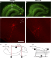
Acute brain slices from control and epileptic rats. A and B: NeuN-labeled sections showing characteristic loss of LIII neurons in medial entorhinal area (MEA) (*) in epileptic (B) but not in control (A) tissue. Arrowheads indicate boundaries of MEA (white) and presubiculum (PrS) (red) along the pial surface. General location of stimulating electrode placement in PrS is indicated by the bulls-eye in A. C and D: gross morphology of a biocytin-labeled pyramidal cell in LIII (C) and a stellate cell in LII (D) used in electrophysiological recordings (L, lateral; M, medial; ab, angular bundle; l.d. lamina dissecans). The cells shown in C and D are both from epileptic tissue. E: preparation of semihorizontal entorhinal cortical slices retaining PrS connectivity. Initial cuts (1–3, solid red lines, left panel) are made with a razor blade. The cut surface of the brain (3, dorsal) is then glued to the vibratome platform as indicated. Horizontal arrows (right panel) indicate cutting direction. Roman numerals (I–III) indicate lamina. Scale bars: 1 mm (A, B); 200 μm (C, D).
Fig. 2.
Fig. 2.
LII stellate cells, but not LIII pyramidal neurons, in MEA have enhanced paired-pulse facilitation under epileptic conditions. A1: superimposed traces of EPSCs evoked in LII stellate cells in response to paired-pulse stimulation of PrS at the indicated interstimulus intervals from control and epileptic rats. Each trace shown in this figure is an average of ≥5 consecutive sweeps. A2: plot of the paired-pulse ratio as a function of interstimulus interval for LII stellate cells from control and epileptic rats. B1: trains of EPSCs evoked in LII stellate cells by repeated stimulation (5 pulses) of PrS at the indicated frequencies in control and epileptic rats. B2: plot of the normalized EPSC amplitude as a function of stimulus number. C: a typical epileptiform discharge recorded in LII stellate cell from an epileptic rat following PrS stimulation (arrows). D–F: data from LIII pyramidal neurons corresponding to experimental manipulations described above for LII stellate cells (A–C). Scale bars in A1 and D1 are applicable to B1 and E1, respectively. *P < 0.05, **P < 0.01, ***P < 0.001, t-test.
Fig. 3.
Fig. 3.
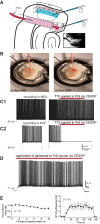
Concomitant ejection and suction of perfusate (CESOP), a technique for focal application of drugs in submerged brain slices. A: schematic of the CESOP system used in this study. The red circle represents the perfusate applied via CESOP while recording from superficial layers of MEA (1) or locally within PrS (2). Inset: an IR-DIC image (magnification: × 10) of the CESOP electrode showing piggy-backed inflow and outflow lines. B: CESOP in action. Note diffusion of dye-laden aCSF (for visualization and calibration) when the outflow line of CESOP is turned off during focal application of perfusate to the PrS in a brain slice submerged in aCSF in a recording chamber (electrodes: S, stimulating; C, CESOP; G, ground; R, recording). C1 and C2: differential effects of focal application of TTX (1 μM) in PrS on action potential discharge of a LIII pyramidal neuron in MEA (C1) and a RS cell in LIII of PrS (C2), triggered by depolarizing current injections. D: sustained action potential discharge triggered in a RS cell in LIII of PrS in response to local application of glutamate (100 μM; 3 min duration) via CESOP. Resting membrane potentials in recorded neurons are indicated juxtaposed to the respective traces. E: there is no significant change in the averaged normalized inflow rate as pressure head decreases in the range indicated (left panel) and focal application of glutamate via CESOP reliably evokes action potential firing in PrS neurons (right panel).
Fig. 4.
Fig. 4.
Assessments of synaptic drive in LII stellate cells. A–D: excitatory synaptic drive is comparable between control and epileptic rats and not greatly influenced by PrS input. E–H: synaptic inhibition is significantly reduced in epileptic rats as is inhibitory synaptic drive from PrS. A and B: voltage-clamp recordings (20 s) of spontaneous excitatory postsynaptic currents (sEPSCs, inward events recorded at −70 mV holding potential) recorded in a LII stellate cell from control (A) and epileptic (B) rats under the indicated conditions (aCSF, top; 100 μM glutamate, middle; 10 μM NBQX, bottom). Insets: averaged composite responses of all events recorded for the representative traces (in aCSF) under the indicated conditions. C: plots of averaged frequency vs. experimental condition for sEPSCs in 1-min-long recordings from LII stellate cells in control and epileptic rats under the indicated conditions. D: plots of averaged sEPSC amplitudes corresponding to data presented in C. E and F: voltage-clamp recordings (20 s) of spontaneous inhibitory postsynaptic currents (sIPSCs, outward events recorded at 0 mV holding potential) recorded in a LII stellate cell from control (E) and epileptic (F) rats under the indicated conditions (aCSF, top; 100 μM glutamate, middle; 10 μM NBQX, bottom). Insets: averaged composite responses of all events recorded for the representative traces (in aCSF) under the indicated conditions. G: plots of averaged frequency vs. experimental condition for sIPSCs in 1-min-long recordings from LII stellate cells in control and epileptic rats under the indicated conditions. H: plots of averaged sIPSC amplitudes corresponding to data presented in G. Error bars, where these are bigger than the size of the symbols used, represent SE. †P < 0.05, paired t-test; *P < 0.05, **P < 0.01, unpaired t-test.
Fig. 5.
Fig. 5.
Assessments of synaptic drive in LIII pyramidal cells. A–D: excitatory synaptic drive is significantly enhanced in epileptic rats despite PrS influence being masked. E–H: synaptic inhibition is not compromised in epileptic rats. A and B: voltage-clamp recordings (20 s) of spontaneous excitatory postsynaptic currents (sEPSCs, inward events recorded at −70 mV holding potential) recorded in a LIII pyramidal cell from control (A) and epileptic (B) rats under the indicated conditions (aCSF, top; 100 μM glutamate, middle; 10 μM NBQX, bottom). Insets: averaged composite responses of all events recorded for the representative traces (in aCSF) under the indicated conditions. C: plots of averaged frequency vs. experimental condition for sEPSCs in 1-min-long recordings from LIII pyramidal cell in control and epileptic rats under the indicated conditions. D: plots of averaged sEPSC amplitudes corresponding to data presented in C. E and F: voltage-clamp recordings (20 s) of spontaneous inhibitory postsynaptic currents (sIPSCs, outward events recorded at 0 mV holding potential) recorded in a LIII pyramidal cell from control (E) and epileptic (F) rats under the indicated conditions (aCSF, top; 100 μM glutamate, middle; 10 μM NBQX, bottom). Insets: averaged composite responses of all events recorded for the representative traces (in aCSF) under the indicated conditions. G: plots of averaged frequency vs. experimental condition for sIPSCs in 1-min-long recordings from LIII pyramidal cell in control and epileptic rats under the indicated conditions. H: plots of averaged sIPSC amplitudes corresponding to data presented in G. Error bars, where these are bigger than the size of the symbols used, represent SE. †P < 0.05, paired t-test; *P < 0.05, **P < 0.01, ***P < 0.001, unpaired t-test.
Fig. 6.
Fig. 6.
Schematic of functional circuitry underlying possible changes in excitatory and inhibitory synaptic drive to LII stellate cells and LIII pyramidal neurons in the MEA under control and epileptic conditions deduced from this study. The PrS provides both glutamatergic and GABAergic projections to superficial layers of the MEA, and RS cells, the predominant cell type in the PrS, have been shown to become hyperexcitable in TLE.

Similar articles

Cited by

References

    1. Abbasi S, Kumar SS. Electrophysiological and morphological characterization of cells in superficial layers of rat presubiculum. J Comp Neurol 521: 3116–3132, 2013. - PubMed
    1. Abbasi S, Kumar SS. Regular-spiking cells in the presubiculum are hyperexcitable in a rat model of temporal lobe epilepsy. J Neurophysiol 112: 2888–2900, 2014. - PMC - PubMed
    1. Bartesaghi R, Di Maio V, Gessi T. Topographic activation of the medial entorhinal cortex by presubicular commissural projections. J Comp Neurol 487: 283–299, 2005. - PubMed
    1. Bartolomei F, Khalil M, Wendling F, Sontheimer A, Regis J, Ranjeva JP, Guye M, Chauvel P. Entorhinal cortex involvement in human mesial temporal lobe epilepsy: an electrophysiologic and volumetric study. Epilepsia 46: 677–687, 2005. - PubMed
    1. Bear J, Fountain NB, Lothman EW. Responses of the superficial entorhinal cortex in vitro in slices from naive and chronically epileptic rats. J Neurophysiol 76: 2928–2940, 1996. - PubMed

Publication types