. 2015 Dec 4;290(49):29531-41.
doi: 10.1074/jbc.M115.680660. Epub 2015 Sep 16.

Structural Basis of Neuronal Nitric-oxide Synthase Interaction with Dystrophin Repeats 16 and 17

Affiliations

Structural Basis of Neuronal Nitric-oxide Synthase Interaction with Dystrophin Repeats 16 and 17

Anne-Elisabeth Molza et al. J Biol Chem. .

Abstract

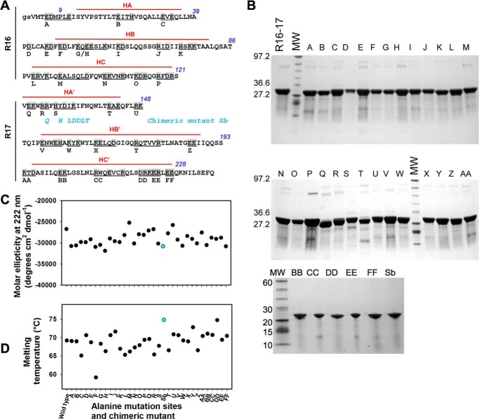
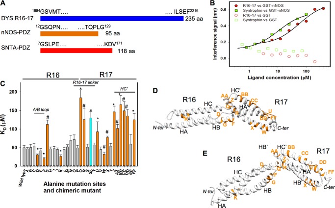
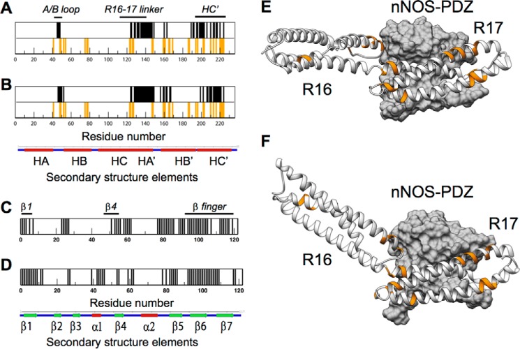
Duchenne muscular dystrophy is a lethal genetic defect that is associated with the absence of dystrophin protein. Lack of dystrophin protein completely abolishes muscular nitric-oxide synthase (NOS) function as a regulator of blood flow during muscle contraction. In normal muscles, nNOS function is ensured by its localization at the sarcolemma through an interaction of its PDZ domain with dystrophin spectrin-like repeats R16 and R17. Early studies suggested that repeat R17 is the primary site of interaction but ignored the involved nNOS residues, and the R17 binding site has not been described at an atomic level. In this study, we characterized the specific amino acids involved in the binding site of nNOS-PDZ with dystrophin R16-17 using combined experimental biochemical and structural in silico approaches. First, 32 alanine-scanning mutagenesis variants of dystrophin R16-17 indicated the regions where mutagenesis modified the affinity of the dystrophin interaction with the nNOS-PDZ. Second, using small angle x-ray scattering-based models of dystrophin R16-17 and molecular docking methods, we generated atomic models of the dystrophin R16-17·nNOS-PDZ complex that correlated well with the alanine scanning identified regions of dystrophin. The structural regions constituting the dystrophin interaction surface involve the A/B loop and the N-terminal end of helix B of repeat R16 and the N-terminal end of helix A' and a small fraction of helix B' and a large part of the helix C' of repeat R17. The interaction surface of nNOS-PDZ involves its main β-sheet and its specific C-terminal β-finger.

Keywords: dystrophin; molecular dynamics; muscular dystrophy; nitric-oxide synthase; site-directed mutagenesis.


Figures

FIGURE 1.
FIGURE 1.
Biochemical characterization of the dystrophin R16–17 fragment and mutants. A, for alanine-scanning mutagenesis, all charged residues of dystrophin R16–17 were changed to alanine in groups of 1 to 5 amino acids (common value of 2–3 amino acids). This grouping encompassed 74 residues (shown in gray), resulting in 32 variants, A through FF. The capital letters are the R16 and R17 sequence according to Koenig and Kunkel (38); the gs italicized letters are residues present due to cloning constraints. The three helices of each repeat are indicated in red lines and noted as HA, HB, and HC for repeat 16 and HA′, HB′, and HC′ for repeat 17. The residues not marked by red lines are those involved in the loops between successive helices. There is no interruption of the HC and HA′ of repeats 16 and 17, respectively, with these two helices forming a common helix with the residues in the junction involved in the so-called linker. The chimeric mutant Sb not corresponding to alanine-scanning mutagenesis is shown: the blue residues are those substituted from the utrophine sequence. B, the native fragment of dystrophin R16–17, the 32 alanine mutants, and the chimeric Sb mutant were produced and purified. The fragments appeared at the expected molecular masses of ∼22,700 Da and at a high degree of purity. MW, molecular weight standards. C, molar ellipticity values at 222 nm as measured from CD spectra for the wild-type dystrophin R16–17 and all the mutants. The chimeric mutant value appears in blue. D, melting temperatures of wild-type dystrophin R16–17 and all the mutants as obtained by heating from 15 to 85 °C and followed by CD at 222 nm. The chimeric mutant value appears in blue.
FIGURE 2.
FIGURE 2.
Consequences of the alanine mutation scan of dystrophin R16–17 on the dissociation constant with nNOS-PDZ. A, schematic representation of the three constructs used for the affinity measurements: dystrophin repeats R16 and R17 (DYS R16–17), nNOS-PDZ domain, and the syntrophin PDZ domain (SNTA-PDZ). The residues at the N- and C-terminal ends are shown together with the residue numbering in the whole protein. The number of amino acids of each fragment is indicated at the extreme right. B, binding curves for nNOS-PDZ-dystrophin R16–17 and nNOS-PDZ-SNTA-PDZ showed a KD of 50 ± 6 and 7.2 ± 1.2 μm, respectively. Controls with GST are also shown. C, dissociation constants (KD values) of the binding of nNOS-PDZ to dystrophin R16–17 and all the mutants. Values for the wild-type are the mean ± S.D. of 12 similar assays. The values are the mean ± S.D. for 3 to 6 similar assays of the mutants. The values that are significantly different from those for the wild-type are shown in orange. *, for p < 0.005; #, for p < 0.05. The value of the chimeric mutant is shown in blue. D and E, the mutated sites with significantly different values are shown in orange on the two three-dimensional dystrophin R16–17 models 1 and 2 represented as a gray schematic. The three helices of each repeat are noted as in Fig. 1A. The models are shown with the N-terminal end (N-ter) on the left and the C-terminal end (C-ter) on the right.
FIGURE 3.
FIGURE 3.
Reciprocal contacts of the dystrophin R16–17 and the nNOS-PDZ domains on each other obtained by rigid docking. Contact frequencies are obtained from the 20 best poses of rigid docking for the two SAXS-based models of dystrophin R16–17 and the homology model of the nNOS-PDZ domain. A and B, interaction sites with a high contact frequency (>60%) projected onto dystrophin R16–17 model 1 (A) and model 2 (B) are shown in purple. The contact frequency and profile of hydrophobic and electrostatic potentials are shown along the primary sequence of dystrophin R16–17 as a color gradient of green to yellow or red to blue, respectively. The secondary structure elements of dystrophin R16–17 are indicated at the bottom of A and B for clarity as follows: α-helices, red; loops, blue. C and D, surface potentials of the two dystrophin R16–17 models as obtained under SAXS volume restraints: platinum hydrophobicity (molecular hydrophobicity potential) is colored from green hydrophilic to yellow hydrophobic (C) and APBS electrostatics with red negative and blue positive for an isosurface of ±50 KT/e (K is Boltzmann constant) (D) with the same orientations as described in the legend to Fig. 2. E and F, surface potentials of the human nNOS-PDZ homology model are shown with similar representations as in C and D. G-I, contact frequency plots between the nNOS-PDZ domain and the two models 1 (G) and 2 (H) of dystrophin R16–17 are shown along the primary sequence of nNOS-PDZ. The hydrophobic and electrostatic potential profiles (I) of the nNOS-PDZ domain are shown. The secondary structural elements of the nNOS-PDZ domain are indicated at the bottom of (I) for clarity as follows: α-helices, red; loops, blue; and β-sheets, green.
FIGURE 4.
FIGURE 4.
Final dystrophin R16–17·nNOS-PDZ complexes obtained after interactive flexible docking. A and B, contact mapping in the final complexes formed by the two SAXS-based models of dystrophin R16–17 with the homology model of the nNOS-PDZ domain are shown along dystrophin sequence. The structural elements of dystrophin R16–17 are shown below the diagram. The sites of alanine-scanning mutagenesis with significant differences in affinity are shown as orange bars below the contact maps of dystrophin R16–17. C and D, contact mapping in the final complexes formed by the two SAXS-based models of dystrophin R16–17 with the homology model of the nNOS-PDZ domain are shown along nNOS-PDZ sequence. E and F, flexible docking led to two three-dimensional nNOS-PDZ·dystrophin R16–17 structural complexes. The two dystrophin R16–17 models 1 (E) and 2 (F) are shown in the gray schematic and the nNOS-PDZ domain as a gray volume. The dystrophin residues inducing significantly different KD for nNOS-PDZ binding are shown in orange as described in the legend to Fig. 2.
FIGURE 5.
FIGURE 5.
In silico dystrophin R16–17 alanine-scanning mutagenesis and the effects of mutations on the interaction with the nNOS-PDZ domain. A, average binding energy changes (ΔΔG, in kcal mol−1) as calculated by FoldX over the two models of R16–17·nNOS-PDZ complexes obtained through interactive flexible docking. Absolute values are provided to dampen the ΔΔG modulations due to conformational changes observed between the two dystrophin R16–17 models. B and C, examples of R16–17 mutation sites producing either an increase or a decrease in the binding affinity for nNOS-PDZ. B, the F site induces an increase in the affinity of binding from KD of 50 ± 6 for the wild-type to KD of 20 ± 2 μm; C, the G site induces a decrease in the binding affinity to KD of 125 ± 30 μm.
FIGURE 6.
FIGURE 6.
Comparison of the native dystrophin R16–17/nNOS binding site with modifications induced by in-frame exon deletions as observed in BMD patients. Five in-frame exon deletions as observed in BMD patients are shown: Δ45–48, Δ45–51, Δ45–53, Δ45–55, and Δ45–57. These deletions permit the reconstitution of a native-like repeat structure by concatenating the N-terminal part of R17 encoded by exon 44 (in black) and the C-terminal part of repeats R19, R20, R21, R22, and R23, denoted R17//19, R17//20, R17//21, R17//22 and R17//23 (in green), respectively. The residues encoded by exon 45 in the native R17 repeat are shown in blue. The blue frame highlights the nNOS-binding site region in wild-type R17, which is replaced after deletions by the corresponding sequences of the concatenated repeats (green frame). Sequence similarities (* for identity and ‡ for similarity) were compared for all residues except those involved in the heptad pattern (a and d positions highlighted in gray), which correspond to core residues not contributing to molecular surface properties.

References

    1. Koenig M., Beggs A. H., Moyer M., Scherpf S., Heindrich K., Bettecken T., Meng G., Müller C. R., Lindlöf M., and Kaariainen H. (1989) The molecular basis for Duchenne versus Becker muscular dystrophy: correlation of severity with type of deletion. Am. J. Hum. Genet. 45, 498–506 - PMC - PubMed
    1. Flanigan K. M., Dunn D. M., von Niederhausern A., Soltanzadeh P., Gappmaier E., Howard M. T., Sampson J. B., Mendell J. R., Wall C., King W. M., Pestronk A., Florence J. M., Connolly A. M., Mathews K. D., Stephan C. M., Laubenthal K. S., Wong B. L., Morehart P. J., Meyer A., Finkel R. S., Bonnemann C. G., Medne L., Day J. W., Dalton J. C., Margolis M. K., Hinton V. J., United Dystrophinopathy Project Consortium, and Weiss R. B. (2009) Mutational spectrum of DMD mutations in dystrophinopathy patients: application of modern diagnostic techniques to a large cohort. Hum. Mutat. 30, 1657–1666 - PMC - PubMed
    1. Tuffery-Giraud S., Béroud C., Leturcq F., Yaou R. B., Hamroun D., Michel-Calemard L., Moizard M. P., Bernard R., Cossée M., Boisseau P., Blayau M., Creveaux I., Guiochon-Mantel A., de Martinville B., Philippe C., Monnier N., Bieth E., Khau Van Kien P., Desmet F. O., Humbertclaude V., Kaplan J. C., Chelly J., and Claustres M. (2009) Genotype-phenotype analysis in 2,405 patients with a dystrophinopathy using the UMD-DMD database: a model of nationwide knowledgebase. Hum. Mutat. 30, 934–945 - PubMed
    1. Fairclough R. J., Wood M. J., and Davies K. E. (2013) Therapy for Duchenne muscular dystrophy: renewed optimism from genetic approaches. Nat. Rev. Genet. 14, 373–378 - PubMed
    1. Wein N., Alfano L., and Flanigan K. M. (2015) Genetics and emerging treatments for Duchenne and Becker muscular dystrophy. Pediatr. Clin. N. Am. 62, 723–742 - PubMed

Publication types

LinkOut - more resources