. 2015 Nov 5;26(22):4109-23.
doi: 10.1091/mbc.E14-06-1114. Epub 2015 Sep 16.

Orchestration of ErbB3 signaling through heterointeractions and homointeractions

Affiliations

Orchestration of ErbB3 signaling through heterointeractions and homointeractions

Meghan McCabe Pryor et al. Mol Biol Cell. .

Erratum in

  • Correction.
    [No authors listed] [No authors listed] Mol Biol Cell. 2016 Feb 15;27(4):731. Mol Biol Cell. 2016. PMID: 26869179 Free PMC article. No abstract available.

Abstract

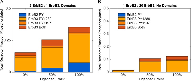
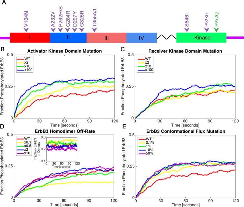
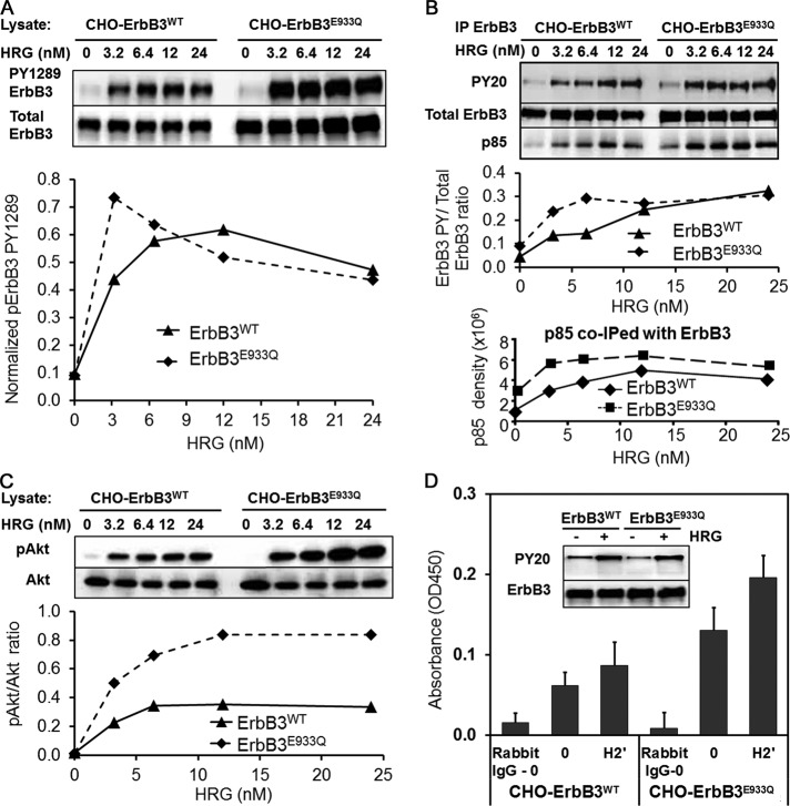
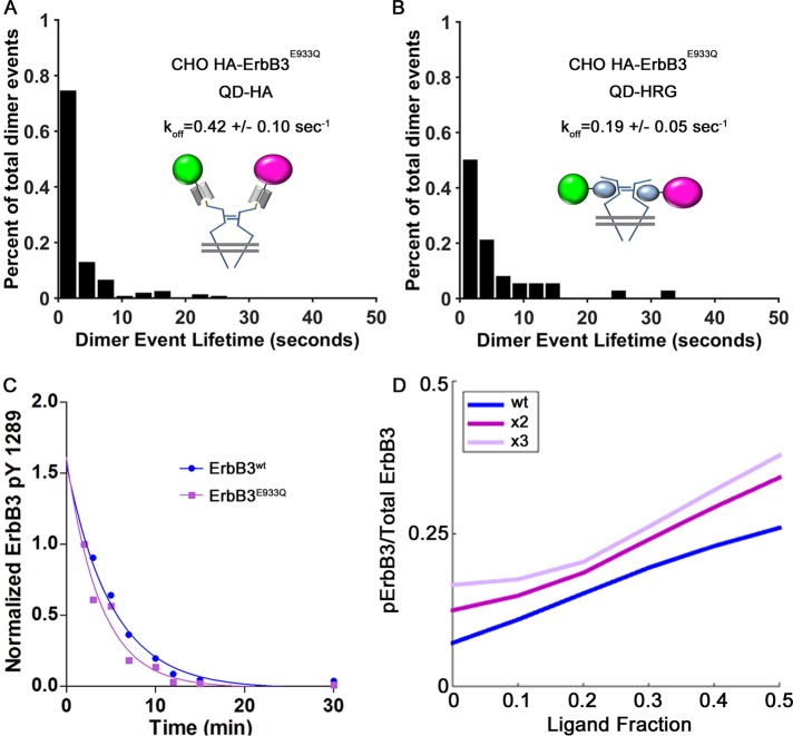
Members of the ErbB family of receptor tyrosine kinases are capable of both homointeractions and heterointeractions. Because each receptor has a unique set of binding sites for downstream signaling partners and differential catalytic activity, subtle shifts in their combinatorial interplay may have a large effect on signaling outcomes. The overexpression and mutation of ErbB family members are common in numerous human cancers and shift the balance of activation within the signaling network. Here we report the development of a spatial stochastic model that addresses the dynamics of ErbB3 homodimerization and heterodimerization with ErbB2. The model is based on experimental measures for diffusion, dimer off-rates, kinase activity, and dephosphorylation. We also report computational analysis of ErbB3 mutations, generating the prediction that activating mutations in the intracellular and extracellular domains may be subdivided into classes with distinct underlying mechanisms. We show experimental evidence for an ErbB3 gain-of-function point mutation located in the C-lobe asymmetric dimerization interface, which shows enhanced phosphorylation at low ligand dose associated with increased kinase activity.


Figures

FIGURE 1:
FIGURE 1:
(A) Cartoon of dimer types and phosphorylation reactions represented in the simulation. The phosphorylation rate (or kinase activity), represented by blue arrows, changes, depending on the dimer type. Unphosphorylated ErbB3 homodimers (orange) have a negligible phosphorylation rate, whereas activated ErbB3 has an intermediate phosphorylation rate after productive encounters with the active kinase ErbB2 (blue). We use the asymmetric kinase domain shuffle mechanism used in Pryor et al. (2013) to permit phosphorylation of activator monomers by receiver monomers within a dimer. (B–E) Three-dimensional (3D) reconstruction of fully hydrated ErbB3 homodimers (extracellular domain only) based on cryo-EM of recombinant protein in the presence of HRG. (B) Electron micrograph displaying fully hydrated, well-separated particles (red boxes). Protein appears dark on light background. Width of the box is 22 nm. (C) Surface representation of the ErbB3-dimer 3D reconstruction. Homology models of ErbB3 monomers (red and blue ribbon representations) were fitted into the density. (D) View turned 45° clockwise from C around the vertical axis. (E) Detail showing potential intradimer interactions.
FIGURE 2:
FIGURE 2:
Characterization of confinement zones using SPT data. (A) ErbB2 and ErbB3 trajectories from SPT data. (B) Compiled ranks of all points for ErbB2 and ErbB3. A bimodal distribution for both species distinguishes points where receptors are confined vs. freely diffusing. Compilation of the ranks for SPT experiments in which ErbB2 was tracked using QD585 and ErbB3 was tracked using QD655. (C) Box plots of characteristic lengths calculated for each SPT data file. The characteristic lengths for ErbB2 and ErbB3 are statistically different, p = 3.46 × 10⁻⁶ (one-way ANOVA test). (D) Average total areas of domains and explored membrane for ErbB2 (blue) and ErbB3 (orange). The explored membrane was calculated using the DRA and setting the characteristic length to the localization error of the SPT experiments. Whereas ErbB2 domains are larger, ErbB3 receptors explore more of the open membrane. (E) Box plots of the ratio of domain area to explored membrane area for ErbB2 and ErbB3. Ratios are statistically different, p = 1.09 × 10⁻¹⁰ (one-way ANOVA test). (F) Reconstructed simulation space for the 2D spatial stochastic model with overlapping ErbB3 domains (orange) and ErbB2 domains (blue) based on the DRA analysis. Domains derived from the SPT data were added to the simulation space until the ratio of domain area to explored membrane area was equal to the ratio calculated for all points in the SPT data file. Receptor densities for ErbB3 (orange stars and dots) and ErbB2 (blue stars and dots) were calculated based on the estimated number of receptors per cell and the average surface area of a CHO cell, scaled to the area of the simulation space.
FIGURE 3:
FIGURE 3:
(A) Comparison of the relative kinase activity of ErbB2 and ErbB3 ± HRG after normalization. ErbB2 kinase levels were set to 1. Data shown are the average of two independent trials ± SD. (B) Blots of lysates from CHO cells stably expressing HAErbB2 and ErbB3-mCitrine stimulated with 12 nM HRG for 2 min at 37°C and then treated with the pan-ErbB kinase inhibitor sapitinib (10 μM) for times ranging from 20 s to 12 min. (C) Plot of the normalized phosphorylation levels for two phosphorylation sites each for ErbB2 and ErbB3 over time. Values are the mean ± SEM based on the quantification of Western blots from three independent experiments. Levels of phosphoreceptors were normalized to β-actin levels. Phosphorylation levels for all sites were set to 1 for the 2-min time point (HRG stimulation with no inhibitor). Values were fitted to a one-phase exponential decay curve to determine the dephosphorylation half-life of each site. (D) Results from the BioNetGen model show good agreement of simulations with experimental measures of site-specific dephosphorylation kinetics.
FIGURE 4:
FIGURE 4:
Receptor states occurring during a 120-s simulation populated with a ratio of 2 ErbB2:1 ErbB3 receptor. (A) Compilation of the dimerization and phosphorylation states for each simulated ErbB3 receptor over time. Individual receptors and their states are represented by vertical lines that switch color with a change in state. (B) Plots illustrating state changes over the simulation time for three representative ErbB3 receptors, two with ligand (receptors 1 and 2) and one without ligand (receptor 3). (C) Compilation of dimerization states over the simulation time for each individual ErbB2 receptor. (D) Receptor state changes over the simulation time for three individual ErbB2 receptors.
FIGURE 5:
FIGURE 5:
Comparison of ErbB3 and ErbB2 phosphorylation at 2 min, with and without HRG. (A) Spatial stochastic simulation results for ErbB3 and ErbB2 phosphorylation, using values of 0, 50, or 100% ligand-bound ErbB3. Ratios of ErbB2:ErbB3 were set at 2:1. (B). BioNetGen results for simulations with a ratio of 1 ErbB2:20 ErbB3. The key indicates ErbB2 (blue) and ErbB3 (orange) phosphorylation sites that were tracked.
FIGURE 6:
FIGURE 6:
Classes of ErbB3 oncogenic mutations can differentially affect receptor phosphorylation. (A) Diagram of the ErbB3 receptor with hot spot mutations from Jaiswal et al. (2013; purple), as well as the E933Q mutation from this study (E952Q based on the alternative numbering system, green). Hot spot mutations occur in different ErbB3 domains and are potentially linked to distinct mechanistic classes. (B–E) Four possible mechanistic classes explored by varying multipliers within the simulation. All simulations were initiated with 50% ligand-bound ErbB3. The fraction of ErbB3 receptors phosphorylated over time is shown for each class of mutations. Shown are the effects of (B) an ErbB3 kinase domain mutation that causes the activator receptor to more efficiently activate its partner kinase, (C) a mutation in the kinase domain that increases kinase activity in the receiver, (D) mutations that increase or decrease the dimer off-rate (inset shows the fraction of ErbB3 receptors in dimers), and (E) a mutation that increases the stability of the extended, active conformation of ErbB3 independent of ligand binding. In E, the reported range is 0.1–50% of unliganded ErbB3 receptors in the extended conformation, by comparison to 0.01% as the original flux rate for wild-type receptors.
FIGURE 7:
FIGURE 7:
The gain-of-function mutant ErbB3 E933Q demonstrates increased sensitivity to ligand and increased kinase activity when expressed in CHO cells. (A) CHO ErbB3E933Q-mCitrine cells have high levels of ErbB3 phosphorylation even at the lowest dose of HRG (3.2 nM). (B) Immunoprecipitated ErbB3 samples show strong phosphorylation detected using a pan- PY antibody, PY20-HRP. ErbB3E933Q coimmunoprecipitates more p85, a PI3K subunit, in the presence of ligand. (C) Increased levels of phosphorylated Akt downstream of PI3K indicate an overall up-regulation of the ErbB3 signaling network in CHO ErbB3E933Q-mCitrine cells. (D) Immunoprecipitated ErbB3E933Q has higher basal and ligand-dependent kinase activity in an in vitro kinase assay compared with ErbB3wt. CHO cells expressing ErbB3wt-mCitrine or ErbB3E933Q-mCitrine were stimulated with low levels of HRG (3.2 nM) before immunoprecipitation.
FIGURE 8:
FIGURE 8:
(A–C) Characterization of the E933Q ErbB3 mutant indicates that changes in off-rate or dephosphorylation do not contribute to gain of function. (A, B) Histograms of dimer lifetimes as revealed by single-particle tracking and HMM analysis of two-color QD-conjugated HA Fab (A) or HRG (B) bound to HAErbB3E933Q receptors on the surface of CHO cells. Unliganded receptor pairs have an off-rate of 0.42/s, which is comparable to the wild-type value. The lifetime of liganded HAErbB3E933Q receptor pairs is also comparable to wild-type receptor at 0.19/s. (C) Normalized ErbB3 PY1289 phosphorylation levels after 2-min stimulation followed by lapatinib treatment (10 μM) in CHO ErbB3wt vs. CHO ErbB3E933Q plotted over time. As in Figure 3, phosphorylation levels of both receptors were set to 1 for the 2-min time point after HRG stimulation. Points were fitted to a one-phase exponential decay curve to determine the dephosphorylation half-life. (D) Simulation in BioNetGen for a gain-of-function mutation applied to the activator function, as a function of ligand occupancy. Ratio of 1 ErbB2:20 ErbB3 is used here, since the data reported in Figure 7 were acquired in cells stably expressing ErbB3 in the background of low, endogenous ErbB2.

References

    1. Andrews SS, Bray D. Stochastic simulation of chemical reactions with spatial resolution and single molecule detail. Phys Biol. 2004;1:137–151. - PubMed
    1. Barretina J, Caponigro G, Stransky N, Venkatesan K, Margolin AA, Kim S, Wilson CJ, Lehar J, Kryukov GV, Sonkin D, et al. The Cancer Cell Line Encyclopedia enables predictive modelling of anticancer drug sensitivity. Nature. 2012;483:603–307. - PMC - PubMed
    1. Baselga J, Swain SM. Novel anticancer targets: revisiting ErbB2 and discovering ErbB3. Nat Rev Cancer. 2009;9:463–475. - PubMed
    1. Berman HM, Westbrook J, Feng Z, Gilliland G, Bhat TN, Weissig H, Shindyalov IN, Bourne PE. The Protein Data Bank. Nucleic Acids Res. 2000;28:235–242. - PMC - PubMed
    1. Blinov ML, Faeder JR, Goldstein B, Hlavacek WS. A network model of early events in epidermal growth factor receptor signaling that accounts for combinatorial complexity. Biosystems. 2006;83:136–151. - PubMed

Publication types