

Clinical Presentation of Ocular Surface Squamous Neoplasia in Kenya
- PMID: 26378395
- PMCID: PMC5321496
- DOI: 10.1001/jamaophthalmol.2015.3335
Clinical Presentation of Ocular Surface Squamous Neoplasia in Kenya
Abstract
Importance: There is a trend toward treating conjunctival lesions suspected to be ocular surface squamous neoplasia (OSSN) based on the clinical impression.
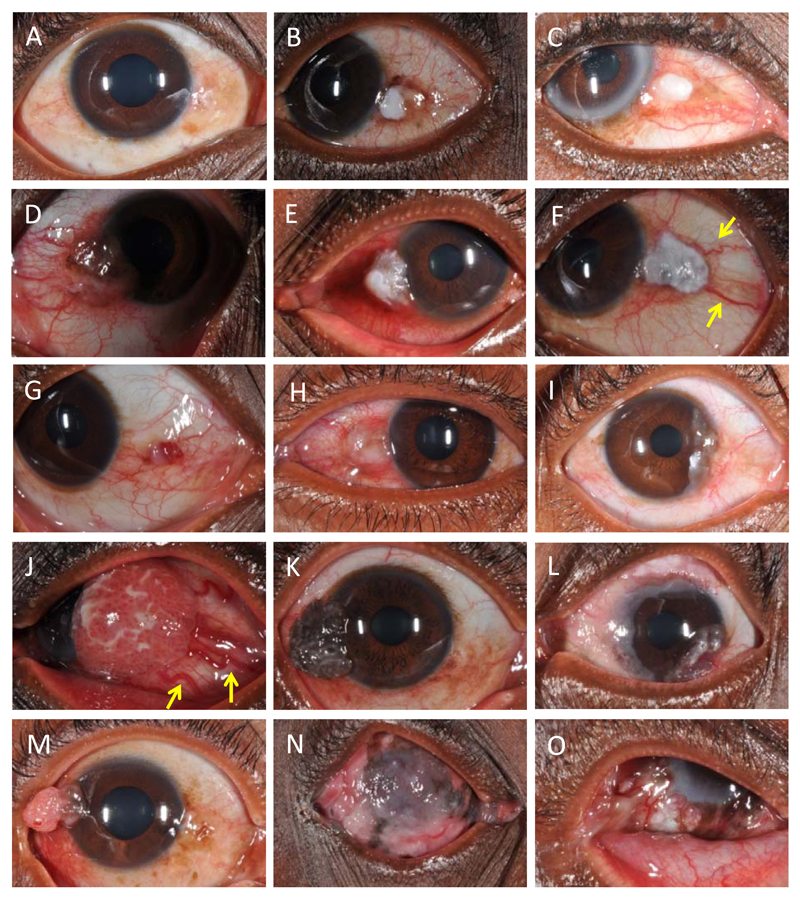
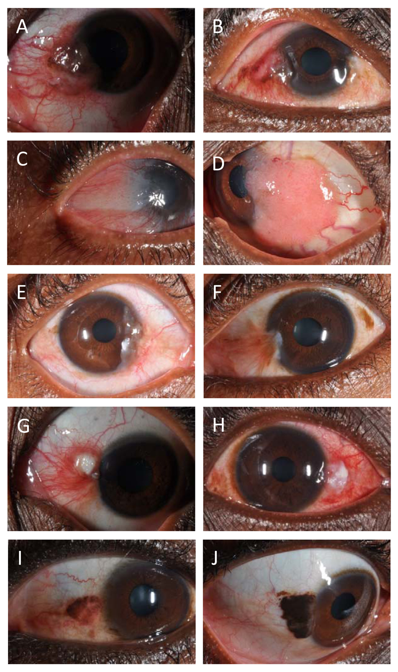
Objective: To describe the presentation of OSSN and identify clinical features that distinguish it from benign lesions and subsequently evaluate their recognizability.
Design, setting, and participants: Prospective multicenter study in Kenya from July 2012 through July 2014 of 496 adults presenting with conjunctival lesions. One histopathologist examined all specimens. Six additional masked ophthalmologists independently examined photographs from 100 participants and assessed clinical features.
Exposures: Comprehensive history, slitlamp examination, and photography before excision biopsy.
Main outcomes and measures: Frequency of clinical features in OSSN and benign lesions were recorded. Proportions and means were compared using χ2, Fisher exact test, or t test as appropriate. Interobserver agreement was estimated using the κ statistic. Examiners' assessments were compared with a reference.
Results: Among 496 participants, OSSN was the most common (38%) histological diagnosis, followed by pterygium (36%) and actinic keratosis (19%). Patients with OSSN were slightly older (mean [SD] age, 41 [11.6] vs 38 [10.9] years; P = .002) and tended to have lower levels of education than patients with benign lesions (P = .001). Females predominated (67% of OSSN vs 64% of benign lesions; P = .65). Human immunodeficiency virus infection was common among patients with OSSN (74%). The most common location was the nasal limbus (61% OSSN vs 78% benign lesions; P < .001). Signs more frequent in OSSN included feeder vessels (odds ratio [OR], 5.8 [95% CI, 3.2-10.5]), moderate inflammation (OR, 3.5 [95% CI, 1.8-6.8]), corneal involvement (OR, 2.7 [95% CI, 1.8-4.0]), leukoplakia (OR, 2.6 [95% CI, 1.7-3.9]), papilliform surface (OR, 2.1 [95% CI, 1.3-3.5]), pigmentation (OR, 1.5 [95% CI, 1.0-2.2]), temporal location (OR, 2.0 [95% CI, 1.2-3.2]), circumlimbal location (6.7% vs 0.3%; P < .001), severe inflammation (6.7% vs 0.3%; P < .001), and larger mean (SD) diameter (6.8 [3.2] vs 4.8 [2.8] mm; P < .001). All OSSN signs were also observed in benign lesions. There was slight to fair interobserver agreement in assessment of most signs and diagnosis (κ, 0.1-0.4). The positive predictive value of clinical appearance in identifying OSSN was 54% (interquartile range, 51%-56%) from photographs in which prevalence was 32%.
Conclusions and relevance: With overlapping phenotypes and modest interobserver agreement, OSSN and benign conjunctival lesions are not reliably distinguished clinically. Point-of-care diagnostic tools may help.
Conflict of interest statement
All authors have completed and submitted the ICMJE Form for Disclosure of Potential Conflicts of Interest and none were reported.
Figures
Comment in
-
Ocular Surface Squamous Neoplasia: From Blue Skies to Blue Dyes--We Still Need Our Ophthalmic Pathologists.JAMA Ophthalmol. 2015 Nov;133(11):1321-2. doi: 10.1001/jamaophthalmol.2015.3359. JAMA Ophthalmol. 2015. PMID: 26378883 No abstract available.
References
-
- Lee GA, Hirst LW. Ocular surface squamous neoplasia. Surv Ophthalmol. 1995;39(6):429–450. - PubMed
-
- Adesina A, Chumba D, Nelson AM, et al. Improvement of pathology in sub-Saharan Africa. Lancet Oncol. 2013;14(4):e152–157. - PubMed
-
- Rambau PF. Pathology practice in a resource-poor setting: Mwanza, Tanzania. Arch Pathol Lab Med. 2011;135(2):191–193. - PubMed
-
- Stone DU, Butt AL, Chodosh J. Ocular surface squamous neoplasia: a standard of care survey. Cornea. 2005;24(3):297–300. - PubMed
Publication types
MeSH terms
Grants and funding
LinkOut - more resources
Full Text Sources
Other Literature Sources
