. 2015 Nov;29(11):1594-607.
doi: 10.1210/me.2014-1375. Epub 2015 Sep 17.

Exploiting Expression of Hippo Effector, Yap, for Expansion of Functional Islet Mass

Affiliations

Exploiting Expression of Hippo Effector, Yap, for Expansion of Functional Islet Mass

Nicholas M George et al. Mol Endocrinol. 2015 Nov.

Abstract

Loss of pancreas β-cell function is the precipitating factor in all forms of diabetes. Cell replacement therapies, such as islet transplantation, remain the best hope for a cure; however, widespread implementation of this method is hampered by availability of donor tissue. Thus, strategies that expand functional β-cell mass are crucial for widespread usage in diabetes cell replacement therapy. Here, we investigate the regulation of the Hippo-target protein, Yes-associated protein (Yap), during development of the endocrine pancreas and its function after reactivation in human cadaveric islets. Our results demonstrate that Yap expression is extinguished at the mRNA level after neurogenin-3-dependent specification of the pancreas endocrine lineage, correlating with proliferation decreases in these cells. Interestingly, when a constitutively active form of Yap was expressed in human cadaver islets robust increases in proliferation were noted within insulin-producing β-cells. Importantly, proliferation in these cells occurs without negatively affecting β-cell differentiation or functional status. Finally, we show that the proproliferative mammalian target of rapamycin pathway is activated after Yap expression, providing at least one explanation for the observed increases in β-cell proliferation. Together, these results provide a foundation for manipulating Yap activity as a novel approach to expand functional islet mass for diabetes regenerative therapy.


Figures

Figure 1.
Figure 1.
Regulation of Yap in pancreas endocrine cells occurs at the transcriptional level and is independent of canonical Hippo signaling. A, Hippo signaling was disrupted in the β-cell compartment through cross of mice conditional for the Hippo kinases, Mst1 and Mst2 (Mst1/2), to a strain with Cre-recombinase driven by the insulin promoter (Ins-Cre). Efficient Mst1/2 deletion in the pancreas was limited to insulin-positive β-cells. Although Yap expression is readily detectible in pancreas ducts (white arrows), it is absent from both control islets and those harboring loss of Mst1/2, suggesting that Hippo signaling is not responsible for Yap regulation in these cells. B, RNA in situ hybridization was used to determine whether Yap mRNA is expressed in islet cells from postnatal day-2 murine offspring. Although the control probe, targeting the ubiquitously expressed cyclophilin B gene (PPIB), was found to be expressed throughout the pancreas, Yap mRNA expression was much more limited. Specifically, Yap was highly expressed in pancreas duct cells (red box, inset) and completely absent from regions harboring insulin-positive β-cells (black box, inset). C, Similar to results obtained in the mouse, Yap mRNA expression is also absent from insulin-positive regions in 72dpc human pancreas. Serial cut sections were used for the in situ hybridization experiments shown in B and C. Scale bars, 50 μm.
Figure 2.
Figure 2.
Onset of Ngn3 expression during development of the endocrine pancreas is sufficient for Yap loss and tightly correlates with proliferative decreases in these cells. A, Immunofluorescent colocalization of Ngn3 and Yap protein during development of the E16.5 mouse (top) and 72dpc human pancreas. Although Yap is highly expressed and localized to the nucleus of nearly all pancreas trunk epithelial cells, its expression is absent from those cells containing robust Ngn3 expression. B, Transduction of the mPAC with an Ngn3-GFP-coexpressing adenovirus was used to determine whether Ngn3 expression is sufficient for Yap down-regulation. Forty-eight hours after transduction, cells were FACS sorted for GFP expression and subsequently used for Western blot analysis (left) or reverse transcription PCR analysis (right). NeuroD1, a well-known Ngn3 target gene was robustly increased after Ngn3 expression, whereas expressions of both Yap and its paralog, Taz, were rapidly down-regulated. C and D, Cell proliferation was analyzed during E16.5 mouse pancreas development and correlated with Yap expression. Although proliferating Ki67-positive cells are frequent throughout the E16.5 pancreas, Ngn3-positive cells are generally not mitotically active (C). Proliferation rates in Yap-positive vs Yap-negative cells were determined by comparing Yap expression with Ki67 immunoreactivity in the E16.5 pancreas. Roughly half of Yap(+) cells were active in the cell cycle compared with only approximately 10% of Yap(minus) cells. We extended this one step further by comparing levels of proliferation in the 3 major pancreas cell lineages (duct [white dotted line], acinar [“A”], and endocrine [“E”]). Although Yap expression is readily detectible in both duct and acinar cells, it is absent from endocrine cells. Proliferation mirrors this phenomenon as both acinar and duct cells display high rates of proliferation, whereas endocrine cells do not. (n = 3 age-matched embryo sections, ***, P ≤ .01). Scale bars, 50 μm.
Figure 3.
Figure 3.
Transduction of human cadaver islets with constitutively active YapS127A is sufficient to drive expression of known Hippo/Yap-target genes. A, Both the control virus (Ad-Con)- and YapS127A-expressing adenovirus (Ad-Yap) readily transduce cells throughout intact human cadaveric islets as monitored by coexpressed GFP. B, Nuclear colocalization of transduced YapS127A with endogenous TEAD transcription factors suggests that transcription of Yap-target genes is likely. Interestingly, TEAD-expressing cells were more often found at the islet exterior and roughly correlated with Yap expression. C, Expression of known Yap-target genes was assessed in islet extracts using Western blot analysis. Although the β-cell-specific protein Pdx1 was found at equal levels in both Ad-Con- and Ad-Yap-transduced islets, expression of the Yap targets, Cyr61 and CTGF, were dramatically increased in only those transduced with YapS127A. Unexpectedly, yet repetitively, levels of one or more TEAD transcription factors were also increased. Scale bars, 50 μm.
Figure 4.
Figure 4.
Human β-cells expressing YapS127A reenter the cell cycle and proliferate. A and B, Expression of YapS127A is sufficient to induce de novo cell proliferation within human cadaveric islets as determined by BrdU incorporation and Ki67 and PCNA expression (72 h after infection). White arrows denote proliferating insulin-positive cells. C, Quantitative analysis of islet cell proliferation after YapS127A expression (***, P ≤ .05). Results are the average (±SD) of 3 independent donor islet preparations (n = 3). D, Proliferation is also evident in glucagon-expressing α-cells. Yellow arrow denotes BrdU(+) Yap-expressing α-cell, white arrows denote Yap-negative BrdU(minus) α-cells. E, Yap transduction of whole-human islets leads to up-regulation of the proproliferative c-Myc and cyclin D1 proteins and proliferation-associated phosphorylation of Rb. Scale bars, 50 μm.
Figure 5.
Figure 5.
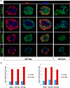
Endocrine differentiation and physiological function is maintained in human islets expressing YapS127A. A, Immunofluorescent colocalization of transduced YapS127A protein with β-cell-specific Nkx6.1 or Pdx1 transcription factors suggests negligible loss of β-cell differentiation in human cadaveric islets, whereas analysis of the endocrine functional marker, synaptophysin, and β-cell functional marker, insulin, suggests overall physiological function remains intact. Ad-Con-transduced islets are shown in the right-hand column for comparison. B, Physiological function of transduced islets was assessed with 2 separate donor islet batches by glucose-stimulated insulin secretion assay. No difference in insulin secretion was observed between mock-transduced islets, Ad-GFP, or Ad-Yap-transduced islets. Each condition was assayed in triplicate with baseline for insulin release being mock infected islets in basal glucose media (2.7mM). Insulin concentration was determined by ELISA and values were subsequently normalized to total islet protein. (Scale bars, 50 μm)
Figure 6.
Figure 6.
Differentiation status is maintained in YapS127A-expressing β-cell lines. A, Min6 or RIN cells were transduced with YapS127A/GFP coexpressing adenovirus and FACS sorted on GFP expression as described in the Materials and Methods. Protein levels of both β-cell markers (Pdx1, Nkx6.1, and Nkx2.2) and β-cell functional markers (chromogranin-A and synaptophysin) remained relatively unchanged compared with mock- or control-transduced cells. Levels of progenitor/duct cell markers (cytokeratin-17/19 and Sox9) were not increased after YapS127A expression, suggesting negligible β-cell transdifferentiation. Functional activity of YapS127A was verified by presence of the TEAD transcription factors and up-regulation of the Yap-TEAD target gene, CTGF. B, Quantitation of Western blot analysis results for the β-cell transcription factors Pdx1, Nkx6.1, and Nkx2.2 indicates trivial changes in expression of these factors. Results are the average (±SD) of 3 individual experiments (n = 3).
Figure 7.
Figure 7.
YapS127A expression activates the mTOR signaling pathway in human β-cells and is necessary for robust cell proliferation. A, Human cadaveric islets were transduced with control (Ad-Con)- or YapS127A-expressing (Ad-Yap) adenovirus and collected 96 hours later. Activated, Ser473-phosphorylated Akt was absent from control islets but was readily detected within YapS127A-expressing islet cells. Phosphorylation of ribosomal S6 protein, an event downstream of activated mTOR signaling, is robustly increased after YapS127A expression. B, Western blot analysis of intact human islets transduced with control or YapS127A-expressing adenovirus and subsequently cultured with either vehicle control (DMSO) or 100nM rapamycin. Signaling through the mTOR pathway, monitored by S6-phosphorylation status, is efficiently decreased with rapamycin treatment. C and D, Yap-induced β-cell proliferation is partially attenuated by cotreatment of human islets with rapamycin (quantitative analysis is presented as side-by-side experiments using separate islet batches; counted cells per condition are noted on each bar).

Similar articles

Cited by

References

    1. Atkinson MA, Eisenbarth GS, Michels AW. Type 1 diabetes. Lancet. 2014;383(9911):69–82. - PMC - PubMed
    1. Pagliuca FW, Melton DA. How to make a functional β-cell. Development. 2013;140(12):2472–2483. - PMC - PubMed
    1. Shapiro AM, Lakey JR, Ryan EA, et al. Islet transplantation in seven patients with type 1 diabetes mellitus using a glucocorticoid-free immunosuppressive regimen. N Engl J Med. 2000;343(4):230–28. - PubMed
    1. Kulkarni RN, Mizrachi EB, Ocana AG, Stewart AF. Human β-cell proliferation and intracellular signaling: driving in the dark without a road map. Diabetes. 2012;61(9):2205–2213. - PMC - PubMed
    1. Bernal-Mizrachi E, Kulkarni RN, Scott DK, Mauvais-Jarvis F, Stewart AF, Garcia-Ocaña A. Human β-cell proliferation and intracellular signaling part 2: still driving in the dark without a road map. Diabetes. 2014;63(3):819–831. - PMC - PubMed

Publication types

MeSH terms