. 2015 Oct 6;87(19):9671-8.
doi: 10.1021/acs.analchem.5b01663.

Integrated glycoprotein immobilization method for glycopeptide and glycan analysis of cardiac hypertrophy

Affiliations

Integrated glycoprotein immobilization method for glycopeptide and glycan analysis of cardiac hypertrophy

Shuang Yang et al. Anal Chem. .

Abstract

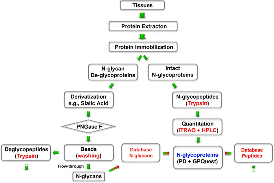
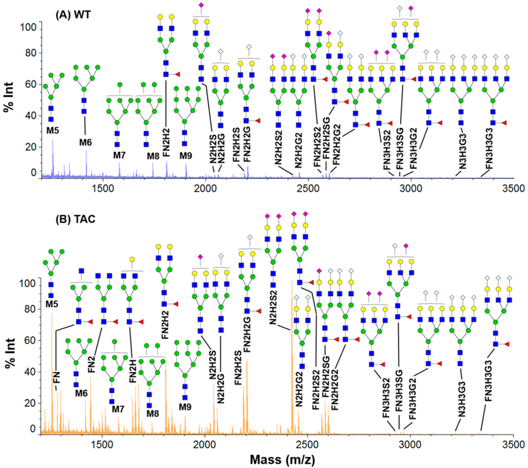
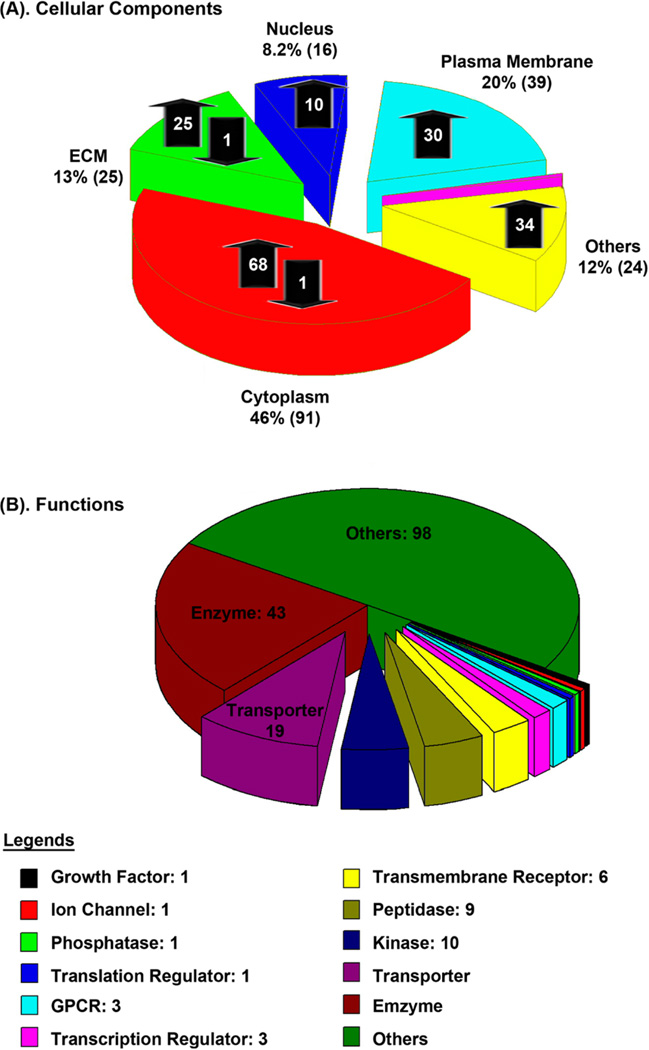
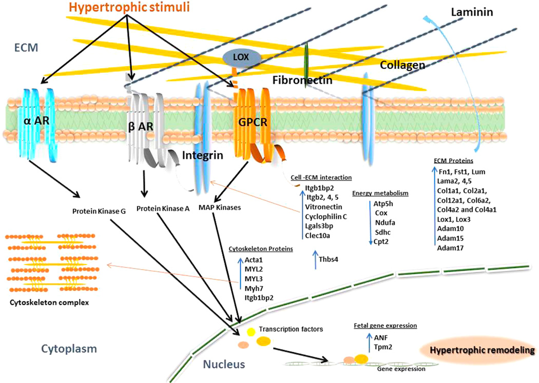
Post-translational modifications of proteins can have a major role in disease initiation and progression. Incredible efforts have recently been made to study the regulation of glycoproteins for disease prognosis and diagnosis. It is essential to elucidate glycans and intact glycoproteins to understand the role of glycosylation in diseases. Sialylated N-glycans play crucial roles in physiological and pathological processes; however, it is laborious to study sialylated glycoproteins due to the labile nature of sialic acid residues. In this study, an integrated platform is developed for the analysis of intact glycoproteins and glycans using a chemoenzymatic approach for immobilization and derivatization of sialic acids. N-Glycans, deglycosylated proteins, and intact glycoproteins from heart tissues of wild type (WT) and transverse aortic constriction (TAC) mouse models were analyzed. We identified 291 unique glycopeptides from 195 glycoproteins; the comparative studies between WT and TAC mice indicate the overexpression of extracellular proteins for heart matrix remodeling and the down-regulation of proteins associated with energy metabolism in cardiac hypertrophy. The integrated platform is a powerful tool for the analysis of glycans and glycoproteins in the discovery of potential cardiac hypertrophy biomarkers.


Figures

Figure 1
Figure 1
Schematic flowchart for analysis of glycoproteins and glycans using an integrated glycoprotein immobilization for glycopeptide and glycan (iGIG). Tissue is sonicated in lysis buffer (RIPA) for protein extraction. Aminolink resin is functionalized with aldehydes for conjugation of the extracted proteins via reductive amination. Sialic acids and acidic amino acids are derivatized via carbodiimide coupling. One-quarter of the proteins are used for identification of N-glycans and deglycopeptides; three-quarters of the proteins are directly digested for analysis of intact glycopeptides. N-Glycans are cleaved off the resin using PNGase F, and the remaining deglycoproteins are digested with trypsin. The intact glycopeptides are quantitated using iTRAQ and fractioned for LC–ESI-MS/MS. N-Glycopeptides are searched by precursor match using a database of N-glycans and deglycopeptides.
Figure 2
Figure 2
MS/MS spectra of the abundant peptides from bovine fetuin serum. Fetuin peptides are digested using trypsin via (A) in solution and (B) iGIG. Peptides that contain E or D, such as HTFSGVASVESSSGEAFHVGK, can be modified by p-toluidine on iGIG; thereby, the retention time differs between the native (42.27 min) and the modified peptide (65.68 min).
Figure 3
Figure 3
MALDI-TOF-MS profiles of the extracted N-glycans using glycoprotein immobilization for glycan extraction (GIG). (A) WT and (B) transverse aortic constriction (TAC) of mice heart tissues using iGIG. An amount of 1 µL out of 40 µL of glycan extraction is analyzed using a Shimadzu Axima Resonance MALDI-MS.
Figure 4
Figure 4
Regulation of glycoproteins in hypertrophic heart and their functions. Quantitative analysis is performed using iGIG and iTRAQ-LC–MS/MS.
Figure 5
Figure 5
Schematic of myocardial protein abnormalities in hypertrophic heart. The diagram depicts how mechanical activation of various hypertrophic stimuli via different receptors activates various downstream signaling cascades leading to hypertrophy. Components of the β-adrenergic (β-AR), α-adrenergic (α-AR), and G protein coupled receptor (GPCR) pathways activate protein kinases (PKA, PKG) and MAP kinases. These pathways are involved in inducing fetal proteins (ANP, Tpm2) expression, overexpression, and modification of extracellular matrix proteins (laminin, collagen, fibronectin, LOX), regulatory thin-filament proteins (Acta1, MYLs), and profibrotic pathways which impact myofilament stiffness and contractile function in hypertrophic cardiomyopathy.

References

    1. Taylor ME, Drickamer K. Introduction to Glycobiology. 3rd ed. Oxford, U.K.: Oxford University Press; 2011.
    1. Haslam SM, North SJ, Dell A. Curr. Opin. Struct. Biol. 2006;16:584–591. - PubMed
    1. Raman R, Raguram S, Venkataraman G, Paulson JC, Sasisekharan R. Nat. Methods. 2005;2:817–824. - PubMed
    1. Zhang H, Li X-j, Martin DB, Aebersold R. Nat. Biotechnol. 2003;21:660–666. - PubMed
    1. Madera M, Mechref Y, Novotny MV. Anal. Chem. 2005;77:4081–4090. - PMC - PubMed

Publication types