. 2015 Sep 17;10(9):e0138360.
doi: 10.1371/journal.pone.0138360. eCollection 2015.

Acinetobacter baumannii Virulence Is Mediated by the Concerted Action of Three Phospholipases D

Affiliations

Acinetobacter baumannii Virulence Is Mediated by the Concerted Action of Three Phospholipases D

Julia Stahl et al. PLoS One. .

Abstract

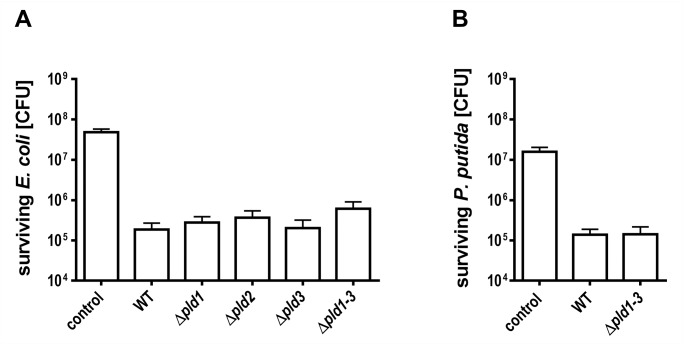
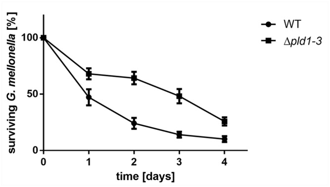
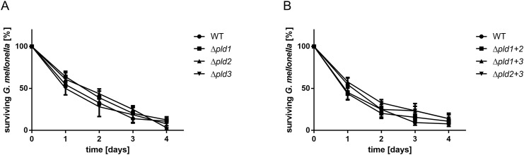
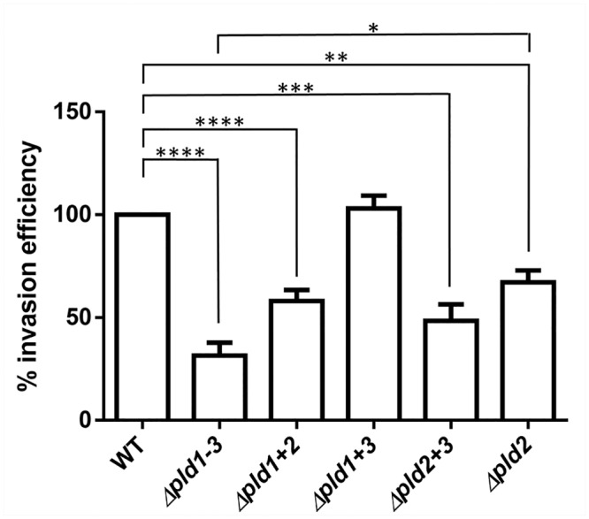
Acinetobacter baumannii causes a broad range of opportunistic infections in humans. Its success as an emerging pathogen is due to a combination of increasing antibiotic resistance, environmental persistence and adaptation to the human host. To date very little is known about the molecular basis of the latter. Here we demonstrate that A. baumannii can use phosphatidylcholine, an integral part of human cell membranes, as sole carbon and energy source. We report on the identification of three phospholipases belonging to the PLD superfamily. PLD1 and PLD2 appear restricted to the bacteria and display the general features of bacterial phospholipases D. They possess two PLDc_2 PFAM domains each encompassing the HxKx4Dx6GS/GGxN (HKD) motif necessary for forming the catalytic core. The third candidate, PLD3, is found in bacteria as well as in eukaryotes and harbours only one PLDc_2 PFAM domain and one conserved HKD motif, which however do not overlap. Employing a markerless mutagenesis system for A. baumannii ATCC 19606T, we generated a full set of PLD knock-out mutants. Galleria mellonella infection studies as well as invasion experiments using A549 human lung epithelial cells revealed that the three PLDs act in a concerted manner as virulence factors and are playing an important role in host cell invasion.


Conflict of interest statement

Competing Interests: The authors have declared that no competing interests exist.

Figures

Fig 1
Fig 1. Growth of A. baumannii ATCC 19606T on PC, palmitate and acetate.
A. baumannii ATCC 19606T was grown in 5 ml MM with either 0.5% PC (triangles), 0.5% palmitate (squares) or 20 mM acetate (circles) as sole carbon source. A culture without carbon source (diamonds) was used as negative control. Growth was followed by plating appropriate dilutions on LB-agar plates at the indicated time points. The amount of colony forming units (CFU) was determined after incubation at 37°C overnight.
Fig 2
Fig 2. Domain architecture of the three A. baumannii ATCC 19606T phospholipase D candidates.
PLD1 and PLD2 each possess PLDc_2 Pfam domains (PF13091). Each domain harbors the conserved motif characteristic for the active center of type D phospholipases. PLD3 has only a single PLD domain encompassing a HKD-like motif (HxKx4Dx6GS/GGxN) where the functionally relevant aspartate is replaced by a tyrosine. A second canonical HKD motif is present in the N-terminal half of the protein and outside the context of a PLDc_2 domain. While both, PLD2 and PLD3, appear membrane bound proteins, PLD1 lacks a transmembrane domain (TM).
Fig 3
Fig 3. Phylogenetic tree combining the orthologous groups of PLD1, -2, and -3.
The tree demonstrates the intertwined evolutionary relationships of orthologs assigned to A. baumannii PLD1 and PLD2, respectively. In contrast, PLD3 sequences form a well separated clade suggesting an evolutionary origin distinct from that of PLD1 and PLD2. The * denotes a local branch support of 0.83.
Fig 4
Fig 4. Evolutionary relationships of Acinetobacter baumannii PLD1 and PLD2.
The tree reveals that PLD1 and PLD2 emerged through a recent gene duplication that occurred after the split of P. aeruginosa and possibly in the last common ancestor of the genus Acinetobacter. Orthologs whose split pre-date the diversification of PLD1 and 2 were renamed to PLD1/2. Branch labels represent percent bootstrap support.
Fig 5
Fig 5. Bacterial competition between A. baumannii ATCC 19606T and E. coli or P. putida.
A. baumannii ATCC 19606T wild-type or pld mutant cells and E. coli DH5α pET28a cells (A) or P. putida 548.C8 cells (B) were mixed at a ratio of 10:1, spotted onto an LB-agar plates and incubated for 4 hours at 37°C. To quantify the number of surviving E. coli or P. putida cells serial dilutions were plated onto kanamycin containing LB-agar and colony forming units were counted after incubation overnight at 37°C or 30°C, respectively. The standard deviation was calculated from three independent experiments. LB medium was used instead of A. baumannii as negative control.
Fig 6
Fig 6. A. baumannii Δpld1-3 mutant is less virulent in G. mellonella infection.
For infection assays 16 caterpillars were injected with approximately 106 bacteria and incubated at 37°C in the dark. Mean difference between the wild-type and the Δpld1-3 mutant in four independent experiments was significant after 1, 2, 3 and 4 days (paired t-test; p<0.05 for day 1, p<0.01 for days 2, 3 and 4). As control served an untreated set of caterpillars as well as a set of caterpillars which was treated with 10 μl of sterile 0.9% NaCl. Tests in which more than 2 caterpillars in one of the control groups died after 4 days were not considered.
Fig 7
Fig 7. Single or double pld mutants display an unaffected virulence towards G. mellonella.
For infection assays 16 caterpillars were injected with approximately 106 bacteria and incubated at 37°C in the dark. Mean difference between wild-type and single (A) or double mutants (B) was not significant. As control served an untreated set of caterpillars as well as a set of caterpillars which was treated with 10 μl of sterile 0.9% NaCl. Tests in which more than 2 caterpillars in one of the control groups died after 4 days were not considered.
Fig 8
Fig 8. PLDs affect invasion efficiency of A. baumannii.
A549 cells were infected with A. baumannii wild-type or mutant cells for 5 hours. Then cells were treated with gentamycin to eliminate all external bacteria, prior to release of internal bacteria by lysis with H2O. Internal bacteria were calculated and the cell count of the wild-type was set to 100%.100% corresponds to 112.5 ± 2.5 CFU/ml Asterisks indicate statistical significance by paired t-test; *p<0.05; **p<0.01; ***p<0.001; ****p<0.0001.

References

    1. Dijkshoorn L, Nemec A, Seifert H. An increasing threat in hospitals: multidrug-resistant Acinetobacter baumannii . Nat Rev Microbiol. 2007;5: 939–951. - PubMed
    1. Antunes LC, Visca P, Towner KJ. Acinetobacter baumannii: evolution of a global pathogen. Pathog Dis. 2014;71: 292–301. 10.1111/2049-632X.12125 - DOI - PubMed
    1. Kempf M, Rolain JM. Emergence of resistance to carbapenems in Acinetobacter baumannii in Europe: clinical impact and therapeutic options. Int J Antimicrob Agents. 2012;39: 105–114. 10.1016/j.ijantimicag.2011.10.004 - DOI - PubMed
    1. Peleg AY, Seifert H, Paterson DL. Acinetobacter baumannii: emergence of a successful pathogen. Clin Microbiol Rev. 2008;21: 538–582. 10.1128/CMR.00058-07 - DOI - PMC - PubMed
    1. Gaynes R, Edwards JR, System NNIS. Overview of nosocomial infections caused by gram-negative bacilli. Clin Infect Dis. 2005;41: 848–854. - PubMed

Publication types

MeSH terms

LinkOut - more resources