. 2015 Jul-Aug;48(4):249-59.
doi: 10.1590/0100-3984.2013.1726.

Magnetic resonance imaging of the vagina: an overview for radiologists with emphasis on clinical decision making

Affiliations

Magnetic resonance imaging of the vagina: an overview for radiologists with emphasis on clinical decision making

Daian Miranda Ferreira et al. Radiol Bras. 2015 Jul-Aug.

Abstract

Magnetic resonance imaging is a method with high contrast resolution widely used in the assessment of pelvic gynecological diseases. However, the potential of such method to diagnose vaginal lesions is still underestimated, probably due to the scarce literature approaching the theme, the poor familiarity of radiologists with vaginal diseases, some of them relatively rare, and to the many peculiarities involved in the assessment of the vagina. Thus, the authors illustrate the role of magnetic resonance imaging in the evaluation of vaginal diseases and the main relevant findings to be considered in the clinical decision making process.

A ressonância magnética é um método com alta resolução de contraste e por isso muito utilizada na avaliação de doenças ginecológicas pélvicas. No entanto, seu potencial para diagnóstico de lesões vaginais ainda é subestimado, provavelmente em razão da escassa literatura referente ao tema, da pouca familiaridade dos radiologistas com doenças vaginais, algumas delas relativamente raras, e das muitas peculiaridades em um exame para avaliação desta víscera oca. Desta forma, ilustraremos neste estudo o papel da ressonância magnética na avaliação das doenças vaginais e os principais achados relevantes para a conduta clínica.

Keywords: Congenital malformations; Magnetic resonance imaging; Tumors; Vagina.


Figures

Figure 1
Figure 1
Normal female pelvis. Axial (A) and sagittal (B) MRI T2-weighted images showing anterior compartment containing the urethral ostium (Ur) and the ostium of the bladder (B), the medial compartment containing the uterus (U), the uterine cervix (C), the vagina distended with gel (V), the anterior wall of the vagina (AW), the posterior wall of the vagina (PW), the vaginal vestibule (VI) and the posterior compartment with the rectum (R).
Figure 2
Figure 2
Uterus didelphys, longitudinal and transverse vaginal septa. MRI T2- weighted (A,B,C) and T1-weighted (D) sequences demonstrating longitudinal septum (thin arrow) dividing the vagina into two parallel cavities. The left hemivagina (asterisks) is obstructed by a transverse septum (thin arrowhead) and distended by hematic contents (high signal on T1- weighted image). Displaced and compressed right hemivagina at right (stars), right uterine horn (bold arrowhead), left uterine horn (bold arrow).
Figure 3
Figure 3
Longitudinal vaginal septum. Contrast-enhanced MRI T2- weighted (A,D) and T1-weighted (B,C) images showing longitudinal vaginal septum (arrows) dividing the vagina into two chambers. Ur, urethra; V, vagina. The identification at MRI may be difficult as the presence of the vaginal septum is not associated with obstruction.
Figure 4
Figure 4
Imperforate hymen (arrow). MRI T2-weighted (A) and T1- weighted, sagittal (B) and coronal (C) sequences demonstrating uterus (U) and vagina (V) distended by hematic contents, which extends inferiorly protruding the ostium.
Figure 5
Figure 5
Partial androgen insensitivity syndrome. Multiplanar MRI T2- weighted sequences (A,B,C) and T2- weighted sequence with fat saturation (D) demonstrating masculine false hermaphroditism (46,XY) in a 21 years old patient with female phenotype and ambiguous genitalia, characterized by a short vagina (V) and presence of a micropenis (thin arrows). The images of the pelvis demonstrate neither uterus nor ovaries, and the testicles are located in the inguinal canals (bold arrows). Observe the hypertrophic rectoabdominal muscles, and the scarcity of subcutaneous fat caused by testosterone activity.
Figure 6
Figure 6
Mayer-Rokitansky-Kuster- Hauser syndrome (complete presentation). Multiplanar T2-weighted sequence (A,B,C) showing absence of the uterus and of the upper third of the vagina (V) between the rectum (R) and the urethra (Ur). The pelvic images confirm the presence of normal ovaries and large cystic mass in the left ovary.
Figure 7
Figure 7
Turner syndrome. Multiplanar MRI T2-weighted sequence (A,B) demonstrate streak uterus and ovaries (arrow), short vagina (V) located between the rectum (R) and the urethra (Ur).
Figure 8
Figure 8
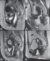
Gartner duct cyst – axial (A1) and sagittal (B1) MRI T2-weighted sequences demonstrating a cyst located in the left lateral vaginal wall, above the level of the pubic symphysis. Bartholin gland cyst – axial (A2) and sagittal (B2) T2-weighted sequences of another patient demonstrating cystic lesion outside the vaginal canal, on the distal posterior wall of the vagina at right.
Figure 9
Figure 9
Cysts of the Skene glands. Multiplanar MRI T2-weighted (A,B) and contrast- enhanced T1-weighted (C,D) sequences identifying distal periurethral cysts (Ur) (arrows) located between the urethra and the vagina.
Figure 10
Figure 10
Giant condyloma acuminatum. Contrast-enhanced, multiplanar MRI T2- weighted (A,B) and T1-weighted (C,D) sequences of the pelvis demonstrating multiple cauliflower-like verrucous lesions in the anogenital region (arrows). After contrast medium injection, marked contrast uptake by the lesion was observed.
Figure 11
Figure 11
Multiplanar MRI T1-weighted (B) and T2- weighted (A,C,D) sequences of the pelvis demonstrating the presence of a focus of endometriosis with low signal intensity in the vaginal dome (bold arrows), with signs of local tissue retraction and extension to the anterior wall of the rectum, characterizing infiltrative endometriosis intermingled with a focus of high signal intensity corresponding to hemorrhagic focus (thin arrow). V, vagina; R rectum.
Figure 12
Figure 12
Spinocellular carcinoma. Axial (A,B), coronal (C) and sagittal (D) multiplanar MRI T2- weighted sequences showing the presence of a solid, lobulated mass in the posterior and right lower vaginal walls (V). The tumor infiltrates the rectovaginal fat plane (arrows).
Figure 13
Figure 13
Vaginal melanoma. Sagittal (A1) and axial (B1) MRI T2-weighted sequences showing the presence of a lobulated mass with low signal intensity (arrows) affecting the anterior and posterior vaginal walls, extending throughout its entire length up to the vaginal ostiuml. MRI of another patient – contrast-enhanced T1-weighted sequence with fat saturation (A2) demonstrates a hypervascular lesion deeply invading the vagina. Diffusion-weighted image (B2) acquired with b = 750 s/mm2 shows significant diffusion restriction (arrows).
Figure 14
Figure 14
Uterine cervix adenocarcinoma with locally invasive tumor. Sagittal (A) and axial (B) MRI T2-weighted sequences demonstrating a heterogeneous and infiltrative lesion extending towards the vaginal dome, rectovaginal septum and mesorectal fascia (arrows). V, vagina; U, uterus.
Figure 15
Figure 15
Vaginal metastasis from ovary adenocarcinoma – Axial (A1) and sagittal (B1) multiplanar MRI T2-weighted sequences demonstrate lymph node enlargement (asterisk) and peritoneal carcinomatosis, including an infiltrating lesion in the vaginal dome (arrows). V, vagina. Vaginal metastasis from endometrial carcinoma – Axial (A2) and sagittal (B2) MRI T2-weighted sequences demonstrating the primary tumor filling the endometrial cavity (U) and a well-defined nodule (skip lesions) with intermediate signal intensity in the right anterior wall of the vagina (arrow). V, vagina. Vaginal metastasis from uterine cervix squamous cell carcinoma – MRI T2-weighted (A3) and contrastenhanced T1-weighted (B3) sequences demonstrating ill-defined mass originating from the uterine cervix and extending towards the lower uterine segment and lower third of the vagina (arrows).
Figure 16
Figure 16
Pelvic floor prolapse. Sagittal (A) and dynamic axial (B) MRI T2-weighted sequences demonstrate large prolapse of the urogenital hiatus characterized by inversion of the vaginal dome, small bowel loops and abdominal fat protrusion.
Figure 17
Figure 17
Post-radiotherapy complications. A1: Fistulous path (thin arrows) between the anterosuperior vaginal wall and the vesicouterine pouch. A small amount of heterogeneous fluid (asterisk) and anterior displacement of the peritoneal fold (bold arrow) are observed. V, vagina; U, uterus; B, bladder. A2: Fistulous path (arrows) between the rectum and the vagina. A3: Large vesicovaginal communication (arrows). Distension of uterine cavity determined by cervix stenosis (U). V, vagina; B, bladder; R, rectum. A4 Stenosis of the upper third of the vagina, 10 months after radiotherapy (late complication).
Figure 18
Figure 18
Surgical evaluation for maleto- female sex reassignment. Axial (A,B) sagittal (C) and coronal (D) MRI T2- weighted sequences demonstrate neovagina (bold arrows) and the remains of the corpora cavernosa and of the corpus spongiosum and urethra (thin arrows).

References

    1. Walker DK, Salibian RA, Salibian AD, et al. Overlooked diseases of the vagina: a directed anatomic-pathologic approach for imaging assessment. Radiographics. 2011;31:1583–1598. - PubMed
    1. Junqueira BL, Allen LM, Spritzer RF, et al. Müllerian duct anomalies and mimics in children and adolescents: correlative intraoperative assessment with clinical imaging. Radiographics. 2009;29:1085–1103. - PubMed
    1. Siegelman ES, Outwater EK, Banner MP, et al. High-resolution MR imaging of the vagina. Radiographics. 1997;17:1183–1203. - PubMed
    1. Chavhan GB, Parra DA, Oudjhane K, et al. Imaging of ambiguous genitalia: classification and diagnostic approach. Radiographics. 2008;28:1891–1904. - PubMed
    1. Giusti S, Fruzzetti E, Perini D, et al. Diagnosis of a variant of Mayer- Rokitansky-Kuster-Hauser syndrome: useful MRI findings. Abdom Imaging. 2011;36:753–755. - PubMed

LinkOut - more resources