

Novel 3-dimensional virtual hepatectomy simulation combined with real-time deformation
- PMID: 26379403
- PMCID: PMC4566391
- DOI: 10.3748/wjg.v21.i34.9982
Novel 3-dimensional virtual hepatectomy simulation combined with real-time deformation
Abstract
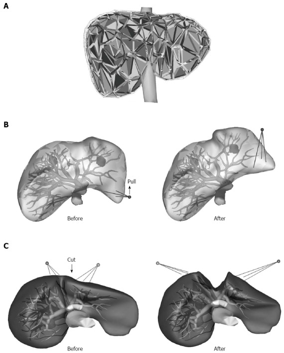
Aim: To develop a novel 3-dimensional (3D) virtual hepatectomy simulation software, Liversim, to visualize the real-time deformation of the liver.
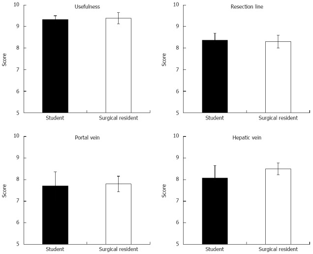
Methods: We developed a novel real-time virtual hepatectomy simulation software program called Liversim. The software provides 4 basic functions: viewing 3D models from arbitrary directions, changing the colors and opacities of the models, deforming the models based on user interaction, and incising the liver parenchyma and intrahepatic vessels based on user operations. From April 2010 through 2013, 99 patients underwent virtual hepatectomies that used the conventional software program SYNAPSE VINCENT preoperatively. Between April 2012 and October 2013, 11 patients received virtual hepatectomies using the novel software program Liversim; these hepatectomies were performed both preoperatively and at the same that the actual hepatectomy was performed in an operating room. The perioperative outcomes were analyzed between the patients for whom SYNAPSE VINCENT was used and those for whom Liversim was used. Furthermore, medical students and surgical residents were asked to complete questionnaires regarding the new software.
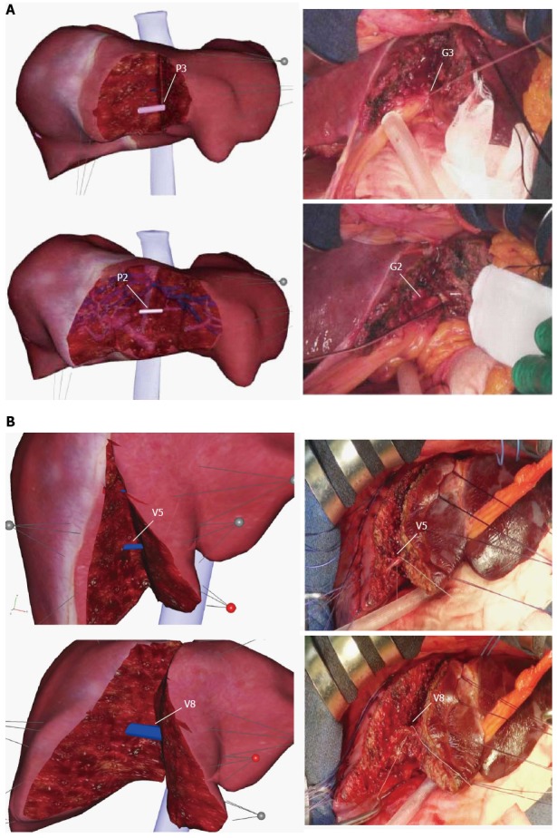
Results: There were no obvious discrepancies (i.e., the emergence of branches in the portal vein or hepatic vein or the depth and direction of the resection line) between our simulation and the actual surgery during the resection process. The median operating time was 304 min (range, 110 to 846) in the VINCENT group and 397 min (range, 232 to 497) in the Liversim group (P = 0.30). The median amount of intraoperative bleeding was 510 mL (range, 18 to 5120) in the VINCENT group and 470 mL (range, 130 to 1600) in the Liversim group (P = 0.44). The median postoperative stay was 12 d (range, 6 to 100) in the VINCENT group and 13 d (range, 9 to 21) in the Liversim group (P = 0.36). There were no significant differences in the preoperative outcomes between the two groups. Liversim was not found to be clinically inferior to SYNAPSE VINCENT. Both students and surgical residents reported that the Liversim image was almost the same as the actual hepatectomy.
Conclusion: Virtual hepatectomy with real-time deformation of the liver using Liversim is useful for the safe performance of hepatectomies and for surgical education.
Keywords: Liver; Real-time deformation; Simulation; Surgery; Surgical education; Virtual hepatectomy.
Figures
References
-
- Sasaki R, Kondo T, Oda T, Murata S, Wakabayashi G, Ohkohchi N. Impact of three-dimensional analysis of multidetector row computed tomography cholangioportography in operative planning for hilar cholangiocarcinoma. Am J Surg. 2011;202:441–448. - PubMed
-
- Oshiro Y, Sasaki R, Nasu K, Ohkohchi N. A novel preoperative fusion analysis using three-dimensional MDCT combined with three-dimensional MRI for patients with hilar cholangiocarcinoma. Clin Imaging. 2013;37:772–774. - PubMed
-
- Takahashi K, Sasaki R, Kondo T, Oda T, Murata S, Ohkohchi N. Preoperative 3D volumetric analysis for liver congestion applied in a patient with hilar cholangiocarcinoma. Langenbecks Arch Surg. 2010;395:761–765. - PubMed
-
- Saito S, Yamanaka J, Miura K, Nakao N, Nagao T, Sugimoto T, Hirano T, Kuroda N, Iimuro Y, Fujimoto J. A novel 3D hepatectomy simulation based on liver circulation: application to liver resection and transplantation. Hepatology. 2005;41:1297–1304. - PubMed
-
- Endo I, Shimada H, Sugita M, Fujii Y, Morioka D, Takeda K, Sugae S, Tanaka K, Togo S, Bourquain H, et al. Role of three-dimensional imaging in operative planning for hilar cholangiocarcinoma. Surgery. 2007;142:666–675. - PubMed
Publication types
MeSH terms
LinkOut - more resources
Full Text Sources
Other Literature Sources
Medical
Miscellaneous
