

Ultrastructure of Placenta of Gravidas with Gestational Diabetes Mellitus
- PMID: 26379710
- PMCID: PMC4561319
- DOI: 10.1155/2015/283124
Ultrastructure of Placenta of Gravidas with Gestational Diabetes Mellitus
Abstract
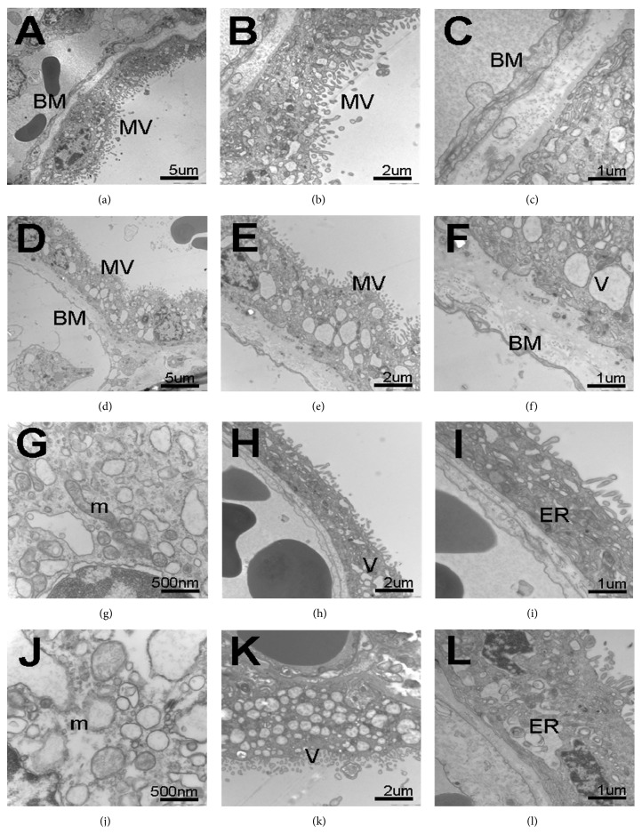
Objectives. Gestational diabetes mellitus (GDM) leads to an abnormal placental environment which may cause some structural alterations of placenta and affect placental development and function. In this study, the ultrastructural appearances of term placentas from women with GDM and normal pregnancy were meticulously compared. Materials and Methods. The placenta tissues of term birth from 10 women with GDM and 10 women with normal pregnancy were applied with the signed informed consent. The morphology of fetomaternal interface of placenta was examined using light microscopy (LM) and transmission electron microscopy (TEM). Results. On LM, the following morphological changes in villous tissues were found in the GDM placentas when compared with the control placentas: edematous stroma, apparent increase in the number of syncytial knots, and perivillous fibrin deposition. On TEM, the distinct ultrastructural alterations indicating the degeneration of terminal villi were found in the GDM placentas as follows: thickening of the basal membrane (BM) of vasculosyncytial membrane (VSM) and the VSM itself, significantly fewer or even absent syncytiotrophoblastic microvilli, swollen or completely destroyed mitochondria and endoplasmic reticulum, and syncytiotrophoblasts with multiple vacuoles. Conclusion. Ultrastructural differences exist between GDM and control placentas. The differences of placenta ultrastructure are likely responsible for the impairment of placental barrier and function in GDM.
Figures


Similar articles
-
Morphological and ultrastructural changes in the placenta of the diabetic pregnant Egyptian women.Acta Histochem. 2018 Jul;120(5):490-503. doi: 10.1016/j.acthis.2018.05.008. Epub 2018 Jun 2. Acta Histochem. 2018. PMID: 29871770
-
Unique Ultrastructural Alterations in the Placenta Associated With Macrosomia Induced by Gestational Diabetes Mellitus.Matern Fetal Med. 2024 Jul 1;6(3):164-172. doi: 10.1097/FM9.0000000000000240. eCollection 2024 Jul. Matern Fetal Med. 2024. PMID: 40406284 Free PMC article.
-
Gestational diabetes mellitus induces placental vasculopathies.Environ Sci Pollut Res Int. 2022 Mar;29(13):19860-19868. doi: 10.1007/s11356-021-17267-y. Epub 2021 Nov 2. Environ Sci Pollut Res Int. 2022. PMID: 34725760
-
Placental pathologic changes in gestational diabetes mellitus.Neuro Endocrinol Lett. 2015;36(2):101-5. Neuro Endocrinol Lett. 2015. PMID: 26071574 Review.
-
Characteristics of histopathological and ultrastructural features of placental villi in pregnant Nepalese women.Med Mol Morphol. 2005 Jun;38(2):92-103. doi: 10.1007/s00795-004-0259-y. Med Mol Morphol. 2005. PMID: 15944816 Review.
Cited by
-
Influence of gestational diabetes mellitus on the cardiovascular system and its underlying mechanisms.Front Endocrinol (Lausanne). 2025 May 16;16:1474643. doi: 10.3389/fendo.2025.1474643. eCollection 2025. Front Endocrinol (Lausanne). 2025. PMID: 40453589 Free PMC article. Review.
-
Low molecular weight heparin (nadroparin) improves placental permeability in rats with gestational diabetes mellitus via reduction of tight junction factors.Mol Med Rep. 2020 Feb;21(2):623-630. doi: 10.3892/mmr.2019.10868. Epub 2019 Dec 6. Mol Med Rep. 2020. PMID: 31974593 Free PMC article.
-
Mechanistic Coupling of a Novel in silico Cotyledon Perfusion Model and a Physiologically Based Pharmacokinetic Model to Predict Fetal Acetaminophen Pharmacokinetics at Delivery.Front Pediatr. 2021 Sep 23;9:733520. doi: 10.3389/fped.2021.733520. eCollection 2021. Front Pediatr. 2021. PMID: 34631628 Free PMC article.
-
The interplay of inflammation and placenta in maternal diabetes: insights into Hofbauer cell expression patterns.Front Immunol. 2024 Mar 25;15:1386528. doi: 10.3389/fimmu.2024.1386528. eCollection 2024. Front Immunol. 2024. PMID: 38590527 Free PMC article.
-
Placental mitochondrial dysfunction with metabolic diseases: Therapeutic approaches.Biochim Biophys Acta Mol Basis Dis. 2021 Jan 1;1867(1):165967. doi: 10.1016/j.bbadis.2020.165967. Epub 2020 Sep 10. Biochim Biophys Acta Mol Basis Dis. 2021. PMID: 32920120 Free PMC article. Review.
References
-
- Dabelea D., Snell-Bergeon J. K., Hartsfield C. L., Bischoff K. J., Hamman R. F., McDuffie R. S. Increasing prevalence of gestational diabetes mellitus (GDM) over time and by birth cohort: Kaiser Permanente of Colorado GDM screening program. Diabetes Care. 2005;28(3):579–584. doi: 10.2337/diacare.28.3.579. - DOI - PubMed
-
- Ferrara A., Kahn H. S., Quesenberry C. P., Riley C., Hedderson M. M. An increase in the incidence of gestational diabetes mellitus: Northern California, 1991–2000. Obstetrics & Gynecology. 2004;103:526–533. - PubMed
-
- Mihu C. M., Şuşman S., Ciucă D. R., Mihu D., Costin N. Aspects of placental morphogenesis and angiogenesis. Romanian Journal of Morphology and Embryology. 2009;50(4):549–557. - PubMed
LinkOut - more resources
Full Text Sources
Other Literature Sources
