

Adenosquamous carcinoma of the pancreas: Molecular characterization of 23 patients along with a literature review
- PMID: 26380056
- PMCID: PMC4569590
- DOI: 10.4251/wjgo.v7.i9.132
Adenosquamous carcinoma of the pancreas: Molecular characterization of 23 patients along with a literature review
Abstract
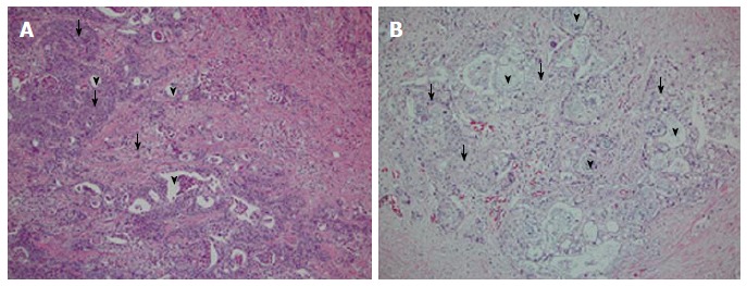
Adenosquamous carcinoma of the pancreas (ASCP) is a rare entity. Like adenocarcinoma of the pancreas, overall survival is poor. Characteristics of ASCP include central tumor necrosis, along with osteoclasts and hypercalcemia. Various theories exist as to why this histological subtype exists, as normal pancreas tissue has no benign squamous epithelium. Due to the rarity of this disease, limited molecular analysis has been performed, and those reports indicate unique molecular features of ASCP. In this paper, we characterize 23 patients diagnosed with ASCP through molecular profiling using immunohistochemistry staining, fluorescent in situ hybridization, chromogenic in situ hybridization, and gene sequencing, Additionally, we provide a comprehensive literature review of what is known to date of ASCP. Molecular characterization revealed overexpression in MRP1 (80%), MGMT (79%), TOP2A (75), RRM1 (42%), TOPO1 (42%), PTEN (45%), CMET (40%), and C-KIT (10%) among others. One hundred percent of samples tested were positive for KRAS mutations. This analysis shows heretofore unsuspected leads to be considered for treatments of this rare type of exocrine pancreas cancer. Molecular profiling may be appropriate to provide maximum information regarding the patient's tumor. Further work should be pursued to better characterize this disease.
Keywords: Adenosquamous carcinoma of the pancreas; Molecular profiling; Review.
Figures
Similar articles
-
Two rare cancers of the exocrine pancreas: to treat or not to treat like ductal adenocarcinoma?J Cancer Metastasis Treat. 2023;9:5. doi: 10.20517/2394-4722.2022.106. Epub 2023 Mar 7. J Cancer Metastasis Treat. 2023. PMID: 37538977 Free PMC article.
-
Loss of chromosome 9p21 is associated with a poor prognosis in adenosquamous carcinoma of the pancreas.Precis Clin Med. 2023 Nov 7;6(4):pbad030. doi: 10.1093/pcmedi/pbad030. eCollection 2023 Dec. Precis Clin Med. 2023. PMID: 38024139 Free PMC article.
-
Recent advances in genomic profiling of adenosquamous carcinoma of the pancreas.J Pathol. 2017 Nov;243(3):271-272. doi: 10.1002/path.4959. Epub 2017 Sep 25. J Pathol. 2017. PMID: 28816351
-
Adenosquamous carcinoma of the pancreas: a distinct clinicopathologic entity.South Med J. 2010 Sep;103(9):903-10. doi: 10.1097/SMJ.0b013e3181ebadbd. South Med J. 2010. PMID: 20697320 Review.
-
Primary cutaneous adenosquamous carcinoma of the penis: the first characterization of HPV status in this rare and diagnostically challenging entity with review of glandular carcinomas of the penis.J Cutan Pathol. 2016 Dec;43(12):1226-1230. doi: 10.1111/cup.12835. J Cutan Pathol. 2016. PMID: 27696488 Review.
Cited by
-
Basic Pancreatic Lesions: Radiologic-pathologic Correlation.J Transl Int Med. 2022 Apr 2;10(1):18-27. doi: 10.2478/jtim-2022-0003. eCollection 2022 Mar. J Transl Int Med. 2022. PMID: 35702187 Free PMC article.
-
Two rare cancers of the exocrine pancreas: to treat or not to treat like ductal adenocarcinoma?J Cancer Metastasis Treat. 2023;9:5. doi: 10.20517/2394-4722.2022.106. Epub 2023 Mar 7. J Cancer Metastasis Treat. 2023. PMID: 37538977 Free PMC article.
-
Decoding the basis of histological variation in human cancer.Nat Rev Cancer. 2024 Feb;24(2):141-158. doi: 10.1038/s41568-023-00648-5. Epub 2023 Dec 22. Nat Rev Cancer. 2024. PMID: 38135758 Review.
-
Loss of chromosome 9p21 is associated with a poor prognosis in adenosquamous carcinoma of the pancreas.Precis Clin Med. 2023 Nov 7;6(4):pbad030. doi: 10.1093/pcmedi/pbad030. eCollection 2023 Dec. Precis Clin Med. 2023. PMID: 38024139 Free PMC article.
-
The origins and consequences of UPF1 variants in pancreatic adenosquamous carcinoma.Elife. 2021 Jan 6;10:e62209. doi: 10.7554/eLife.62209. Elife. 2021. PMID: 33404013 Free PMC article.
References
-
- Siegel R, Ma J, Zou Z, Jemal A. Cancer statistics, 2014. CA Cancer J Clin. 2014;64:9–29. - PubMed
-
- Fitzgerald TL, Hickner ZJ, Schmitz M, Kort EJ. Changing incidence of pancreatic neoplasms: a 16-year review of statewide tumor registry. Pancreas. 2008;37:134–138. - PubMed
-
- Chen J, Baithun SI. Morphological study of 391 cases of exocrine pancreatic tumours with special reference to the classification of exocrine pancreatic carcinoma. J Pathol. 1985;146:17–29. - PubMed
-
- Cihak RW, Kawashima T, Steer A. Adenoacanthoma (adenosquamous carcinoma) of the pancreas. Cancer. 1972;29:1133–1140. - PubMed
Publication types
LinkOut - more resources
Full Text Sources
Other Literature Sources
Research Materials
Miscellaneous
