. 2015 Sep 8;2(1):e000114.
doi: 10.1136/lupus-2015-000114. eCollection 2015.

Significantly reduced lymphadenopathy, salivary gland infiltrates and proteinuria in MRL-lpr/lpr mice treated with ultrasoluble curcumin/turmeric: increased survival with curcumin treatment

Affiliations

Significantly reduced lymphadenopathy, salivary gland infiltrates and proteinuria in MRL-lpr/lpr mice treated with ultrasoluble curcumin/turmeric: increased survival with curcumin treatment

Biji T Kurien et al. Lupus Sci Med. .

Abstract

Objectives: Commercial curcumin (CU), derived from food spice turmeric (TU), has been widely studied as a potential therapeutic for a variety of oncological and inflammatory conditions. Lack of solubility/bioavailability has hindered curcumin's therapeutic efficacy in human diseases. We have solubilised curcumin in water applying heat/pressure, obtaining up to 35-fold increase in solubility (ultrasoluble curcumin (UsC)). We hypothesised that UsC or ultrasoluble turmeric (UsT) will ameliorate systemic lupus erythematosus (SLE) and Sjögren's syndrome (SS)-like disease in MRL-lpr/lpr mice.

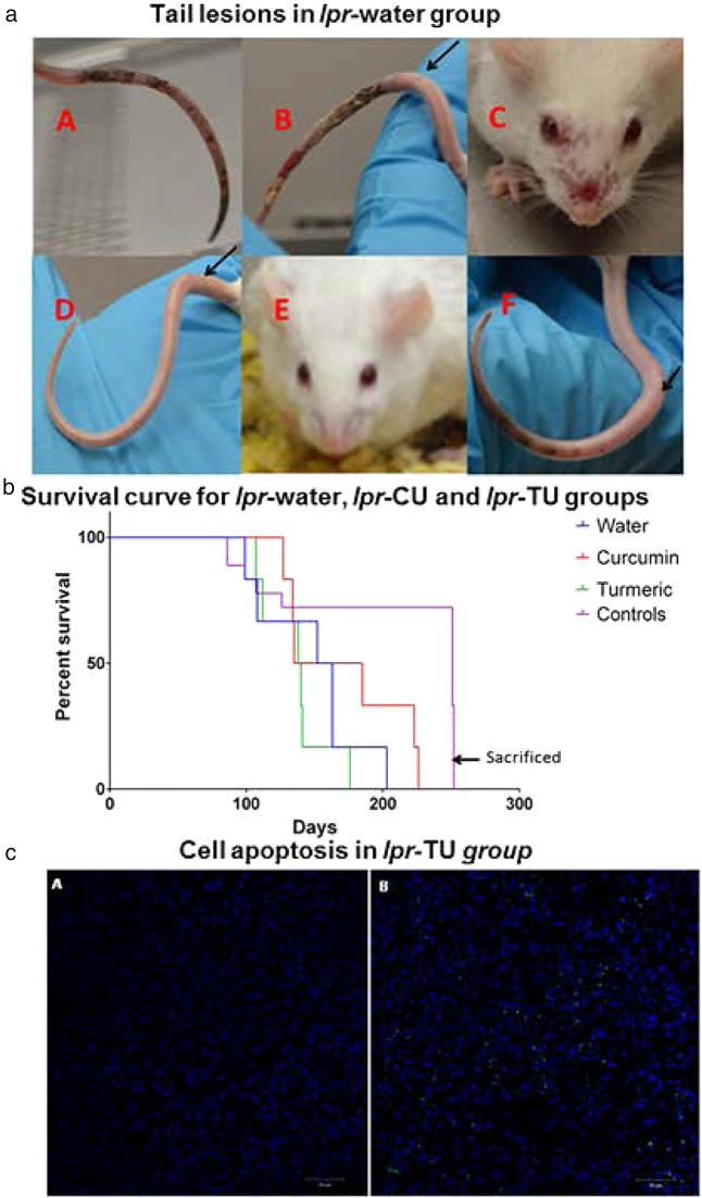
Methods: Eighteen female MRL-lpr/lpr (6 weeks old) and 18 female MRL-MpJ mice (6 weeks old) were used. Female MRL-lpr/lpr mice develop lupus-like disease at the 10th week and die at an average age of 17 weeks. MRL-MpJ mice develop lupus-like disease around 47 weeks and typically die at 73 weeks. Six mice of each strain received autoclaved water only (lpr-water or MpJ-water group), UsC (lpr-CU or MpJ-CU group) or UsT (lpr-TU or MpJ-TU group) in the water bottle.

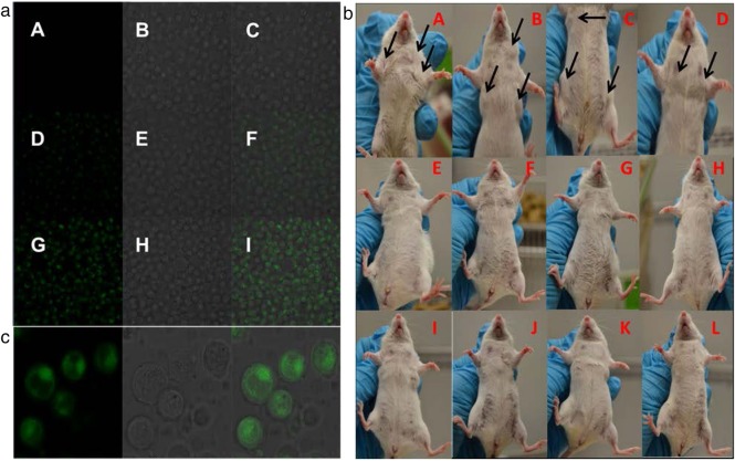
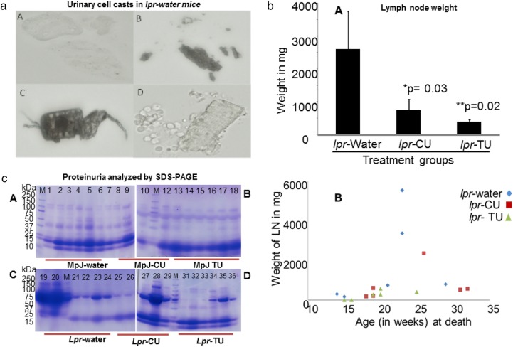
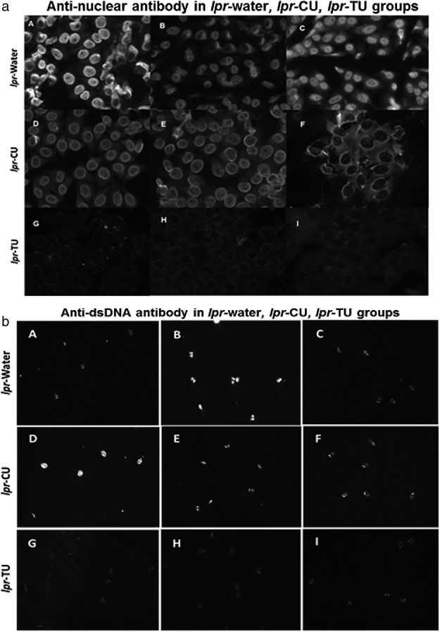
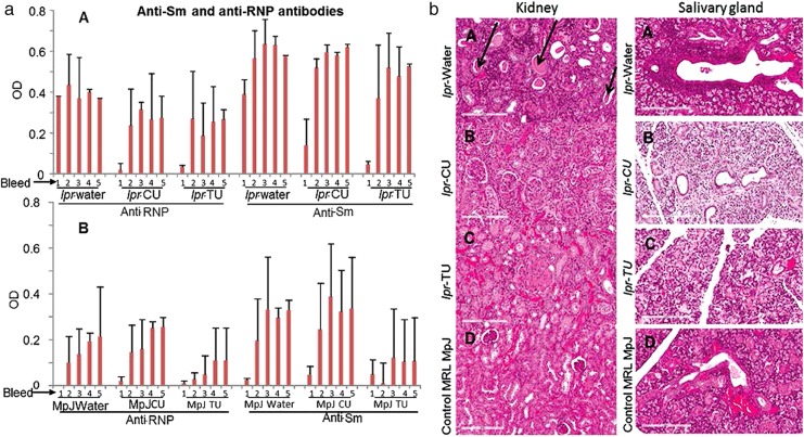
Results: UsC or UsT ameliorates SLE in the MRL-lpr/lpr mice by significantly reducing lymphoproliferation, proteinuria, lesions (tail) and autoantibodies. lpr-CU group had a 20% survival advantage over lpr-water group. However, lpr-TU group lived an average of 16 days shorter than lpr-water group due to complications unrelated to lupus-like illness. CU/TU treatment inhibited lymphadenopathy significantly compared with lpr-water group (p=0.03 and p=0.02, respectively) by induction of apoptosis. Average lymph node weights were 2606±1147, 742±331 and 385±68 mg, respectively, for lpr-water, lpr-CU and lpr-TU mice. Transferase dUTP nick end labelling assay showed that lymphocytes in lymph nodes of lpr-CU and lpr-TU mice underwent apoptosis. Significantly reduced cellular infiltration of the salivary glands in the lpr-TU group compared with the lpr-water group, and a trend towards reduced kidney damage was observed in the lpr-CU and lpr-TU groups.

Conclusions: These studies show that UsC/UsT could prove useful as a therapeutic intervention in SLE/SS.

Keywords: Autoantibodies; Autoimmunity; Sjøgren's Syndrome; Systemic Lupus Erythematosus; Treatment.


Figures

Figure 1
Figure 1
Lymphadenopathy (at week 14) in the lpr-water, lpr-CU and lpr-TU groups of mice and ultrasoluble curcumin (UsC) uptake by Epstein–Barr virus (EBV) transformed human PBMCs. (a, A–C) EBV-transformed human PBMCs incubated with UsC for 1 min; (a, D–F) EBV-transformed human PBMCs incubated with UsC for 2 min; (a, G–I) EBV-transformed human PBMCs incubated with UsC for 4 min; (A, D, G) fluorescence; (B, E, H) transmitted light overlay; (C, F, I) merged image (fluorescence+transmitted light overlay); (b) lymphadenopathy in the lpr-water group (A–D), lpr-CU group (E–H) and lpr-TU group (I–L). Arrows point to enlarged lymph nodes; (c) enlarged image of UsC uptake by EBV-transformed human PBMCs. (Left panel) fluorescence; (middle panel) transmitted light overlay; (right panel) merged image (fluorescence+transmitted light overlay). CU, curcumin; TU, turmeric.
Figure 2
Figure 2
Urinary cell casts, lymph node weights and proteinuria in the lpr-water, lpr-CU and lpr-TU groups of mice. (a) Urinary cell casts in the lpr-water group. (A–D) Red or white cell casts in four individual mice belonging to the lpr-water group at week 14. None of the mice in the lpr-CU or lpr-TU group had urinary cell casts at week 14; (bA) average lymph node weights in the lpr-water, lpr-CU and lpr-TU groups of mice. Average lymph node weights: lpr-water group= 2606 mg; lpr-CU= 743 mg; lpr-TU=385 mg. *p=0.03, lpr-water group versus lpr-CU group; **p=0.02, lpr-water group versus lpr-TU group (Mann–Whitney U test). Values are given as means±SE for each group. Statistical analyses were done by Mann–Whitney U test and the p-value was arrived at to assess the statistical significance of the changes observed. p<0.05 was considered to be significant; (bB) Weight of lymph nodes at death for the lpr-water, lpr-CU and lpr-TU groups of mice; (c) proteinuria in the MRL-lpr/lpr and MRL-MpJ mice provided with water, CU or TU analysed by sodium dodecyl sulfate polyacrylamide gel electrophoresis (SDS PAGE) (third urine collection; week 11). In total, 20 µL urine from each mouse was electrophoresed on 4–20% SDS-PAGE gels and stained with Coomassie brilliant blue. Lanes 1–6: MpJ-water group; lanes 7–12: MpJ-CU group; lanes 13–18: MpJ-TU group; lanes 19–24: lpr-water group; lanes 25–29: lpr-CU group; lanes 31–36: lpr-TU group. Increased protein excretion can be seen in the lpr-water group compared with the lpr-CU, lpr-TU and control MpJ mice. CU, curcumin; TU, turmeric.
Figure 3
Figure 3
Antinuclear antibody (ANA) patterns (using HEp-2 ANA assay kit) and anti-dsDNA (analysed by Crithidia luciliae assay) in sera of the lpr-water, lpr-CU and lpr-TU groups of mice. (a) ANA patterns observed in sera (third bleed) (1:80 dilution) of the lpr-water group (A–C), lpr-CU group (D–F) and lpr-TU group (G–I); (b) anti-dsDNA observed in sera (first bleed) (1:100 dilution) of the lpr-water group (A–C), lpr-CU group (D–F) and lpr-TU group (G–I). Only results obtained from three animals are shown for each experimental group. CU, curcumin; TU, turmeric.
Figure 4
Figure 4
Anti-RNP/anti-Sm antibodies in sera (bleeds 1–5) and H&E stained sections of kidney/salivary gland of the MRL-lpr/lpr and MRL-MpJ mice provided with water, CU or TU. (aA) Anti-RNP and anti-Sm in sera of the lpr-water, lpr-CU and lpr-TU groups of mice. (aB) Anti-RNP and anti-Sm in sera of the MpJ-water, MpJ-CU and MpJ-TU groups of mice. Values for each bleed were normalised to an anti-Sm or anti-RNP-positive control. Values are given as means±SD for each group. Statistical analyses were done by Student's t test and the p-value was arrived at to assess the statistical significance of the changes observed. p<0.05 or lower was considered to be significant; (b) kidney and salivary gland histopathology. (A) lpr-water, (B) lpr-CU (C) lpr-TU and (D) control MpJ mice. Protein casts in kidney and cellular infiltrates in salivary gland are indicated by arrows. CU, curcumin; RNP, ribonucleoprotein; TU, turmeric.
Figure 5
Figure 5
Lesions, survival and cell apoptosis in the lpr-water, lpr-CU and lpr-TU groups of mice. (a) Lesions in the lpr-water (A–C), lpr-CU (D and E) and lpr-TU (F) groups. Circumferential erythematous lesions, unrelated to tail vein bleeding, seen in the lpr-water group. Arrows point to cuts made for tail bleeding. (b) Survival curve for the lpr-water, lpr-CU and lpr-TU groups of mice. There was no difference in the survival between the MRL-MpJ mice (used as control mice for the MRL-lpr/lpr mice) fed with water, curcumin or turmeric. The control MpJ mice provided water (MpJ-water), curcumin (MpJ-CU) or turmeric (MpJ-TU) were sacrificed at 260 days. (c) Terminal deoxynucleotidyl transferase dUTP nick end labelling (TUNEL) of axillary lymph node section of the lpr-TU group mouse. Apoptotic cells are indicated by dUTP incorporation, shown by FITC labelling. (cA) Overlay of 4′,6-diamidino-2-phenylindole (DAPI) and FITC staining in the absence of terminal deoxynucleotidyl transferase. (cB) Overlay of DAPI and FITC staining in the presence of the enzyme. CU, curcumin; TU, turmeric.

References

    1. Arbuckle MR, McClain MT, Rubertone MV et al. Development of autoantibodies before the clinical onset of systemic lupus erythematosus. N Engl J Med 2003;349:1526–33. doi:10.1056/NEJMoa021933 - DOI - PubMed
    1. Alarcon-Segovia D. The pathogenesis of immune dysregulation in systemic lupus erythematosus. A troika. J Rheumatol 1984;11:588–90. - PubMed
    1. Danchenko N, Satia JA, Anthony MS. Epidemiology of systemic lupus erythematosus: a comparison of worldwide disease burden. Lupus 2006;15:308–18. doi:10.1191/0961203306lu2305xx - DOI - PubMed
    1. Ainiala H, Dastidar P, Loukkola J et al. Cerebral MRI abnormalities and their association with neuropsychiatric manifestations in SLE: a population-based study. Scand J Rheumatol 2005;34:376–82. doi:10.1080/03009740510026643 - DOI - PubMed
    1. Aisen AM, Gabrielsen TO, McCune WJ. MR imaging of systemic lupus erythematosus involving the brain. AJR Am J Roentgenol 1985;144:1027–31. doi:10.2214/ajr.144.5.1027 - DOI - PubMed

LinkOut - more resources